Online Gather.town Pitches
Genitourinary & Women's Imaging III
Joint Annual Meeting ISMRM-ESMRMB & ISMRT 31st Annual Meeting • 07-12 May 2022 • London, UK

Online Gather.town Pitches
Genitourinary & Women's Imaging III
| Booth # | ||||
|---|---|---|---|---|
 |
4007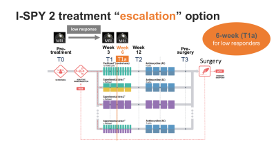 |
1 | Early treatment re-direction based on functional tumor volume at 3 and 6-week MRI for breast cancer patients undergoing neoadjuvant chemotherapy
Natsuko Onishi1, Jessica Gibbs1, Wen Li1, David C. Newitt1, Elissa R. Price1, Barbara LeStage2, William F. Symmans3, Angela DeMichele4, Christina Yau5, The I-SPY 2 Imaging Working Group6, The I-SPY 2 Consortium6, Laura J. Esserman5, and Nola M. Hylton1
1Department of Radiology & Biomedical Imaging, University of California, San Francisco, San Francisco, CA, United States, 2I-SPY 2 Advocacy Group, San Francisco, CA, United States, 3MD Anderson Cancer Center, Houston, TX, United States, 4University of Pennsylvania, Philadelphia, PA, United States, 5Department of Surgery, University of California, San Francisco, San Francisco, CA, United States, 6Quantum Leap Healthcare Collaborative, San Francisco, CA, United States
In the I-SPY2 breast cancer trial, functional tumor volume (FTV) derived from dynamic contrast-enhanced MRI is used as a longitudinal measure of response to adjust patient randomization and evaluate drug efficacy. I-SPY2 is introducing a treatment “escalation” option, giving patients the opportunity to re-direct to potentially more effective treatment based on early indication of inferior response by MRI. We retrospectively investigated the ability of FTV reduction to predict inferior outcome. Combined criteria using FTV reduction at 3 and 6 weeks of treatment showed high PPV and high sensitivity in early detection of non-responders.
|
|
4008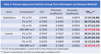 |
2 | Histological validation of prostate tissue composition measurement using Hybrid Multidimensional MRI: Agreement with pathologists’ measures
Aritrick Chatterjee1,2, Tatjana Antic3, Alexander J Gallan4, Gladell P Paner3, Lawrence I-Kuei Lin5, Gregory S Karczmar1,2, and Aytekin Oto1,2
1Department of Radiology, University of Chicago, Chicago, IL, United States, 2Sanford J. Grossman Center of Excellence in Prostate Imaging and Image Guided Therapy, Chicago, IL, United States, 3Department of Pathology, University of Chicago, Chicago, IL, United States, 4Department of Pathology, Medical College of Wisconsin, Medical College of Wisconsin, WI, United States, 5JBS Consulting Services Inc., Carlsbad, CA, United States
We validated prostate tissue composition measured using HM-MRI by comparing with reference standard results from pathologists’ interpretation of clinical histopathology slides following whole mount prostatectomy. We are 95% confident that 90% of absolute paired differences (TDI0.9) between HM-MRI and consensus results of pathologists were within 20.6% and 24.2% in measuring epithelium and lumen fractional volumes, respectively. These were less than our criterion of 30% and inter-pathologists’ agreement (22.3% for epithelium and 24.2% for lumen). Therefore, we accept the agreement performance of HM-MRI in measuring tissue composition measurement and consensus of pathologists is on par with the inter-raters (pathologists) agreement.
|
||
|
4009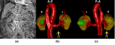 |
3 | Renal mass assessment using Ultrashort Time-to-Echo sequences with ferumoxytol
Tianyi Zhou1, Liam Timms1, Aya Hamadeh2, David Drew3, Mukesh Harisinghani2, and Srinivas Sridhar1
1Department of Physics, Northeastern University, Boston, MA, United States, 2Department of Radiology, Massachusetts General Hospital, Boston, MA, United States, 3Tufts Medical Center, Boston, MA, United States A ferumoxytol-enhanced MRA technique using Ultrashort Time-to-Echo (UTE) sequences is demonstrated for kidney vasculature mapping and renal mass assessment. The blood volume (BV) map is calculated by scaling the post- and pre-contrast subtraction image from 0 to 1. BV is measured in one cystic mass (-6±7)% and one complex mass (17±8)% with sub-regions BV up to (32±10)% and (30±7)%. The quantification of BV provides a measurement of the distribution of micro-vessel density, reflecting the vascularity of the kidneys and masses. This technique potentially benefits surgical planning, renal mass characterization, and disease progression in patients with impaired renal function. |
|
4010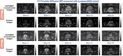 |
4 | Improving Ultra-High b-value Prostate Diffusion Image Reconstruction at 3T
Neha Koonjoo1, Bo Zhu1,2, Danyal Bhutto1,2,3, Arnaud Guidon4, Matthew Christensen1,2, Mukesh Harisinghani1, and Matthew S Rosen1,2,5
1Department of Radiology, A.A Martinos Center for Biomedical Imaging / MGH, Charlestown, MA, United States, 2Harvard Medical School, Boston, MA, United States, 3Department of Biomedical Engineering, Boston University, Boston, MA, United States, 4GE Healthcare, Boston, MA, United States, 5Department of Physics, Harvard University, Cambridge, MA, United States
Ultra-high b-value Diffusion-weighted Prostate MR is gaining more attention in medical imaging, due to the non-invasiveness of imaging and to better contrast of malignant tissues as the lower diffusivity of water molecules can enable early diagnosis of cancer. The main drawback of MR at ultra-high b-value is the poor resultant SNR of the reconstructed images. We propose to improve the image quality of the prostate data acquired at b=2000 s/mm2 using a machine learning based reconstruction approach. Significant increase in signal intensities in the central gland and peripheral zone of the prostate was observed in healthy subjects.
|
||
4011 |
5 | Quantification of bilateral whole-organ renal metabolic rate of O2 by exploiting conservation of flow and mass principle: a preliminary study
Rajiv S Deshpande1,2, Michael C Langham2, and Felix W Wehrli2
1Dept. of Bioengineering, University of Pennsylvania, Philadelphia, PA, United States, 2Dept. of Radiology, University of Pennsylvania, Philadelphia, PA, United States
Type 2 diabetes mellitus is the leading cause of chronic kidney disease. Clinical measures of kidney function often detect dysfunction after irreversible damage has occurred. This highlights an unmet need for a biomarker that can enable earlier detection of disease. Quantification of renal metabolic rate of oxygen is a promising metric because it increases by 40-65% during the early stages of diabetic kidney disease. Here, we propose a “difference method” to quantify bilateral renal metabolic rate of oxygen by imaging above and below the renal vessels and exploiting conservation of blood flow rate and mass.
|
||
4012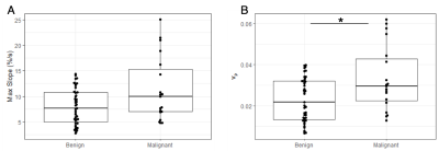 |
6 | Quantitative analysis of ultrafast DCE-MRI to distinguish benign and malignant breast lesions in a clinical setting
Anum S. Kazerouni1, Yun An Chen1, Callie Lind2, Inyoung Youn3, Savannah C. Partridge1, and Habib Rahbar1
1Radiology, University of Washington, Seattle, WA, United States, 2Bioengineering, University of Washington, Seattle, WA, United States, 3Radiology, Sungkyunkwan University School of Medicine, Seoul, Korea, Republic of
We investigate the ability of quantitative measures derived from ultrafast DCE-MRI to improve breast MRI specificity for lesion diagnosis in a clinical setting. Sixty-nine women with BI-RADS 3, 4, or 5 lesions identified on clinical breast MRI exam, with subsequent diagnostic biopsy, were retrospectively identified. Ultrafast DCE-MRI was used to calculate semi-quantitative (bolus arrival time, max slope) and quantitative (Ktrans, vp) features for each lesion visible on the ultrafast series. Malignant lesions showed significantly higher vp compared to benign lesions. Combined with qualitative assessment of visibility on ultrafast DCE-MRI (“yes”/“no”), vp showed an AUC=0.73 for lesion diagnosis.
|
||
4013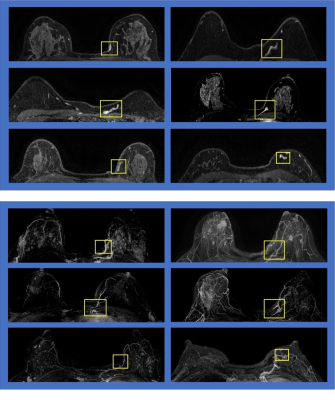 |
7 | Evaluation of the Combined Deep Learning Networks Using Mask R-CNN and ResNet50 Classification for Detection and Diagnosis of Breast Cancer on MRI
Yang Zhang1,2, Yan-Lin Liu1, Ke Nie2, Jiejie Zhou3, Zhongwei Chen3, Jeon-Hor Chen1, Meihao Wang3, and Min-Ying Su1
1Department of Radiological Sciences, University of California, Irvine, CA, United States, 2Department of Radiation Oncology, Rutgers-Cancer Institute of New Jersey, Robert Wood Johnson Medical School, New Brunswick, NJ, United States, 3Department of Radiology, The First Affiliated Hospital of Wenzhou Medical University, Wenzhou, China
We developed two deep learning methods for breast MRI evaluation, first using Mask R-CNN for detection of suspicious areas, and then using ResNet50 for estimating the malignancy probability. These two networks were combined to test its diagnostic validity in two datasets. In Dataset-1, sensitivity=96.1% and specificity=78.1%. In Dataset-2, sensitivity=81.1% and specificity= 80.6%. We further characterized all false positives (FPs), and found other than confirmed benign lesions, FPs may come from vessels and asymmetric parenchymal enhancements, which can be further eliminated by other algorithms. The results suggest the potential of combined deep learning networks as a fully-automatic breast MRI CAD.
|
||
4014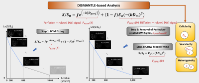 |
8 | Integrated DWI for Pre-treatment Prediction of Response to Neoadjuvant Chemotherapy in Breast Cancer
Muge Karaman1,2, Shunan Che3, Guangyu Dan1,2, Zheng Zhong1,2, Han Ouyang3, Xiaohong Joe Zhou1,2,4, and Xinming Zhao3
1Center for MR Research, University of Illinois at Chicago, Chicago, IL, United States, 2Department of Bioengineering, University of Illinois at Chicago, Chicago, IL, United States, 3Department of Radiology, Cancer Hospital, Chinese Academy of Medical Sciences, Beijing, China, 4Departments of Radiology and Neurosurgery, University of Illinois at Chicago, Chicago, IL, United States
Neoadjuvant chemotherapy (NAC) has been shown to improve the outcome in patients with locally advanced inoperable or operable breast cancer. An early imaging assessment of response to NAC is critical for managing breast cancer to minimize the toxic side effects of ineffective chemotherapy. In this study, we have used an integrated diffusion-weighted imaging approach for simultaneous assessment of tissue cellularity, vascularity, and heterogeneity – DISMANTLE – from a full b-value spectrum to predict breast cancer’s response to NAC. Histogram features of pre-treatment DISMANTLE parameters were evaluated for predicting tumor response to NAC in terms of pathological complete response.
|
||
4015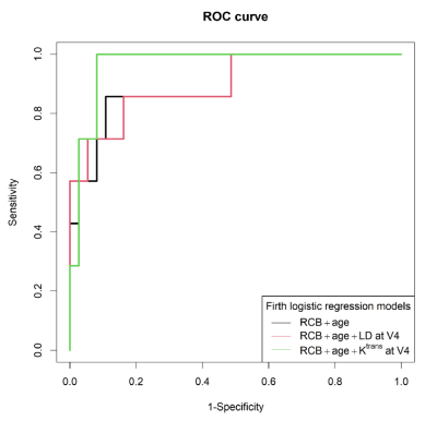 |
9 | Quantitative DCE-MRI Prediction of Breast Cancer Recurrence Following Neoadjuvant Chemotherapy
Rajat Thawani1, Lina Gao2, Ajay Mohinani2, Alina Tudorica2, Xin Li2, Zahi Mitri1, and Wei Huang2
1Division of Hematology and Oncology, Oregon Health and Science University, Portland, OR, United States, 2Oregon Health and Science University, Portland, OR, United States
Neoadjuvant chemotherapy (NAC) is considered standard of care for locally advanced breast cancer. Pre- and post-NAC MRI is routinely used to assess response. This study aims to investigate pre- and post-NAC quantitative DCE-MRI parameters, alone and in combination with clinico-pathologic variables, for prediction of breast cancer recurrence following NAC. 47 patients underwent DCE-MRI studies pre- and post-NAC. The results show that quantitative pharmacokinetic DCE-MRI parameters, whether alone or in combination with clinico-pathologic variables, outperformed tumor size measurement by conventional imaging in prediction of recurrence. Furthermore, DCE-MRI parameters provided added value in predictive performance when combined with clinico-pathologic variables.
|
||
4016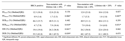 |
10 | A Propensity Score Matched Analysis of Quantitative Background Parenchymal Enhancement on MRI for Improved Breast Cancer Risk Stratification
Ran Yan1,2, Wakana Murakami1, Shabnam Mortazavi1, Tiffany Yu1, Stephanie Lee-Felker1, and Kyunghyun Sung1,3
1Department of Radiological Sciences, David Geffen School of Medicine, University of California, Los Angeles, Los Angeles, CA, United States, 2Department of Bioengineering, University of California, Los Angeles, Los Angeles, CA, United States, 3Physics and Biology in Medicine IDP, David Geffen School of Medicine, University of California, Los Angeles, Los Angeles, CA, United States
High background parenchymal enhancement (BPE) on breast MRI is related to increased breast cancer risk. In this study, we investigate possible differences in BPE among different breast cancer risk groups stratified based on BRCA 1/2 mutation status and lifetime risks. We use propensity score matching to adjust for potential confounders. We performed a quantitative BPE analysis on the 3D volume of the breast and fibroglandular tissue based on the enhancement maps and principal component analysis. The results showed a significant difference in BPE among different risk groups, indicating that BPE may improve breast cancer risk stratification.
|
||
4017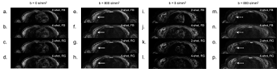 |
11 | Multi-Shot Diffusion-Weighted Imaging of the Breast in the Supine Position
Catherine J. Moran1, Matthew J. Middione1, Valentina Mazzoli1, Jessica McKay-Nault1, Arnaud Guidon2, Uzma Waheed1, Eric L. Rosen1, Steven P. Poplack1, Jarrett Rosenberg1, Daniel B Ennis1, Brian A. Hargreaves1, and Bruce L. Daniel1
1Radiology, Stanford University, Stanford, CA, United States, 2General Electric Healthcare, Botson, MA, United States Diffusion-Weighted Imaging (DWI) in the supine position may provide a non-invasive, comfortable, and efficient MRI for breast cancer screening. The strong performance of multi-shot DWI in prone breast MRI has been established. However, supine breast MRI has increased motion, specific B0-inhomogeneity, and cardiac artifacts, all of which may affect multi-shot DWI. This work investigates supine multi-shot DWI of the breast with a flexible array coil. High image quality is achieved and the effects of number of shots and respiratory-gating are delineated. B0-inhomogeneity and ADCs in the supine position are reported including the potential effect of cardiac artifact on ADC measurement. |
||
4018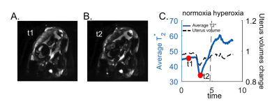 |
12 | Change in T2* measurements of placenta during Braxton Hicks contractions
Esra Abaci Turk1,2, Jeffrey N. Stout1, Borjan Gagoski1,2, Mary Katherine Manhard3, Elfar Adalsteinsson4,5,6, Polina Golland4,7, Drucilla J. Roberts8, William H. Barth Jr9, and P. Ellen Grant1,2
1Fetal‐Neonatal Neuroimaging & Developmental Science Center, Boston Children's Hospital, Boston, MA, United States, 2Harvard Medical School, Boston, MA, United States, 3Department of Radiology, Cincinnati Children's Hospital Medical Center, Cincinnati, OH, United States, 4Department of Electrical Engineering and Computer Science, Massachusetts Institute of Technology, Cambridge, MA, United States, 5Harvard-MIT Health Sciences and Technology, Massachusetts Institute of Technology, Cambridge, MA, United States, 6Institute for Medical Engineering and Science, Massachusetts Institute of Technology, Cambridge, MA, United States, 7Computer Science and Artificial Intelligence Laboratory (CSAIL), Massachusetts Institute of Technology, Cambridge, MA, United States, 8Department of Pathology, Massachusetts General Hospital, Boston, MA, United States, 9Maternal-Fetal Medicine, Obstetrics and Gynecology, Massachusetts General Hospital, Boston, MA, United States
Maternal-placental perfusion can be temporarily compromised by Braxton Hicks uterine contractions. The effect of Braxton Hicks contractions on placental function has not been well characterized or understood. In this study, we investigated the effect of Braxton Hicks contractions on placental T2* values across gestation together with the outcome measures. We observed positive correlation between the change in T2* during contraction, gestational age at delivery, and fetal birth weight. Additionally we observed differences in the placental T2* response in the regions closer to the maternal surface compared to regions closer to the fetal surface.
|
||
4019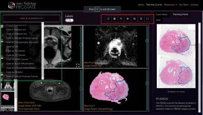 |
13 | An Interactive App with Multi-parametric MRI - Whole Mount Histology Correlation for Enhanced Prostate MRI Training of Radiologists
Aritrick Chatterjee1,2, Teodora Szasz3, Milson Munakami3, Ibrahim Karademir1, Mohamed Shaif Yusufishaq1, Spencer Martens1, Christina Wheeler4, Stephen Thomas1, Gregory S Karczmar1,2, and Aytekin Oto1,2
1Department of Radiology, University of Chicago, Chicago, IL, United States, 2Sanford J. Grossman Center of Excellence in Prostate Imaging and Image Guided Therapy, Chicago, IL, United States, 3Research Computing Center, University of Chicago, Chicago, IL, United States, 4C. Wheeler Studios, Chicago, IL, United States
We created an interactive application Learn Radiology with multi-parametric MRI - whole mount histology correlation for enhanced prostate MRI training of radiologists and validated whether the use of a newly created learning application can enhance prostate MRI training of radiologists using an observer study. Sensitivity (R1: 54%→64%, R2: 44%→59%, R3: 62%→72%), PPV (R1: 68%→76%, R2: 52%→79%, R3: 48%→65%) and confidence score (R1: 4.0±1.0→4.3±0.8, R2: 3.1±0.8→4.0±1.1, R3: 2.8±1.2→4.1±1.1, p<0.05) for prostate cancer diagnosis using mpMRI improved for all three radiologists along with inter-observer agreement (α 0.78→0.85) after being exposed to our developed teaching app.
|
||
 |
4020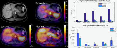 |
14 | Whole-Abdomen Metabolic Imaging in Healthy Volunteers Using Hyperpolarized [1-13C]pyruvate MRI
Philip Meng-en Lee1, Hsin-Yu Chen1, Jeremy W Gordon1, Zhen J Wang1, Robert A Bok1, Kiersten Cheung1, Francesca De Las Alas1, Heather Daniel1, Peder EZ Larson1, Cornelius von Morze2, Daniel B Vigneron1, and Michael A Ohliger1
1Radiology & Biomedical Imaging, University of California, San Francisco, San Francisco, CA, United States, 2Radiology, Washington University in St. Louis, St. Louis, MO, United States Whole-abdomen imaging with hyperpolarized 13C is challenging due to B0 and B1 inhomogeneities, respiratory motion, and broad spatial coverage. There is also little baseline data about healthy metabolism in abdominal organs. We develop and describe a reliable imaging method to overcome these challenges, enabling metabolic imaging of the entire abdomen in a series of healthy volunteers. We establish normal values for conversion of HP [1-13C]pyruvate to lactate and alanine in key organs such as the liver, kidneys, pancreas, and spleen. Methods established here set a firm foundation for investigating a broad spectrum of metabolic and neoplastic abnormalities in the liver. |
|
4021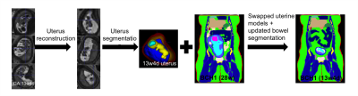 |
15 | Assessment of SAR variability across early gestational age subjects at 3T fetal MRI
Esra Abaci Turk1,2, Filiz Yetisir1, Cindy Zhou1, Jeffrey N. Stout1, Judy A. Estroff3,4, Carol Barnewolt3,4, Elfar Adalsteinsson5,6,7, P. Ellen Grant1,2, and Lawrence L. Wald2,6,8
1Fetal‐Neonatal Neuroimaging & Developmental Science Center, Boston Children's Hospital, Boston, MA, United States, 2Harvard Medical School, Boston, MA, United States, 3Department of Radiology, Boston Children’s Hospital, Boston, MA, United States, 4Maternal Fetal Care Center, Boston Children’s Hospital, Boston, MA, United States, 5Department of Electrical Engineering and Computer Science, Massachusetts Institute of Technology, Cambridge, MA, United States, 6Harvard-MIT Health Sciences and Technology, Massachusetts Institute of Technology, Cambridge, MA, United States, 7Institute for Medical Engineering and Science, Massachusetts Institute of Technology, Cambridge, MA, United States, 8Athinoula A. Martinos Center for Biomedical Imaging, Massachusetts General Hospital, Charlestown, MA, United States
Despite the clinical and research potential of fetal MRI, there is no consensus on MRI safety in the 1st and early 2nd trimesters. Prior simulation studies at early gestational ages (GA) used only a single or unrealistic pregnant body models. To address this gap, we built 2 realistic uterine models for the early 2nd trimester and placed them into 3 existing pregnant body models at a later GA to investigate differences in RF exposure. Lower pSAR10g in both the fetus and amniotic fluid was observed in the early 2nd trimester models compared to later GA models.
|
||
 Back to Meeting Home
 Back to the Program-at-a-Glance
The International Society for Magnetic Resonance in Medicine is accredited by the Accreditation Council for Continuing Medical Education to provide continuing medical education for physicians.
 Back to Meeting Home
 Back to the Program-at-a-Glance
 View the Presentation