Online Gather.town Pitches
Pediatrics, Normal Development & Aging II
Joint Annual Meeting ISMRM-ESMRMB & ISMRT 31st Annual Meeting • 07-12 May 2022 • London, UK

Online Gather.town Pitches
Pediatrics, Normal Development & Aging II
| Booth # | ||||
|---|---|---|---|---|
4029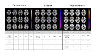 |
1 | Sex-differences in resting-state brain activity in children with concussion in comparison to matched healthy controls.
Bhanu Sharma1,2,3, Carol DeMatteo4,5, Michael D Noseworthy1,3,6,7, and Brian W Timmons2,5
1Department of Electrical and Computer Engineering, McMaster University, Hamilton, ON, Canada, 2Child Health and Exercise Medicine Program, Department of Pediatrics, McMaster University, Hamilton, ON, Canada, 3Imaging Research Centre, St. Joseph's Healthcare, Hamilton, ON, Canada, 4School of Rehabilitation Science, McMaster University, Hamilton, ON, Canada, 5CanChild Centre for Childhood Disability Research, McMaster University, Hamilton, ON, Canada, 6School of Biomedical Engineering, McMaster University, Hamilton, ON, Canada, 7Department of Radiology, McMaster University, Hamilton, ON, Canada
There are known sex-differences with respect to the clinical presentation of pediatric concussion, with girls reporting more symptoms and symptoms with greater severity than boys. This is the first study to show that there are also sex-differences in resting state brain activity in children with concussion, suggesting that the sex-specific clinical presentation may have neurological underpinnings. Specifically, girls with concussion had resting state disturbances that were not present in boys, which is consistent with the variable symptom presentation of the injury. Continued research into these resting state differences is encouraged.
|
||
4030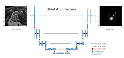 |
2 | Deep Learning Model for Automatic Fetal Brain Landmark Localization in MRI
Jie Deng1,2, Xuchu Liu2, Jubril O Adepoju2, and Sharon E Byrd2
1Radiation Oncology, UT Southwestern Medical Center, Dallas, TX, United States, 2Diagnostic Radiology and Nuclear Medicine, Rush University Medical Center, Chicago, IL, United States
Fetal MRI provides great anatomic details for diagnosis of perinatal disorders when ultrasound is inconclusive in assessing fetal anatomy during all phases of gestation. Immediate interventions can be attempted when abnormal anatomic changes of fetal brain structures are identified early during gestation. Size measurements of a fetal brain structure can be realized by identifying several landmarks of the structure accurately and calculating the distance between the landmarks. We developed a deep learning model that exploits U-net as a ‘transforming’ function to learn imaging features adjacent to a landmark point and predict the landmark location automatically.
|
||
4031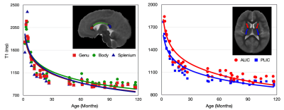 |
3 | Mapping Brain Development in Infants and Young Children Using MPnRAGE T1 Relaxometry
Douglas Dean1,2,3, Leela Shah3, Marissa DiPiero3,4, Colleen Pletcher3, Lauren Heinrich3, Elizabeth Planalp3, Andrew Alexander2,3,5, and Steven Kecskemeti3
1Pediatrics, University of Wisconsin–Madison, Madison, WI, United States, 2Medical Physics, University of Wisconsin–Madison, Madison, WI, United States, 3Waisman Center, University of Wisconsin–Madison, Madison, WI, United States, 4Neuroscience Training Program, University of Wisconsin–Madison, Madison, WI, United States, 5Psychiatry, University of Wisconsin–Madison, Madison, WI, United States
Quantitative magnetic resonance imaging during the first years of life can provide novel insights into brain maturation, yet acquisition of these data are limited by challenges in imaging pediatric populations. Here, we assessed the self-navigated, motion robust MPnRAGE technique to acquire high-resolution T1-weighted structural brain images and quantitative T1 relaxometry maps in infants and young children. Our results demonstrate the ability to utilize MPnRAGE as a robust quantitative technique to assess early brain development.
|
||
4032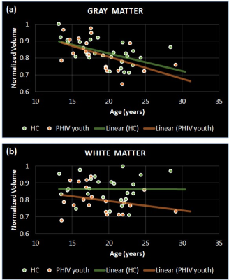 |
4 | Regional Brain Volumes, Cortical Thickness, Folding and Sulcal Depth in Perinatally HIV-Infected Youths
Ajin Joy1, Manoj K Sarma1, Jhelum Paul1, Andres Saucedo1, Paul M Macey2, Dieter J Meyerhoff3, Ebrahim Haroon4, Margaret A Keller5, and M Albert Thomas1
1Radiological Sciences, University of California-Los Angeles, Los Angeles, CA, United States, 2School of Nursing and Brain Research Institute, University of California-Los Angeles, Los Angeles, CA, United States, 3Radiology and Biomedical Imaging, University of California-San Francisco, San Francisco, CA, United States, 4Psychiatry and Behavioral Sciences, Emory University, Atlanta, GA, United States, 5Pediatrics, The Lundquist Institute, Harbor-UCLA Medical Center, Torrance, CA, United States
The human immunodeficiency virus (HIV) can affect the morphometry of the developing brain of perinatally HIV (PHIV)-infected youths. Surface-based morphometric analysis can reveal if local effects on gray and white matter volumes is significant in the regions with alterations in cortical thickness (CT), gyrification index (GI), or sulcal depth (SD). So, we compared these morphometric parameters of 19 PHIV-youths on combination antiretroviral therapy with those in 26 uninfected healthy controls. PHIV-youths had altered gray and white matter regional volume, CT, GI, and SD, partly in overlapping regions. The findings survived multiple comparisons using both false discovery rate and Holm-Bonferroni corrections.
|
||
4033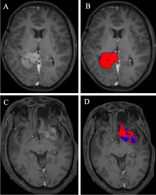 |
5 | Novel persistent tumor index to predict survival in pediatric high-grade gliomas treated with immunovirotherapy
Asim K Bag1, Joseph Holtrop1, Silu Zhang1, Matthew A Scoggins1, Yimei Li2, Tushar Patni2, Richard J Whitley3, G Yancey Gillespie4, James M Markert4, John B Fiveash5, Rong Li6, Joshua D Bernstock7,8,9, James Johnston4, and
Gregory K Friedman3,4
1Department of Diagnostic Imaging, St. Jude Children's Research Hospital, Memphis, TN, United States, 2Department of Biostatistics, St. Jude Children's Research Hospital, Memphis, TN, United States, 3Department of Pediatrics, University of Alabama at Birmingham, Birmingham, AL, United States, 4Department of Neurosurgery, University of Alabama at Birmingham, Birmingham, AL, United States, 5Department of Radiation Oncology, University of Alabama at Birmingham, Birmingham, AL, United States, 6Department of Pathology, University of Alabama at Birmingham, Birmingham, AL, United States, 7Department of Neurosurgery, Brigham and Women's Hospital, Boston, MA, United States, 8Department of Neurosurgery, Boston Children's Hospital, Boston, MA, United States, 9Department of Neurosurgery, Harvard Medical School, Boston, MA, United States
Early indicators of the response to imunovirotherapy are currently limited due to the novelty of the therapies and unpredictability of responses. We propose using the persistent tumor index (PTI) derived from diffusion and perfusion MRI as a predictor of treatment response. In 10 high grade glioma patients treated with imunovirotherapy, only the baseline PTI measure was associated with progression free survival whereas other known imaging biomarkers of survival were not.
|
||
4034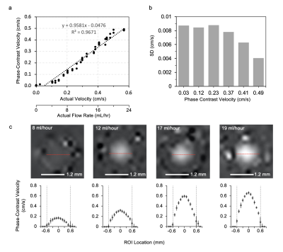 |
6 | Phase-Contrast MRI for Quantifying Cerebrospinal Flow in Patients with Shunted Hydrocephalus
Joseph Ha1, Eamon K Doyle, PhD2,3,4, Matthew T. Borzage, PhD3,5, and Peter A Chiarelli, MD, DPhil1,3,4
1Division of Neurosurgery, Children's Hospital of Los Angeles, Los Angeles, CA, United States, 2Radiology, Children's Hospital of Los Angeles, Los Angeles, CA, United States, 3Keck School of Medicine, University of Southern California, Los Angeles, CA, United States, 4Shared last author, Los Angeles, CA, United States, 5Fetal and Neonatal Institute, Division of Neonatology, Department of Pediatrics, Children's Hospital of Los Angeles, Los Angeles, CA, United States
We investigate a new approach to assess ventricular shunt function using phase-contrast magnetic resonance imaging (PC MRI). A flow phantom was constructed to assess CSF flows between 0-24 ml/hour. The PC MRI sequence showed strong flow correlation (R2 = .9671) even for flow velocities below 1 cm/s. The lowest non-zero CSF flow we measured was 1 ml/hour (0.02 cm/s), 67% lower than previously reported shunt flow measurements. The sequence was tested in a human subject after phantom calibration. With a 96 second duration, PC SHUNT shows promise as a practical means for determining shunt failure clinically.
|
||
4035 |
7 | Brain Iron Deposition in Healthy Aging and association with Alzheimer’s Disease Pathology Revealed by Quantitative Susceptibility Mapping Video Not Available
Qixiang Lin1, Junjie Wu2, Shuai Huang2, Aditya Bisht1, Allan Levey1,3, James Lah1,3, and Deqiang Qiu2,3,4
1Department of Neurology, School of Medicine, Emory University, Atlanta, GA, United States, 2Department of Radiology and Imaging Sciences, School of Medicine, Emory University, Atlanta, GA, United States, 3Goizueta Alzheimer’s Disease Research Center, Emory University, Atlanta, GA, United States, 4Joint Department of BioMedical Engineering, Emory University and Georgia Institute of Technology, Atlanta, GA, United States
This study aims to evaluate brain iron depositions in healthy aging and asymptomatic Alzheimer’s Disease using Quantitative Susceptibility Mapping (QSM) MR technique. 3D-GRE scans were performed in 146 healthy old and 58 healthy young participants. Whole-brain voxel-wise analysis showed increased susceptibility value in bilateral basal ganglia and widespread cortical regions. Significant correlations were found between CSF biomarkers of AD and susceptibility value in the caudate and left orbital frontal area. Together these results suggest microstructural alterations associated with healthy aging and AD-related CSF biomarkers. QSM measurements might provide sensitive neuroimaging biomarkers for iron deposition during normal aging and neurodegeneration diseases.
|
||
4036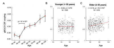 |
8 | Gender-specific age-related changes in glymphatic function as measured by resting-state fMRI
Feng Han1, Yifan Yang1, and Xiao Liu1
1the Pennsylvania State University, State College, PA, United States
The glymphatic system responsible for brain waste clearance may play an important role in aging and dementias. However, a lack of non-invasive tools for gauging glymphatic function in human subjects hindered the research on the age-related glymphatic changes. The global component of low-frequency (<0.1 Hz) resting-state fMRI signals was recently found coupled with CSF flow, and their coupling was used for quantifying the glymphatic function and found reduced significantly in neurodegenerative patients. Here we used the gBOLD-CSF coupling to evaluate glymphatic function change along healthy aging, and found it starts to decrease around age 55, particularly in females.
|
||
4037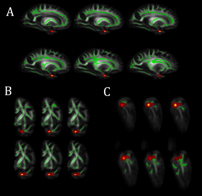 |
9 | Limbic Predominant Age-related TDP-43 Encephalopathy Neuropathological Change (LATE-NC) is Associated with Lower Diffusion Anisotropy
Mahir Tazwar1, Arnold M. Evia2, Ashish A. Tamhane2, Abdur Raquib Ridwan1, David A. Bennett2, Julie A. Schneider2, and Konstantinos Arfanakis1,2
1Department of Biomedical Engineering, Illinois Institute of Technology, Chicago, IL, United States, 2Rush Alzheimer's Disease Center, Rush University Medical Center, Chicago, IL, United States
The MRI signature of limbic predominant age-related TDP-43 encephalopathy neuropathological change (LATE-NC) has not been fully determined. In this work, we investigated the association of diffusion anisotropy with LATE-NC in autopsied brains of community-based older adults (N=148). Voxel-wise analysis revealed an independent association of lower fractional anisotropy (FA) with greater LATE-NC burden in medial temporal lobe white matter, controlling for other neuropathologies. The white matter connections implicated include fibers connecting cortical and subcortical regions where LATE-NC is typically found. Comparison of FA values across LATE-NC stages revealed that FA may allow detection of LATE-NC in later stages of the disease.
|
||
4038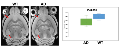 |
10 | Diffusion MRI in a transgenic mouse model of Alzheimer's disease Video Permission Withheld
Surendra Maharjan1, Kaitlyn Marie Dybing1, and Nian Wang1
1Department of Radiology and Imaging Sciences, Indiana University School of Medicine, Indianapolis, IN, United States
Alzheimer’s disease (AD) is common neurodegenerative dementia. Though the disease has no cure, early intervention can help to maintain normal mental function and slow down the progression of the disease. Brain imaging, especially Magnetic Resonance Imaging (MRI) has been extensively used to characterize the location and progression of the disease. Diffusion MRI is a novel imaging technique that exploits the microscopic mobility of water molecules. However, a comprehensive study incorporating high-resolution diffusion tensor imaging (DTI), diffusion kurtosis imaging (DKI), and Neurite Orientation Dispersion and Density Imaging (NODDI) is still lacking. So, we aim to apply DTI, DKI and NODDI.
|
||
4039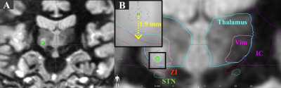 |
11 | Is the tremor relief in Parkinson’s disease ancillary benefits during the ablation of thalamic Vim using MR guided focused ultrasound (MRgFUS)?
Hongchae Baek1,2, Sean Nagel2, Daniel Lockwood1, Emmanuel C. Obusez1, and Stephen E. Jones1
1Imaging Institute, Cleveland Clinic, Cleveland, OH, United States, 2Center for Neurological Restoration, Cleveland Clinic, Cleveland, OH, United States Essential tremor (ET) and tremor dominant Parkinson’s disease (PD) treatment using MRgFUS share the same therapeutic target and the thermal dose threshold to create a thalamic lesion at ViM. However, PD patients respond to treatment typically only during the last few sonications, whereas ET patients respond earlier, typically within the first few sonications. Furthermore, we confirmed that zona incerta (ZI) is commonly affected due to proximity to ViM, by using postoperative T2-weighted images fused with auto-segmented ViM and ZI, demonstrating a possible causal relationship between the lesion overlap with ZI that primarily reduces tremor reduction in PD patients. |
||
4040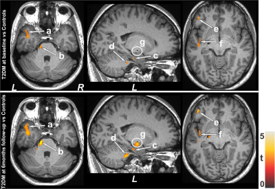 |
12 | Non-Gaussian Brain Diffusion Cross-sectional and Longitudinal Changes in Type 2 Diabetes Mellitus Adults
Bhaswati Roy1, Daisy Mercado2, Matthew J Freeby3, Sarah Choi2, and Rajesh Kumar1,4,5,6
1Anesthesiology, University of California Los Angeles, Los Angeles, CA, United States, 2UCLA School of Nursing, Los Angeles, CA, United States, 3Medicine, University of California Los Angeles, Los Angeles, CA, United States, 4Radiological Sciences, University of California Los Angeles, Los Angeles, CA, United States, 5Bioengineering, University of California Los Angeles, Los Angeles, CA, United States, 6Brain Research Institute, University of California Los Angeles, Los Angeles, CA, United States
Patients with Type 2 diabetes mellitus (T2DM) show brain changes in mood and cognitive control sites, functions that are deficient in the condition, examined by Gaussian diffusion-based diffusion tensor imaging (DTI). However, the majority of brain areas with complex fibers, follow non-Gaussian diffusion, and DTI-based measures may not show adequate diffusion changes. We examined brain changes at baseline and 6 months in T2DM patients using the diffusion kurtosis imaging-based mean kurtosis procedures and showed wide-spread acute and chronic tissue changes in the condition, with continued progression after 6 months in areas involved in mood and cognitive regulatory functions.
|
||
4041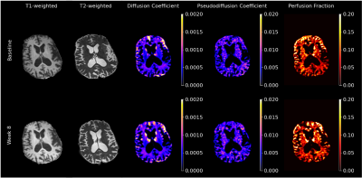 |
13 | Preliminary Evaluation of Risperidone in Huntington’s Disease: An Intravoxel Incoherent Motion (IVIM) Study
Alan Finkelstein1, Md Nasir Uddin2, Jianhui Zhong1,3,4, Ruth Schneider2, and Giovanni Schifitto2,3
1Biomedical Engineering, University of Rochester, Rochester, NY, United States, 2Neurology, University of Rochester, Rochester, NY, United States, 3Imaging Sciences, University of Rochester, Rochester, NY, United States, 4Physics and Astronomy, University of Rochester, Rochester, NY, United States
Huntington’s disease (HD) is a neurodegenerative movement disorder that negatively impacts quality of life. Atypical antipsychotics are used for treatment, though with limited evidence from controlled trials. In this preliminary study we investigated the effects of risperidone, an atypical antipsychotic for the treatment of chorea in HD using intravoxel incoherent motion diffusion weighted imaging (IVIM-DWI) and volumetric analysis. Preliminary findings suggest that early effects of risperidone may present as changes in the microcirculation within the basal ganglia prior to any anatomical changes. However, further evaluation in a larger sample size is needed to better assess the clinical response to risperidone.
|
||
4042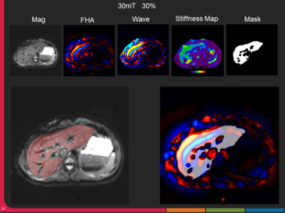 |
14 | MR Elastography: Clinical Protocol Optimization for Liver Stiffness Estimation
Manjunathan Nanjappa1, Brian Raterman1, Bradley D. Bolster, Jr2, Kannengiesser Stephan3, Ning Jin2, and Arunark Kolipaka1
1Department of Radiology, The Ohio State University, Columbus, OH, United States, 2MR Collaborations, Siemens Medical Solutions USA, Malvern, PA, United States, 3Siemens Healthineers, Erlangen, Germany
Magnetic resonance elastography (MRE) has become a standard clinical tool to stage liver fibrosis and it is important to optimize the imaging protocols to achieve robust stiffness measurement. Imaging was performed using a 3T MRI scanner on 20 healthy adult volunteers with varied driver powers and acquisition parameters. The stiffness value at 60Hz across the settings was found to be stable for all the subjects. 40-50% driver power with 20-35mT motion encoding gradient amplitude would deliver adequate shear waves to achieve diagnostic quality of liver stiffness maps.
|
||
4043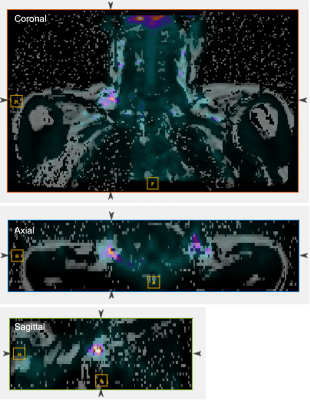 |
15 | Evaluation of the Effects of Capsinoids on Brown Adipose Tissue Recruitment and Activation in Obesity Using PET/MRI
Jie Deng1, Lisa M Neff2, and Bin Zhang3
1Radiation Oncology, UT Southwestern Medical Center, Dallas, TX, United States, 2Chicago, IL, United States, 3Department of Pediatrics, Cincinnati Children's Hospital Medical Center, Cincinnati, OH, United States
In this randomized, placebo-controlled trial in subjects with obesity, we implemented multi-parametric MRI including fat fraction (FF), R2*, and T1 values on PET-MRI in both thermoneutral and cold-activated non-shivering thermogenesis conditions to investigate whether brown adipose tissue property changes measured by MRI can reflect its activation secondary to cold-activated stimulation and chronic capsinoids ingestion before and after weight loss. In subjects taking capsinoids, FF decreased and R2* increased significantly after weight loss as compared with baseline, and these changes were significantly larger than the changes seen after weight loss in the group taking placebo.
|
||
 Back to Meeting Home
 Back to the Program-at-a-Glance
The International Society for Magnetic Resonance in Medicine is accredited by the Accreditation Council for Continuing Medical Education to provide continuing medical education for physicians.
 Back to Meeting Home
 Back to the Program-at-a-Glance
 View the Presentation