Online Gather.town Pitches
White Matter & Nervous System I
Joint Annual Meeting ISMRM-ESMRMB & ISMRT 31st Annual Meeting • 07-12 May 2022 • London, UK

Online Gather.town Pitches
White Matter & Nervous System I
| Booth # | ||||
|---|---|---|---|---|
3011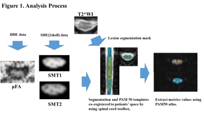 |
1 | Microscopic Fractional Anisotropy Estimation of Spinal Cord in Multiple Sclerosis and Neuromyelitis Optica Spectrum Disorder
Masaaki Hori1,2, Kouhei Kamiya1,2,3, Akifumi Hagiwara2, Kazumasa Yokoyama4, Issei Fukunaga5, Sano Katsuhiro2, Koji Kamagata2, Murata Katsutoshi6, Shohei Fujita2, Christina Andica2, Akihiko Wada2, Julien Cohen-Adad7, and Shigeki Aoki2
1Radiology, Toho University Omori Medical Center, Tokyo, Japan, 2Radiology, Faculty of Medicine, Juntendo University, Tokyo, Japan, 3Radiology, Tokyo University, Tokyo, Japan, 4Neurology, Faculty of Medicine, Juntendo University, Tokyo, Japan, 5Radiological Technology,, Faculty of Health Science, Juntendo University, Tokyo, Japan, 6Siemens Japan K.K., Tokyo, Japan, 7NeuroPoly Lab, Polytechnique Montreal, Montreal, QC, Canada
We investigated the microstructural changes in the spinal cords of patients with multiple sclerosis (MS) and neuromyelitis optica spectrum disorder (NMOSD) using micro fractional anisotropy (μFA) derived from both double diffusion encoding (DDE) and 2-shell single diffusion encoding data with spherical mean techniques (SMT). There was no significant difference in μFA between MS and NMO. SMTs were not correlated with μFA derived from DDE. Therefore, SMT may be better treated as a separate diffusion MRI metric from μFA to investigate the microstructural alterations of spinal cord in patients with MS and NMOSD in vivo.
|
||
3012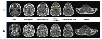 |
2 | Test-retest repeatability of MRI radiomics features in head and neck region using 3D-T1w-TSE and 3D-T2w-TSE: a prospective multi-scan study
Cindy xue1,2, Jing Yuan1, Yihang Zhou1, Oilei Wong1, Kin Yin Cheung3, and Siu Ki Yu3
1Research Department, Hong Kong Sanatorium and Hospital, Hong Kong, Hong Kong, 2Department of Imaging and Interventional Radiology, The Chinese University of Hong Kong, Hong Kong, Hong Kong, 3Medical Physics Department, Hong Kong Sanatorium and Hospital, Hong Kong, Hong Kong
Radiomics has been increasingly used as potential quantitative imaging biomarkers for head and neck cancer diagnosis and treatment. However, the test-retest repeatability of radiomics features in the head and neck (HN) region is rarely investigated. This study aims to investigate the test-retest repeatability of MRI radiomics features in HN. Radiomics features were extracted from 15 volunteers receiving 4 re-positioned MRI scans with thermoplastic mask immobilization and flexible coils using 3D-T1w-TSE and 3D-T2w-TSE. The results found that test-retest repeatability of MRI radiomics features varied dependent on tissues and pulse sequences. Only a small percentage of radiomics features showed excellent test-retest repeatability.
|
||
3013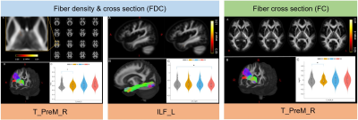 |
3 | Micro- and macro-structural alterations of white matter following sleep deprivation: a longitudinal fixel-based analysis
Chunxiang Jiang1,2,3, Marco Bigica3, Ilenia D’Onofrio3, Zhishan Liu3, Lijuan Zhang1, and Chen Song3,4
1Shenzhen Institutes of Advanced Technology, Chinese Academy of Sciences, Shenzhen, China, 2University of Chinese Academy of Sciences, Beijing, China, 3Cardiff University Brain Research Imaging Centre, Cardiff, United Kingdom, 4University of Wisconsin-Madison, Madison, WI, United States
We examined the effect of sleep deprivation (SD) on human brain white matter(WM) using advanced fixel-based analysis. MRI data collected at four morning time points (7-8am, before and after SD, one night recovery and 3 days recovery). Compared to the session before SD, several WM tracts involving in sleep and wakefulness regulation, memory consolidation and emotion response showed varied fiber density (FD), fiber cross-section (FC) and the combined measure of FD and FC (termed FDC) at different time points. These findings demonstrated the heterogeneous sensitivity of WM tracts in response to SD and the microstructural homeostasis.
|
||
3014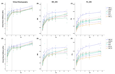 |
4 | Exploring Development Trajectories of White Matter through MR imaging-based Virtual Elastography in Children
Congcong Liu1, Miaomiao Wang1, Xianjun Li1, Yao Ge1, and Jian Yang1
1the Department of Radiology, the First Affiliated Hospital of Xi'an Jiaotong University, Xi'an, China
Characterizing development trajectories of white matter (WM) is vital for identifying causes of neurodevelopmental disorders. Besides DKI, MRE depicted mechanical properties of brain as a sensitive technique. But WM development trajectories of children were lack of researches on MRE. Therefore, we aimed to investigate age-related development of WM based on virtual elastography, comparing with DKI. We found virtual shear stiffness of WM was positively correlated with age, particularly presenting rapid-growth period of WM before 4-years old, consistent with changes of MK, FA. Virtual elastography may be potentially a valuable technique for depicting WM development trajectories, complementary to diffusion metrics.
|
||
3015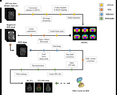 |
5 | Application of Deterministic Tractography Analysis Using SIGMA atlas: Stroke Model
Sang-Jin Im1, Ji-Yeon Suh1, Jae hyuk Shim1, and Hyeon-Man Baek1
1Lee Gil Ya Cancer and Diabetes Institute, Incheon, Korea, Republic of
Unlike image analysis studies with human subjects, where various programs have been developed and validated to provide a complete assessment and step-by-step work procedure, methods for analyzing image data using MRI in preclinical research settings have not been agreed upon. Therefore, in this study, for the high-accuracy SIGMA atlas, we present a deterministic tractographic analysis pipeline that can perform detailed structural segmentation of the rat brain and confirm structural connectivity based on the segmented regions.
|
||
3016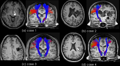 |
6 | Pyramidal tract visualization ability of automatic brain white matter extraction software in patients with brain arteriovenous malformations
Yuichi Suzuki1, Shinya Yuki2, Tsuyoshi Ueyama1, Kentaro Sakata1, Takahiro Iwasaki1, Nobuhito Saito2, Hideyuki Iwanaga1, and Osamu Abe1,3
1Radiology Center, The University oh Tokyo Hospital, Tokyo, Japan, 2Department of Neurosurgery, The University oh Tokyo Hospital, Tokyo, Japan, 3Department of Radiology, The University oh Tokyo Hospital, Tokyo, Japan
There are no reports on the use of TractSeg in patients with brain arteriovenous malformations (BAVMs). This study aimed to investigate the effect of the presence of BAVMs on TractSeg findings of the corticospinal tract (CST) and their clinical usefulness. In case of BAVMs through which the CST runs, the visualization was affected, but in most cases, tractography reconstruction was possible. In case of BAVMs through which the CST does not run, the same results as that of normal volunteers were obtained.
|
||
3017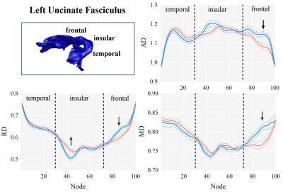 |
7 | Disruption on specific segments of uncinate fasciculus in first-episode drug-naïve adolescent depression
Kaili Liang1, Ruohan Feng1, Xuan Bu1, Suming Zhang1, Yingxue Gao1, Weijie Bao1, Lingxiao Cao1, Jing Liu1, Hailong Li1, Lianqing Zhang1, and Xiaoqi Huang1
1Huaxi MR Research Center (HMRRC), Functional and molecular imaging Key Laboratory of Sichuan Province, Department of Radiology, West China Hospital of Sichuan University, Chengdu, China
We aimed to measure diffusion parameters along the uncinate fasciculus and examine the specific and focal alterations of the uncinate fasciculus in first-episode and drug-naïve patients with major depressive disorder in adolescents by using a tract-based quantitative technique. Notably, we found the opposite alterations of the diffusion metrics in the insular and frontal segments of left uncinate fasciculus in adolescents with depression. It will help us to better understand the neurobiological mechanisms of adolescent depression and optimize adolescent-specific treatments for depression.
|
||
3018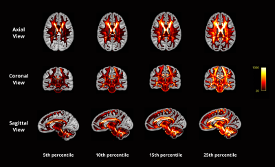 |
8 | Characterising the Common Wiring of the Human Brain
Mai Phuong Ho1, Fernando Calamante1,2,3, and Jinglei Lv1,2
1School of Biomedical Engineering, University of Sydney, Sydney, Australia, 2Brain and Mind Centre, University of Sydney, Sydney, Australia, 3Sydney Imaging, University of Sydney, Sydney, Australia
To compare white matter fibre tracks between individuals or within the same individual over time, a diffusion MRI tractography template is essential. Tractography template describes the location and orientation of fibre bundles that build a representative organization of human white matter. Despite numerous recent advances in methods to map human brain connectivity, tractography suffers from several limitations, including the over- and the under-representation of certain fibre populations. By integrating multimodal registration and SIFT2 quantification approach, we explore the most consistent bundles across subjects, which could help us advance our understanding of the common wiring in the human brain.
|
||
3019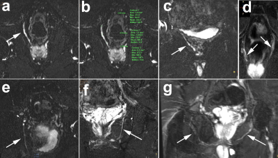 |
9 | Pudendal nerve involvement in young men with erectile dysfunction: demonstration by 3T MR neurography
Tao Gong1,2, Guoquan Huang3, Liangjie Lin4, and Guangbin Wang1
1Departments of Radiology, Shandong Provincial Hospital Affiliated to Shandong First Medical University, Jinan, Shandong, 250021, China, Jinan, China, 2Departments of Radiology, Shandong Provincial Hospital, Cheeloo College of Medicine, Shandong University, Jinan, Shandong, 250021, China, Jinan, China, 3Department of Radiology, Wuhu City Second People's Hospital, No. 259 Jiuhua Rd, Wuhu, 241000, Anhui, China., Wuhu, China, 4MSC Clinical & Technical Solutions, Philips Healthcare, Beijing, China., Beijing, China
More evidences have been identified that pudendal nerve was involved in the pathogenesis of erectile dysfunction (ED) in young men. This prospective study aimed to detect and quantify pudendal nerve alterations in young men with ED using 3T MR neurography protocol. Results certified that pudendal nerve could be visualized and quantified. T2 signal contrast ratio and nerve diameter of pudendal nerve were increased significantly in ED patients compared with healthy controls, suggesting the involvement of pudendal nerve in young men with ED and thus offering a new insight into the pathophysiology and treatment of ED.
|
||
3020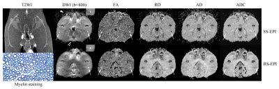 |
10 | Performance of single-shot echo-planar imaging DTI in rat sciatic nerve compared with readout-segmented echo-planar imaging Video Not Available
Yueyao Chen1, Zhongxian Pan1, Fanqi Meng1, Zhujing Li1, Jinyun Gao1, Yuanming Hu1, Yihao Guo2, Qian Xu1, Leyu Huang1, and Hanqing Lv*1
1Department of Radiology, Shenzhen Traditional Chinese Medicine Hospital (The Fourth Clinical Medical College of Guangzhou University of Chinese Medicine), Shenzhen, China, 2MR Collaboration, Siemens Healthineers Ltd., Guangzhou, China This study compared single-shot echo-planar imaging (SS-EPI) with readout-segmented echo-planar imaging (RS-EPI) diffusion tensor imaging (DTI) in vivo anesthetized rat sciatic nerve at 3T. The result showed that the image quality scores of SS-EPI were higher than those of RS-EPI in terms of the nerve morphology and homogeneity of the neuromuscular region. The coefficients of FA and RD obtained with SS-EPI were higher than those obtained with RS-EPI SS-EPI. This suggests that in rat sciatic nerve DTI, SS-EPI showed higher image quality and offered more sensitive and stable parameters to detect the histopathological change in the rat sciatic nerve. |
||
3021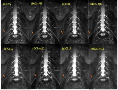 |
11 | 3D NerveView Neurography of the lumbosacral plexus with a deep learning constrained Compressed SENSE Reconstruction
Caijuan Zhang1, Dong Dong1, Shiyu Guo1, Lishan Wu1, Yi Zhu2, and Ke Jiang2
1Radiology, First Hospital of Jilin University, changchun, China, 2Philips Heathcare, Beijing, China
High resolution three dimensional (3D) sequences of MR Neurography always takes a very long scan time. However, using very high acceleration factors leads to degradation of image quality due to insufficient noise removal. In this study, we used Compressed-SENSE Artificial Intelligence (CS-AI) framework to acquire highly accelerated 3D NerveView imaging of lumbosacral plexus. The result showed that CS-AI reconstruction can significantly decrease scan time with sufficient image quality compared to Compressed SENSE(CS) and might be clinically useful in assessment of lumbosacral plexus.
|
||
3022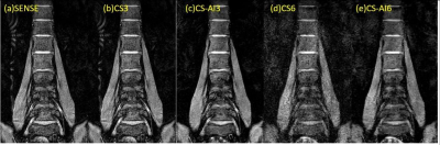 |
12 | 3D T2-FFE MR Neurography of lumbosacral plexus with a deep learning constrained Compressed SENSE reconstruction
Caijuan Zhang1, Dong Dong1, Shiyu Guo1, Lishan Wu1, Yi Zhu2, Ke Jiang2, and Wenxin Wang2
1Radiology, First Hospital of Jilin University, changchun, China, 2Philips Heathcare, Beijing, China
Three-dimensional (3D) T2 Fast Field Echo (FFE) sequence of MR Neurography always takes a very long scan time. However, using very high acceleration factors (AF) will result in degradation of image quality due to insufficient noise removal. In this study, we used Compressed-SENSE Artificial Intelligence (CS-AI) framework to acquire highly accelerated T2 FFE imaging in lumbosacral plexus. The results showed that CS-AI reconstruction might significantly decrease scan time with sufficient image quality compared with conventional SENSE, and might be clinically useful for assessment of lumbosacral plexus.
|
||
3023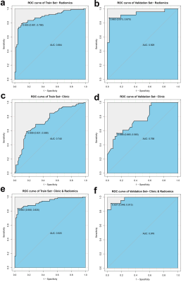 |
13 | Radiomics Model to Predict Radiation-induced Temporal Lobe Injury in Nasopharyngeal Carcinoma
Dan Bao1, Yanfeng Zhao1, and Dehong Luo2
1Department of Radiology, National Cancer Center/National Clinical Research Center for Cancer/Cancer, Beijing, China, 2National Cancer Center/National Clinical Research Center for Cancer/Cancer, Beijing, China
In order to early identify and predict RTLI after IMRT in patients with NPC, we developed and validated a radiomics model based on pretreatment MRI of temporal lobe. The radiomics model demonstrated excellent predictive performance in the validation set (AUC, 0.93; sensitivity, 87%; specificity, 97%). The clinical-radiomics nomogram outperformed clinical nomogram, and showed excellent predictive performance of RTLI in patients within different clinical-pathologic subgroups. These findings suggest that the identified radiomics signature has the potential as a biomarker for risk stratification in RTLI, which may potentially improve the quality of life and prognosis in NPC patients.
|
||
3024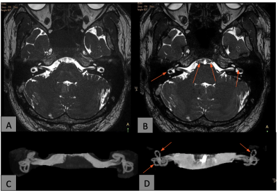 |
14 | Evaluation of banding artifacts suppression for inner ear MR imaging by 3D b-FFE-XD technique Video Not Available
Qiaoling Wu1 and Geli Hu2
1Department of Radiology, Peking Union Medical College Hospital,Chinese Academy of Medical Sciences, Beijing, China, 2Philips Healthcare, Beijing, China
High resolution inner ear MR imaging plays an important role for preoperative plan of hearing system, such as cochlear implant surgery etc. Nowadays, 3D balanced fast-field echo sequence (3D b-FFE) is an effective technique to deliver high-resolution images up to a spatial resolution 0.5 0.5 0.5mm. However, black banding artifacts are frequently observed in the images of inner ear anatomic structures at 3.0T due to magnetic field inhomogeneity. In this study, the 3D b-FFE-XD technique which combines a cycling RF phase with the conventional b-FFE sequence to suppress the banding artifacts and improve the image quality.
|
||
 Back to Meeting Home
 Back to the Program-at-a-Glance
The International Society for Magnetic Resonance in Medicine is accredited by the Accreditation Council for Continuing Medical Education to provide continuing medical education for physicians.
 Back to Meeting Home
 Back to the Program-at-a-Glance
 View the Presentation