Online Gather.town Pitches
Gastrointestinal & Lungs II
Joint Annual Meeting ISMRM-ESMRMB & ISMRT 31st Annual Meeting • 07-12 May 2022 • London, UK

Online Gather.town Pitches
Gastrointestinal & Lungs II
| Booth # | ||||
|---|---|---|---|---|
4247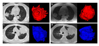 |
1 | The feasibility of ZTE MR lung imaging in the assessment of pulmonary nodules or masses: a prospective head-to-head comparison with CT Video Not Available
Qianyun LIU1, Zhichao Feng2, Wenming Zhou1, and Weiyin Vivian LIU3
1Medical Imaging, Yueyang Central Hospital, Medical Imaging, Yueyang Central Hospital, Yueyang, Hunan, China, China, 2Department of Radiology, The Third Xiangya Hospital, Central South University, Changsha, Hunan, China, Department of Radiology, The Third Xiangya Hospital, Central South University, Changsha, Hunan, China, Changsha, Hunan, China, China, 3GE Healthcare, MR Research China, Beijing, Beijing, China, China
The feasibility of zero echo time MR lung imaging in the assessment of pulmonary nodules or masses: a prospective head-to-head comparison with CT.
|
||
4248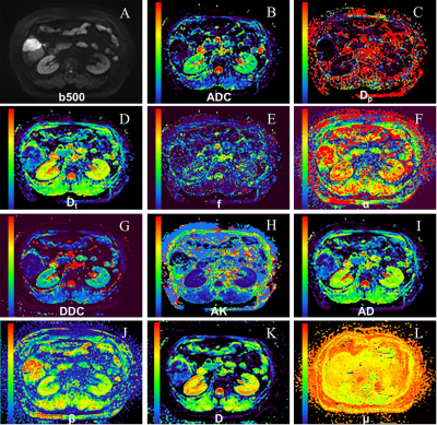 |
2 | Differentiating Cytokeratin 19 Expression of HCC by Using Multi-b-value Diffusion-Weighted MR Imaging Video Permission Withheld
Jiejun Chen1, Yixian Guo1, Shujie Zhang1, Wei Sun1, Yinglong Guo1, Yunfei Zhang2, Yongming Dai2, Dongmei Wu3, and Xiuzhong Yao1
1Zhongshan Hospital affiliated to Fudan University, Shanghai, China, 2Central Research Institute, United Imaging Healthcare, Shanghai, China, 3Shanghai Key Laboratory of Magnetic Resonance, School of Physics and Electronics Science, East China Normal University, Shanghai, China
Diffusion-weighted imaging (DWI) is a powerful functional MRI technique. However, scarcely has the DWI been used to identify the expression of Cytokeratin 19 (CK19) in hepatocellular carcinoma (HCC). In this study, we aim to differentiate CK19 positive and negative HCC with diffusion parameters derived from different DWI techniques. Our results showed that diffusion parameters can be used as noninvasive quantitative imaging markers for preoperatively predicting the CK19 expression of HCCs. More importantly, the integration of different diffusion parameters could better describe the characteristics of tumor tissues,thus reaching better diagnostic performance.
|
||
4249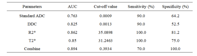 |
3 | The value of IVIM and enhanced T2star weighted angiography for preoperatively predicting Ki-67 status in hepatocellular carcinoma
Qihao Xu1, Ying Zhao1, Wenjing Qi2, Xue Ren1, Tao Lin1, Qingwei Song1, and Ailian Liu1,3
1Department of Radiology, The First Affiliated Hospital, Dalian Medical University, Dalian, Liaoning, China, China, 2Department of Pathology, The First Affiliated Hospital, Dalian Medical University, Dalian, Liaoning, China, China, 3Dalian Engineering Research Center for Artificial Intelligence in Medical Imaging, Dalian, Liaoning, China
In hepatocellular carcinoma (HCC) patients, high Ki-67 was closely related with tumor size, vein invasion, histological grade and the metastasis. This worked aimed at investigating the value of intravoxel incoherent motion (IVIM) diffusion-weighted magnetic resonance (MR) imaging and enhanced T2star weighted angiography(ESWAN) for predicting tumor Ki-67 status in HCC. The results showed that Standard ADC, DDC, R2*, T2* and combine had promising performances in predicting Ki-67 status. This research suggested that the IVIM and ESWAN held great potential in predicting the Ki-67 status of HCC, which is valuable for noninvasively, preoperatively and accurately predicting the prognostic factors during clinical practice.
|
||
4250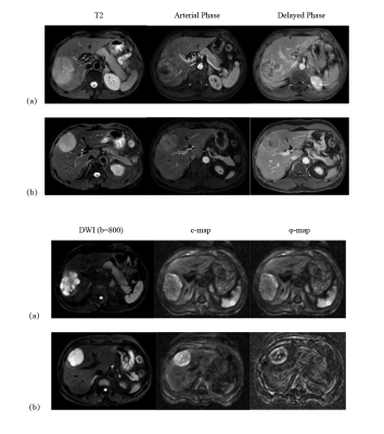 |
4 | Multifrequency MR elastography for the detection of Glypican 3-positive hepatocellular carcinoma
Yihuan Wang1, Ruokun Li1, Jiahao Zhou1, Jing Guo2, Ingolf Sack2, and Fuhua Yan1
1Department of Radiology, Department of Radiology, Ruijin Hospital,Shanghai Jiao Tong University School of Medicine, Shanghai, China, 2Department of Radiology, Charité–Universitätsmedizin Berlin, Berlin, Germany, Berlin, Germany
Glypican-3 (GPC3) expression in hepatocellular carcinoma (HCC) is often associated with a poor prognosis. GPC3 is a promising target for tumor-specific immunotherapy in HCC. We investigated the diagnostic performance of viscoelastic properties quantified by tomoelastography, a multifrequency MR-elastography (MRE) technique, for the detection of GPC3-positive HCC. Preliminary results showed that reduced stiffness quantified by tomoelastography is a mechanical signature of positive GPC3 expression in HCC. Combining stiffness and serum alpha-fetoprotein (AFP) level could be considered as a viable biomarker for detecting GPC3-positive HCC as well as for predicting the outcome of GPC3-targeted immunotherapy.
|
||
4251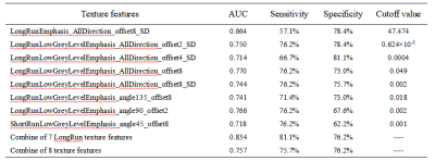 |
5 | Texture analysis of R2* maps for evaluating the pathological grade of hepatocellular carcinoma
Qihao Xu1, Ying Zhao1, Dahua Cui1, Qingwei Song1, Xue Ren1, Tao Lin1, Xin Li2, Yan Guo2, Tingfan Wu2, and Ailian Liu1,3
1Department of Radiology, The First Affiliated Hospital, Dalian Medical University, Dalian, Liaoning, China, 2GE Healthcare, Shanghai, China, 3Dalian Engineering Research Center for Artificial Intelligence in Medical Imaging, Dalian, Liaoning, China
Hepatocellular carcinoma (HCC) is a common malignant tumor with a high mortality. The higher the pathological grade represents lower the differentiation degree of HCC. Patients with low differentiation have a higher postoperative recurrence rate and the worse prognosis. Texture analysis is a post-processing method that highlights the difference between the brightness of pixel features and the intensity of background signals by analyzing the distribution and spatial relationship of gray values, and quantify and evaluate the heterogeneity of tumors at the pixel level. This research suggested that R2* maps texture analysis held great potential in evaluating the pathological grade of HCC.
|
||
4252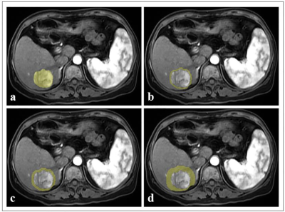 |
6 | Predicting treatment response using intratumoral and peritumoral radiomics based on CE-MRI for patients with HCC treated with TACE
Ying Zhao1, Ailian Liu1,2, Yu Yao3,4, Han Wen3,4, Qingwei Song5, Tao Lin1, Xin Li6, Yan Guo6, and Tingfan Wu6
1The First Affiliated Hospital of Dalian Medical University, Dalian, China, 2Dalian Engineering Research Center for Artificial Intelligence in Medical Imaging, Dalian, China, 3Chengdu Institute of Computer Application, Chinese Academy of Sciences, Chengdu, China, 4University of Chinese Academy of Sciences, Beijing, China, 5Department of Radiology, The First Affiliated Hospital of Dalian Medical University, Dalian, China, 6GE Healthcare (China), Beijing, China
This work aimed at exploring the role of radiomics assessment of hepatocellular carcinoma (HCC) intratumoral and peritumoral regions based on contrast-enhanced magnetic resonance imaging (CE-MRI) to predict treatment response to transarterial chemoembolization (TACE) in patients with HCC. The results showed that the radiomics approach based on intratumoral and peritumoral regions has potential to improve performance for response prediction using preoperative CE-MRI.
|
||
4253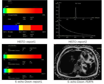 |
7 | Application of Multi-Echo DIXON and HISTO quantitative techniques for staging of liver fibrosis in patients with chronic hepatitis B
Laiyang Ma1, Yanli Jiang1, Jie Zou1, Fengxian Fan1, Jing Zhang1, and Shaoyu Wang2
1Lanzhou University 2 Hospital, Lanzhou, China, 2MR Scientific Marketing, Siemens Healthineers, ShangHai, China
The purpose of this study was to evaluate the value of Multi-echo DIXON(6 echo) and HISTO techniques for staging of liver fibrosis in patients with chronic hepatitis B. We found that PDFF% value and R2* value had a certain correlation with liver fibrosis, Multi-echo DIXON and HISTO quantitative techniques may be helpful for staging liver fibrosis.
|
||
4254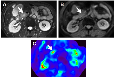 |
8 | The feasibility of using magnetic resonance elastography for differentiation of benign and malignant solid pancreatic masses Video Permission Withheld
Jiejun Chen1, Dingxia Liu1, Shujie Zhang1, Wei Sun1, Yinglong Guo1, Yunfei Zhang2, Yongming Dai2, and Xiuzhong Yao1
1Zhongshan Hospital affiliated to Fudan University, Shanghai, China, 2Central Research Institute, United Imaging Healthcare, Shanghai, China
Magnetic resonance elastography (MRE) is an emerging functional MR technique. Pancreatic ductal adenocarcinoma (PDAC), as one of the most lethal diseases, accounts for 85–90% of all malignant pancreatic tumors. Histologically, PDAC is a hard mass characterized by a marked desmoplastic reaction and build-up of fibrotic tissues. In this study, we aim to evaluate the usefulness of 2D spin-echo echo-planar-imaging (SE-EPI) MRE for differentiation of solid pancreatic masses. Our results showed that compared with benign pancreatic masses, PDAC demonstrated higher mass stiffness and stiffness.
|
||
4255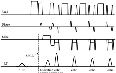 |
9 | Robust fat saturation by combination of SPIR with gradient reversal for TSE at large FOV and coverage
Zhigang Wu1, Yajing Zhang2, Xiuquan Hu1, Jing Zhang1, Guangyu Jiang3, Fei Zeng1, Xiaofang Xu1, Yan Zhao3, and Jiazheng Wang1
1Philips Healthcare, Beijing, China, 2MR Clinical Science, Philips Healthcare (Suzhou), Suzhou, China, 3MR R&D, Philips Healthcare (Suzhou), Suzhou, China
Robust fat suppression remains essential in clinical MRI to improve tissue signal contrast, minimize fat-related artifacts, and enhance image quality. It’s still a challenge to suppress the fat signal when the FOV and coverage is large, especially for abdomen imaging, where uneven fat suppression become common owing to both B0 and B1 field inhomogeneity. We propose a new solution that combines the optimized gradient reversal technique and Spectral Presaturation with Inversion Recovery (SPIR) simultaneously to overcome these challenges. This framework allows to suppress the fat signal robustly in large FOV with whole liver and kidney coverage.
|
||
4256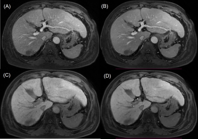 |
10 | Comparison between image quality of 3D T1 GRE sequence with and without deep learning reconstruction at gadoxetic acid-enhanced liver MRI Video Permission Withheld
Jeong Hee Yoon1, Joonsung Lee2, Ersin Bayram3, and Jeong Min Lee1
1Radiology, Seoul National University Hospital, Seoul, Korea, Republic of, 2GE Healthcare, Seoul, Korea, Republic of, 3GE Healthcare, Houston, TX, United States
Liver magnetic resonance imaging (MRI) has been widely performed for liver lesion detection and characterization. There have been attempts to improve the image quality of T1-weighted images at liver MRI. Recently, deep learning (DL)-based reconstruction gains attention as a tool for improving the image quality without substantial computational power or difficult sequence modification.
|
||
4257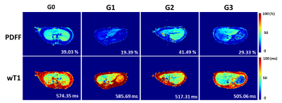 |
11 | Water specific T1 mapping for characterization of inflammation grades at early stage in Rats with Non-alcoholic fatty liver disease (NAFLD)
Qian Wan1, Hao Peng 1, Jianxun Lyu1, Yi Wang2, Feng Liu2, Chuanli Cheng1, Yangzi Qiao1, Hairong Zheng1, Xin Liu1, and Chao Zou1
1Shenzhen Institutes of Advanced Technology, Chinese Academy of Sciences, Shenzhen, China, 2Peking University People's Hospital, Beijing, China
Detection of inflammation is important to the NAFLD patients. Liver biopsy is limited by the sampling error. T1 quantification from MRI is believed to be one of promising imaging method to detect inflammation. However, intracellular hepatocyte lipid is a confounding factor for the accurate T1 quantitation in the case of the NAFLD. This study established a variable flip-angle multi-echo GRE sequence to produce the T1 value of water component (wT1) in a NAFLD rat model who underwent biopsy. The results indicate that wT1 could discriminate between moderate and severe inflammation stages (G2 +G3) from the no and mild stages (G0+G1).
|
||
4258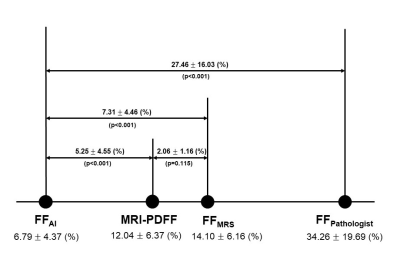 |
12 | The spectrum of MRI-PDFF, MRS, and two different histopathologic methods (AI vs. pathologist) in the quantification of hepatic steatosis
Jeong Woo Kim1 and Chang Hee Lee2
1Radiology, Korea University Guro Hospital, Seoul, Korea, Republic of, 2Korea University Guro Hospital, Seoul, Korea, Republic of The aim of our study was to 1) provide spectrum of values of multi-echo mDixon MRI-PDFF, FFMRS, and FFs measured by two different histopathologic methods (pathologist and automatic fat vacuole segmentation), and 2) to evaluate the correlation among them. The values of MRI-PDFF and FFMRS were significantly higher than FFAI and significantly lower than FFpathologist. Except for one case (97.9%,46/47), FFMRS always showed higher value than MRI-PDFF and the average difference was 2.06 %. MRS and MRI-PDFF showed strong correlation with each other and with each histopathologic method. MRI-PDFF or MRS could be used as an alternative non-invasive reference standard. |
||
4259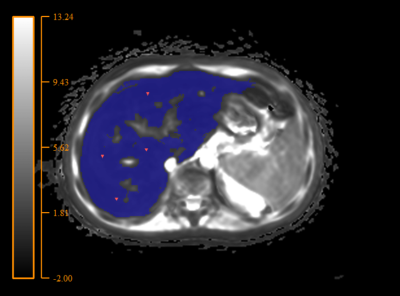 |
13 | The ultrashort echo time (UTE) sequence quantifies high liver iron more accurately than the multiecho gradient-recalled echo (GRE).
Petr Bulanov1, Evelina Manzhurtseva2, Petr Menshchikov3, Artem Tolkalov4, Dmitriy Kupriyanov3, Galina Tereshchenko2, and Galina Novichkova2
1Lomonosov Moscow State Univesity, Moscow, Russian Federation, 2Dmitry Rogachev National Research Center of Pediatric Hematology, Oncology and Immunology, Moscow, Russian Federation, 3Philips Healthcare, Moscow, Russian Federation, 4National Research Nuclear University MEPhI (Moscow Engineering Physics Institute), Moscow, Russian Federation
The most common MRI sequence for iron overload assessment is Cartesian multiecho gradient-recalled echo (GRE) sequence typically measured in a single breath-hold. However, Breath-holding, is not possible for people who are sedated and is difficult for those who have difficulties with holding their breath for a long period. Ultrashort TE (UTE) imaging is one of the solutions for this challenge. In this method radial data acquisition utilized for free breath scanning. Thus, the main goal of this study is to compare the GRE and UTE sequences on a sample of patients with varying degrees of liver iron overload.
|
||
4260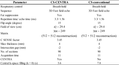 |
14 | Optimal breath-hold dynamic liver MRI using a combination of compressed sensing with contrast-enhanced timing robust angiography technique
Nobuyuki Kawai1, Yoshifumi Noda1, Tetsuro Kaga1, Kimihiro Kajita2, and Masayuki Matsuo1
1Gifu University, Gifu, Japan, 2Gifu University Hospital, Gifu, Japan
Rapid dynamic liver MRI with compressed-sensing sensitivity encoding (Compressed SENSE) can reduce respiratory motion artifacts. However, Since the central k-space filling is random or posterior half of the acquisition in Compressed SENSE, optimal acquisition timing is missed only using a conventional scan delay method for hepatic arterial dominant phase (HAP). We assessed a combination of Compressed SENSE with contrast enhanced robust angiography (CENTRA) for dynamic liver MRI. CS-CENTRA sequence achieved significantly higher arterial enhancement with comparable CNR compared to CS-conventional one in HAP. CS-CENTRA contributed to optimal acquisition timing for HAP.
|
||
4261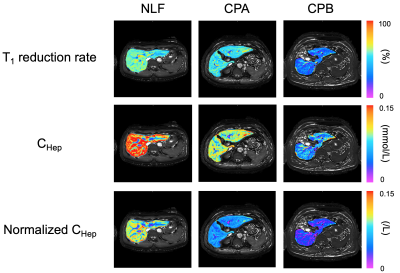 |
15 | Quantitative Evaluation of Liver Function using Normalized Gadoxetic Acid Concentration in Hepatobiliary Phase
Shoma Eitoku1, Yasuhiro Fujiwara2, and Motohira Mio3
1Graduate School of Health Sciences, Kumamoto University, Kumamoto, Japan, 2Department of Medical Image Sciences, Faculty of Life Sciences, Kumamoto University, Kumamoto, Japan, 3Department of Radiology, Fukuoka University Chikushi Hospital, Fukuoka, Japan
The purpose of this study was to clarify whether gadoxetic acid concentration in the hepatic focal area at hepatobiliary phase (CHep), normalized by dose (normalized CHep), could be used as an index for quantitative liver function assessment. In severe liver dysfunction, a moderate correlation was observed between gadoxetic acid concentration and dose. Normalized CHep with bias correction based on the dose of gadoxetic acid may be used as an index of liver function, especially in the stratification of severe liver dysfunction.
|
||
4262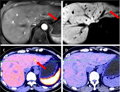 |
16 | Diagnostic values and agreement of Gadoxetic Acid-enhanced MRI and Dual Tracer PET/CT (18F-FDG and 11C-Acetate) in Hepatocellular carcinoma
Solomon Yig-Joon Ka1, Gladys Goh Lo1, Christine Shing Yen Lo1, and Cindy Xue2,3
1Department of Diagnostic & Interventional Radiology, Hong Kong Sanatorium and Hospital, Hong Kong, Hong Kong, 2Research Department, Hong Kong Sanatorium and Hospital, Hong Kong, Hong Kong, 3Department of Imaging and Interventional Radiology, The Chinese University of Hong Kong, Hong Kong, Hong Kong
The diagnoses of hepatocellular carcinoma (HCC) are mostly made at advanced stage, which is associated with unfavorable prognosis and suboptimal survival. While dual-tracer PET/CT (dt-PET/CT) and gadoxetic acid-enhanced MRI are effective in detecting liver lesions, diagnostic performance of gadoxetic acid-enhanced MRI in identifying small HCC lesions compared with dt-PET/CT remains unclear. Here, we compare the sensitivity and specificity of each imaging modality to diagnose HCC. Gadoxetic acid-enhanced MRI has higher sensitivity in detecting HCC lesions than using dt-PET/CT, especially in smaller HCC lesions. Thus, gadoxetic acid-MRI could provide clinical value in the early diagnosis of HCC.
|
||
 Back to Meeting Home
 Back to the Program-at-a-Glance
The International Society for Magnetic Resonance in Medicine is accredited by the Accreditation Council for Continuing Medical Education to provide continuing medical education for physicians.
 Back to Meeting Home
 Back to the Program-at-a-Glance
 View the Presentation