Online Gather.town Pitches
RF Coils, Technologies & Sequences III
Joint Annual Meeting ISMRM-ESMRMB & ISMRT 31st Annual Meeting • 07-12 May 2022 • London, UK

Online Gather.town Pitches
RF Coils, Technologies & Sequences III
| Booth # | ||||
|---|---|---|---|---|
 |
4101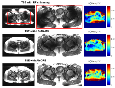 |
1 | Improved TSE imaging at Ultra-High Field using Non-localized Efficiency Shimming and Acquisition of Modes Optimized for Refocused Echo (AMORE)
Xiaoxuan He1, Simon Schmidt1, Stefan Zbyn1, Tobey Haluptzok1, Steen Moeller1, and Gregory J. Metzger1
1Center for Magnetic Resonance Research, University of Minnesota, Minneapolis, MN, United States
To mitigate B1+ inhomogeneity at ultra-high field (UHF), we propose a novel non-localized efficiency shim and utilize this to optimize for T2W TSE contrast in a technique termed Acquisitions of Modes Optimized for Refocused Echo (AMORE). A non-localized efficiency shim is ideal for turbo spin echo (TSE) or fast spin echo (FSE) imaging of smaller anatomical targets such as brain and knee, while AMORE is an optimized TIAMO for improved TSE imaging in the torso by ensuring the desired B1+ is achieved. With AMORE two phase-only modes appear sufficient to mitigate the B1+ shadings and nulls in the interior torso.
|
|
4102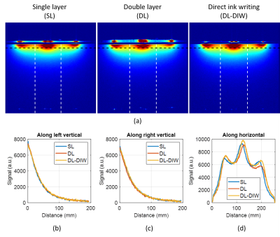 |
2 | Fabrication Methods for Stretchable, Self-tuning Multi-element Liquid Metal Coil Arrays (LiquiTune)
Elizaveta Motovilova1,2, Terry Ching3,4, Jana Vincent5, James Shin1, Ek Tsoon Tan2, Victor Taracila5, Fraser Robb5, Michinao Hashimoto3, Darryl Sneag2, and Simone Angela Winkler1
1Department of Radiology, Weill Cornell Medicine, New York, NY, United States, 2Department of Radiology and Imaging, Hospital for Special Surgery, New York, NY, United States, 3Pillar of Engineering Product Development, Singapore University of Technology and Design, Singapore, Singapore, 4Department of Biomedical Engineering, National University of Singapore, Singapore, Singapore, 5GE Healthcare, Aurora, OH, United States
We recently proposed a self-tuning stretchable receive coil concept in a single element. Here, we expand the technique to a multi-element array and compare 3 fabrication techniques: 1) single layer, 2) double layer, 3) direct ink writing (DIW). Numerical simulations are used to find the critical overlap for decoupling. In vitro experiments demonstrate almost identical sensitivity between the 3 techniques and between coil elements. DIW is most suitable due to its mechanical stability and thinner/less MR-visible coil elements with a frequency stability of 128+/-0.6 MHz (0 to 30% of stretch) and an SNR improvement of 50% over a commercial coil.
|
||
4103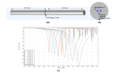 |
3 | A Dielectric Material Coated Half-Wave Dipole antenna for Ultrahigh Field MRI at 7T/300MHz
Aditya Ashok Bhosale1, Divya Gawande1, and Xiaoliang Zhang1
1Biomedical Engineering, University at Buffalo, Buffalo, NY, United States The potential of half-wave dipole antennas in MRI is primarily recognized. This study presents the dielectric material-coated half-wave dipole antenna design and compares it with the conventional half-wave dipole antenna. The design includes a high dielectric material coating around the length of each conductor. The dipole length primarily determines the resonant frequency of a dipole antenna; therefore, it is difficult to tune small half-wave dipole antennas less than 50 cm at 300MHz. Our design provides a solution to this problem and can tune smaller half-wave dipole antennas at 300MHz without meandering the dipole conductors. |
||
4104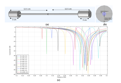 |
4 | B1 field flattening and length control of half-wave dipole antenna with discrete dielectric coating
Aditya Ashok Bhosale1, Divya Gawande1, and Xiaoliang Zhang1
1Biomedical Engineering, University at Buffalo, Buffalo, NY, United States This study presents a method for the B1 field flattening and length control of the half-wave dipole antenna for MR imaging with discrete dielectric material coating. To demonstrate this method, we designed a half-wave dipole antenna to operate at 300MHz, including coating the ends of the dipole conductors with a high dielectric material, serving as a way to control the dipole length and electromagnetic field distribution. We also compare the proposed technique with the conventional half-wave dipole antenna and show how the new technique effectively tunes the shorter half-wave dipole antennas at specific frequencies and produces efficient field distributions. |
||
4105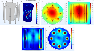 |
5 | An 8-Channel High-permittivity Dielectric Material-Coated Half-Wave Dipole Antenna Array for Knee Imaging at 7T
Aditya Ashok Bhosale1, Leslie L Ying1, and Xiaoliang Zhang1
1Biomedical Engineering, University at Buffalo, Buffalo, NY, United States Magnetic resonance imaging for Musculoskeletal imaging plays a vital role in the early detection of various abnormalities. New array systems designed explicitly for Musculoskeletal imaging at Ultra-high field strengths are being developed rapidly. In this study, we propose a new 8-channel array system consisting of High-permittivity dielectric material-coated half-wave dipole antennas for knee imaging at Ultra-high field 7 Tesla. |
||
4106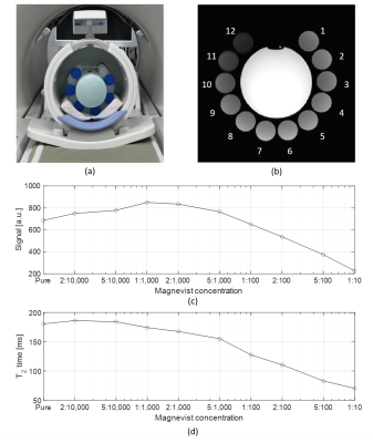 |
6 | Rendering MRI Coils Less Visible in MR images
Elizaveta Motovilova1,2, Frida Maria Galaviz Huerta3, Jana Vincent4, James Shin1, Ek Tsoon Tan2, Fraser Robb4, Victor Taracila4, Darryl Sneag2, and Simone Angela Winkler1
1Department of Radiology, Weill Cornell Medicine, New York, NY, United States, 2Department of Radiology and Imaging, Hospital for Special Surgery, New York, NY, United States, 3Department of Psychology, New York University, New York, NY, United States, 4GE Healthcare, Aurora, OH, United States
It is commonly undesirable to see an RF coil in MR images. Novel research into innovative coil designs often involves the use of new materials that can be visible in MR images. In this abstract, we address this limitation by doping the coil substrate with Magnevist, drawing from a little-known phenomenon of signal hypointensity via T2-shortening for large concentrations of gadolinium-based (Gd) contrast agents that is especially applicable to hydrogen-rich materials. We show successful reduction of MR signal of >60% using our previously developed stretchable, liquid-metal based Ecoflex coil (6) using relaxometry measurements and in vitro experiments.
|
||
4107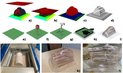 |
7 | A 24-channel vacuum formed pediatric pTx knee coil for 7T
Matt Waks1, Karthik Gopalan2, Stefan Zbyn1, Lance DelaBarre1, Abdul Wahed Kajabi1, Julian Maravilla2, Gregor Adriany1, Michael (Miki) Lustig2, Ana Claudia Arias2, and Jutta Ellermann1
1University of Minnesota, Minneapolis, MN, United States, 2UC Berkely, Berkeley, CA, United States
A pediatric focused 24-channel prototype pTx 7T knee coil consisting of an 8 channel stripline transceiver and a 16-channel close fitting receiver array is described. Initial comparison with an adult size knee coil indicates peripheral SNR gains and more modest - yet notable- gains for central SNR due to the favorable filling factor and form fitting receivers of the pediatric knee coil prototype.
|
||
4108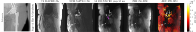 |
8 | A High RF Bandwidth Diffusion Preparation for Diffusion Weighted Imaging Near Metallic Implants
Philip Kenneth Lee1,2, Daehyun Yoon2, Jesse Kerr Sandberg2, Shreyas Vasanawala2, and Brian Andrew Hargreaves1,2,3
1Electrical Engineering, Stanford University, Stanford, CA, United States, 2Radiology, Stanford University, Stanford, CA, United States, 3Bioengineering, Stanford University, Stanford, CA, United States There is no widely adopted clinical protocol for diffusion weighted imaging near metal since the commonly used EPI trajectory fails completely due to distortion from off-resonance. We combine the magnetization prepared, stimulated echo diffusion preparation with 3D MSI encoding to enable volumetric diffusion weighted imaging near metal in clinically feasible times. Imaging time is reduced by a factor of 2× with high RF bandwidth root-flipped Shinnar-Le Roux refocusing pulses since spectral coverage can be maintained with fewer excitations. Joint design of the excitation and refocusing pulses reduces B1 sensitivity. |
||
4109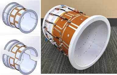 |
9 | A 16-channel splittable non-overlapped self-decoupled loop transmitter for 10.5 Tesla human head imaging
Matt Waks1, Nader Tavaf1, Russell Lagore1, Steve Jungst1, Jerahmie Radder1, Andrea Grant1, Lance DelaBarre1, Pierre-Francois Van de Moortele1, Gregor Adriany1, and Kamil Ugurbil1
1University of Minnesota, Minneapolis, MN, United States
A 16-channel dual row loop transmitter utilizing self-decoupling principles has been designed and prototyped for human head imaging at 10.5 Tesla. The coil employs an intentional asymmetry of impedances within each coil element in order to reduce the interaction between coil elements. Electromagnetic simulations provide an estimate for achievable decoupling and the expected loop current distribution with both a phantom load and a human head model. Bench measurements confirmed the simulation results and indicated coil to coil decoupling on the order of -10 dB or better. MR experimental results indicated minimal interaction with our 63 channel receiver array.
|
||
4110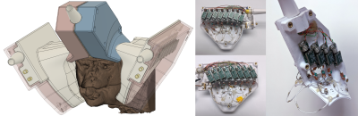 |
10 | A 24-channel Array for Awake NHP Imaging at 10.5 Tesla
Steve Jungst1, Russell Lagore1, Matt Waks1, Jan Zimmermann2, and Gregor Adriany1
1Radiology, University of Minnesota, Minneapolis, MN, United States, 2Neuroscience, University of Minnesota, Minneapolis, MN, United States
A 24-channel receiver – dual channel transmitter array for awake monkey imaging at 10.5 Tesla is presented. This was achieved by adding an 8-channel extension to a previously built 16-channel receive array. The new layout does not require any user adjustments and fully supports monkey imaging.
|
||
4111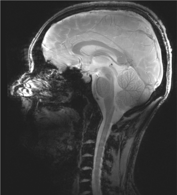 |
11 | 7 Tesla 16-channel transceiver loop array for simultaneous head and cervical spinal cord imaging
Bei Zhang1, Daniel Lowrance1,2, Ole Geldschläger3, and Anke Henning1,3
1Advanced Imaging Research Center, UTSouthwestern Medical Center, Dallas, TX, United States, 2Graduate school, University of Texas Dallas, Richardson, TX, United States, 3Max Planck Institute for Biological Cybernetics, Tuebingen, Germany
Simultaneous assessment of the brain and the cervical spinal cord is of great importance in clinical decision making in areas such as head & neck cancer, traumatic injury, multiple sclerosis or stroke. The small diameter of the cervical spinal cord necessitates high spatial resolution, so there is a growing need to provide 7T simultaneous head and cervical spinal cord imaging, to greatly benefit from increased signal-to-noise ratio and contrast at ultrahigh field. In this work, we developed a 7T 16ch transceiver array that is capable for simultaneous and high-resolution brain and cervical spinal cord imaging.
|
||
4112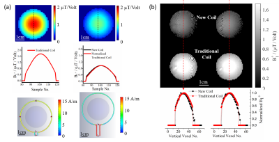 |
12 | B1+ efficiency of a single-loop dual-frequency surface coil for proton and deuterium or oxygen-17 magnetic resonance imaging at 16.4T
Guangle Zhang1, Wei Zhu1, Xin Li1, Tao Wang1, Xiao-Hong Zhu1, and Wei Chen1
1University of Minnesota, Minneapolis, MN, United States We compared the B1 efficiency of a newly developed dual-frequency surface coil operating at proton and low-g X-nuclei frequencies at 16.4T with that of traditional proton coil and X-nuclei coil, respectively. Both simulation and experimental results revealed that the B1 field strength of the new coil was similar to that of traditional coil at deuterium or oxygen-17 frequency though was significantly reduced at proton frequency, while their B1 distributions were identical for both proton and deuterium/oxygen-17 frequencies. The efficacy of the new coil for in-vivo deuterium metabolic imaging applications was demonstrated in normal and tumor rat brains. |
||
4113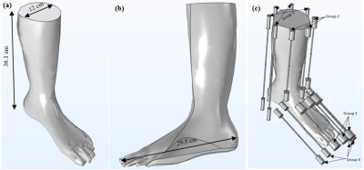 |
13 | A 15-channel End-coated Half-wave Dipole Antenna Array System for Foot/Ankle/Calf Imaging at 7T
Aditya Ashok Bhosale1, Leslie L Ying1, and Xiaoliang Zhang1
1Biomedical Engineering, University at Buffalo, Buffalo, NY, United States This study proposes a new 15-channel end-coated half-wave dipole antenna array system for foot/ankle/calf imaging at 7T. The ends of the dipole antennas, operating at 300MHz, are coated with a high-permittivity dielectric material to decrease their length and flatten the B1 field simultaneously. The end-coated dipole antennas are placed around the region of interest to gain the required B1 coverage in the imaging target. |
||
4114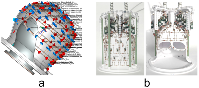 |
14 | Comparison of 64-channel and 32-channel head arrays at 7T and 10.5T in full-wave simulation
Bei Zhang1, Jerahmie Radder2, Andrea Grant2, Russell Lagore2, Matt Waks2, Nader Tavaf2, Pierre-francois Van De Moortele2, Gregor Adriany2, Riccardo Lattanzi3, and Kamil Ugurbil2
1Advanced Imaging Research Center, UTSouthwestern Medical Center, Dallas, TX, United States, 2Center for Magnetic Resonance Research (CMRR), University of Minnesota, Minneapolis, MN, United States, 3Center for Advanced Imaging Innovation and Research (CAI2R), Department of Radiology, New York University Grossman School of Medicine, New York, NY, United States
Idealized analytical electromagnetic (EM) models predict a spatially non-uniform signal-to-noise-ratio (SNR) gain with increasing coil elements and magnetic field strength. Using realistic EM models, we calculated the performance of prototype 32- and 64-channel receive head arrays at 7T and 10.5T using full-wave simulation with a head-mimicking gel phantom. We obtained SNR and g-factor results in agreement with predictions from ultimate intrinsic calculations, showing ~2-fold SNR gains for a large central region for 64 channels at 10.5T vs. 7T, and lower g-factors with the higher field and higher channel counts; peripheral SNR depended on both field magnitude and channel count.
|
||
4115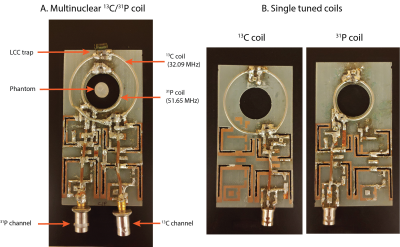 |
15 | Characterizing the relative performance of a 13C/31P surface coil for simultaneously studying metabolism and energetics
Manushka Vaidya1, Bei Zhang2, DongHyun Hong1, Ryan Brown3, Georgios Batsios1, Pavithra Viswanath1, Jan Paska3, Gerburg Wulf4, Aaron Grant5, Sabrina Ronen1, and Peder Larson1
1Department of Radiology, UCSF, San Francisco, CA, United States, 2Advanced Imaging Research Center, UT Southwestern Medical Center, Dallas, TX, United States, 3Center for Advanced Imaging Innovation and Research, and Center for Biomedical Imaging, Department of Radiology, New York Univerisity School of Medicine, New York City, NY, United States, 4Department of Hematology-Oncology, Beth Israel Deaconess Medical Center, Boston, MA, United States, 5Department of Radiology, Beth Israel Deaconess Medical Center, Boston, MA, United States
We evaluated the performance of a 13C/31P surface coil designed to detect metabolism and energetics data in the same scan session. Individually tuned coils were constructed to compare the performance of both channels of the multinuclear coil. Performance metrics such as Q ratio, SNR, flip angle maps, and transmit efficiency were measured. Our results demonstrate a decrease in performance of the 13C channel of the multinuclear coil. Removing the LCC trap circuit, used to decouple the channels, improved the Q ratio and SNR efficiency of the 13C channel. For future work, designs without a LCC trap circuit should be considered.
|
||
 Back to Meeting Home
 Back to the Program-at-a-Glance
The International Society for Magnetic Resonance in Medicine is accredited by the Accreditation Council for Continuing Medical Education to provide continuing medical education for physicians.
 Back to Meeting Home
 Back to the Program-at-a-Glance
 View the Presentation