Online Gather.town Pitches
Cancer I
Joint Annual Meeting ISMRM-ESMRMB & ISMRT 31st Annual Meeting • 07-12 May 2022 • London, UK

| Booth # | ||||
|---|---|---|---|---|
3405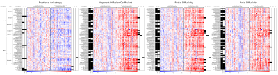 |
1 | Cerebellar damage, by medulloblastoma and related surgery, induces global cerebral microstructural
Joseph Holtrop1, John R Glass1, Stuart S McAfee1, Silu Zhang1, Matthew A Scoggins1, Amar Gajjar2, Giles W Robinson2, Wilbur E Reddick1, and Asim K Bag1
1Department of Diagnostic Imaging, St. Jude Children's Research Hospital, Memphis, TN, United States, 2Department of Pediatrics, St. Jude Children's Research Hospital, Memphis, TN, United States Survivors of medulloblastoma patients frequently have cognitive impairment, which is currently thought to be due to radiation and chemotherapy. In this work, we have showed that there is widespread changes in diffusion tensor parameters in cerebral white and gray matters, suggestive of brain microstructure damage, even before start of radiation and chemotherapy and not related to hydrocephalus. |
||
3406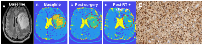 |
2 | Investigating spatial heterogeneity in diffuse gliomas with ultra-high gradient diffusion MRI and a model-free diffusion tensor distribution
Ina Ly1, Yiqiao Song1, Qiuyun Fan2, Aapo Nummenmaa1, Maria Martinez-Lage Alvarez1, William Curry1, Brian Nahed1, Daniel Cahill1, Pamela Jones1, Jorg Dietrich1, Deborah Forst1, Scott Plotkin1, Tracy Batchelor3, Bruce Rosen1,
Susie Huang1, and Elizabeth Gerstner1
1Massachusetts General Hospital, Boston, MA, United States, 2Department of Biomedical Engineering, College of Precision Instruments and Optoelectronics Engineering, Tianjin University, Tianjin, China, 3Brigham and Women's Hospital, Boston, MA, United States
Diffuse gliomas demonstrate significant spatial heterogeneity which is not captured on standard anatomical MRI or with existing diffusion MRI (dMRI) methods. This is because current dMRI approaches provide voxel-averaged information about tissue composition and are based on a priori assumptions about underlying tissue microstructure, thus providing a simplified model of tissue properties. Here, we apply an unbiased, model-agnostic dMRI method (model-free diffusion tensor distribution (FDTD) and K-means clustering) to seven subjects with diffuse gliomas. We confirm the presence of intratumoral heterogeneity and find distinct differences in diffusion properties between gliomas of different histologic grades and pre- and post-treatment.
|
||
3407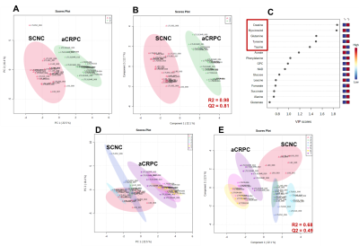 |
3 | Identifying Metabolic Differentiators of Small Cell Neuroendocrine versus Adenocarcinoma subtypes of Castration-Resistant Prostate Cancer Video Permission Withheld
Deepti Upadhyay1, Jinny Sun1, Shubhangi Agarwal1, Robert Bok1, Romelyn DeLos Santos1, Donna Peehl1, John Kurhanewicz1, and Renuka Sriram1
1Department of Radiology and Biomedical Imaging, University of California, San Francisco, San Francisco, CA, United States NMR-based stable isotope resolved metabolomics was used to characterize small cell neuroendocrine (SCNC) and adenocarcinoma (aCRPC) subtypes of castration-resistant prostate cancer patient-derived xenografts (PDXs). Targeted metabolomics indicated distinct upregulation of metabolic pathways in SCNC relative to aCRPC PDXs. Specifically, [U-13C]glucose and [U-13C]glutamine labeling demonstrated that SCNC PDXs had increased glycolytic rate, alanine aminotransferase and tricarboxylic acid cycle activity, including anaplerotic sources. Further, the metabolic differences observed among the aCRPC and SCNC PDXs exhibited a continuous range as predicted by clinical genomic data of these subtypes of CRPC. |
||
3408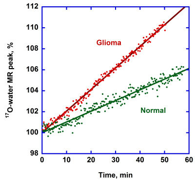 |
4 | Comparison of [6-17O]glucose metabolism in tumor and normal brain using 9L rat glioma model
Victor D. Schepkin1, Cathy W. Levenson2, Shannon Helsper3, and Dean Sherry4
1CIMAR, NHMFL/FSU, Tallahassee, FL, United States, 2College of Medicine, FSU, Tallahassee, FL, United States, 3NHMFL/FSU, Tallahassee, FL, United States, 4Southwestern Medical Center, UT Dallas, Dallas, TX, United States Labeled glucose[6-17O] was used to evaluate differences in glucose metabolism between normal and tumor brain tissue. Experiments were performed using an intracranial 9L glioma model at 21.1 T magnet by observing changes of 17O-water and 17O-glucose MR peaks. The estimated rate of the total glucose metabolism in 9L glioma was approximately 11 times more than in the normal brain. Correspondingly, glucose metabolism rate is 7.5 times more in glioma at the enolase step. 17O MRI/MRS is an encouraging tool for assessing the differences in glucose metabolism in various cancer cells. |
||
3409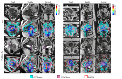 |
5 | Tracking acute & chronic RT-induced fibrosis in cervical cancer using native & Late-Gadolinium-Enhancement Short-Inversion-Time (STIR) UTE
Khadija Sheikh1, Junghoon Lee2, Marc Morcos2, Mohammad Rezae2, Ravi T. Seethamraju3, Thomas Benkert4, Himanshu Bhat3, Henry R Halperin5, Bruce L. Daniel6, Akila N Viswanathan2, and Ehud J Schmidt2,5
1Johns Hopkins School of Medicine, Washington, DC, United States, 2Radiation Oncology and Molecular Radiation Sciences, Johns Hopkins School of Medicine, Baltimore, MD, United States, 3Siemens Medical Solutions, Boston, MA, United States, 4MR Applications Predevelopment, Siemens Healthcare GmbH, Erlangen, Germany, 5Cardiology, Johns Hopkins School of Medicine, Baltimore, MD, United States, 6Radiology, Stanford University, Stanford, CA, United States
Imaging the temporal evolution of post-radiation fibrosis, which forms in irradiated tissues, supports quantifying tumor response and estimating surrounding-tissue injury. Five cervical cancer patients were scanned pre-external-beam-radiotherapy (EBRT), during and after EBRT, and 3-months post-EBRT and High-Dose-Rate (HDR) brachytherapy. A novel stack-of-spirals STIR-UTE sequence (TI=60ms) was used to image acute-fibrosis, utilizing collagen-bound-water’s short T2* and T1, and Late-Gadolinium-Enhancement (11-15min post-contrast) IR-UTE sequence (TI=200ms) to image chronic-fibrosis, utilizing scar’s slow-contrast-perfusion. Acute-fibrosis spatial-distribution followed EBRT and brachytherapy dose distributions. Over time, acute-fibrosis was converted into chronic-fibrosis. This is important as it forms the basis to guide radiation-therapy for re-irradiation and adaptive-planning.
|
||
3410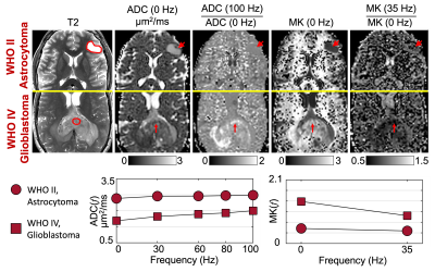 |
6 | Characterizing Restricted Diffusion in Pre-/Post- treatment Gliomas Using Time-dependent Diffusion MRI at Ultra-high-gradient Human 3.0T
Ante Zhu1, Robert Y. Shih2,3, James Kevin DeMarco2,3, Herman Douglas Morris2,3, Radhika Madhavan1, Gail Kohls2,3, Tim Sprenger4, Maureen Hood2,3, Luca Marinelli1, Jason A. Gregory2,3, Brett J. Theeler2,3, Vincent B. Ho2,3, and Thomas K.F. Foo1
1Biology and Applied Physics, GE Research, Niskayuna, NY, United States, 2Uniformed Services University of the Health Sciences, Bethesda, MD, United States, 3Walter Reed National Military Medical Center, Bethesda, MD, United States, 4GE Healthcare, Stockholm, Sweden
Time-dependent diffusion MRI has shown high specificity to non-Gaussian restricted diffusion, which improves assessing cytoarchitectural alternations in pre-/post- treatment glioma. However, the characterization of time-dependent diffusivity ADC(f) and kurtosis MK(f) at short diffusion time using oscillating gradient spin echo (OGSE) diffusion encoding is limited. In our preliminary studies at ultra-high-gradient 3.0T, larger changes of ADC(f) and MK(f) were shown in glioblastoma compared to a low-grade astrocytoma, indicating highly restricted microenvironment as confirmed on histopathology. OGSE-based time-dependent diffusion showed promise for evaluating cytoarchitectural alternations in gliomas. ADC(f) and MK(f) of contrast-enhancing lesions in three post-glioblastoma-treatment patients showed strong time-dependence.
|
||
3411 |
7 | Test-retest Repeatability of High-resolution Whole-brain DCE-MRI in High-grade Glioma Patients
Zhibo Zhu1, Yannick Bliesener1, Frances Chow2, Jay Acharya3, and Krishna S. Nayak4
1Department Of Electrical and Computer Engineering, University of Southern California, Los Angeles, CA, United States, 2Department of Neurosurgery, University of Southern California, Los Angeles, CA, United States, 3Department of Radiology, University of Southern California, Los Angeles, CA, United States, 4Department of Electrical and Computer Engineering, University of Southern California, Los Angeles, CA, United States We perform a prospective test-retest repeatability study of one such approach, in patients with high-grade glioma. Two separate scans were performed within 3-7 days apart. Tumor, scalp, normal white matter, pons, cerebellum and muscle regions were segmented manually. The coefficients of variation of median Kt were 50.4% to 66.6% and of median vp were 23.2% to 80.0%, based on our first 5 subjects. |
||
3412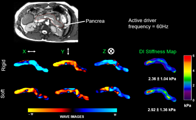 |
8 | Magnetic Resonance Elastography of Pancreas: Comparison between different drivers and driving frequencies. Video Permission Withheld
Manjunathan Nanjappa1 and Arunark Kolipaka1
1Department of Radiology, The Ohio State University, Columbus, OH, United States
Magnetic Resonance Elastography is an emerging alternative diagnostic tool for palpation and it is known that stiffness is altered in chronic pancreatitis and as well as in pancreatic tumors. In this study we compared performances of a drum-shaped rigid driver with a flat soft-pad passive driver in 10 volunteers at 40Hz and 60Hz diver frequencies. The variations of mean stiffness values measured between the drivers were found to be insignificant at a given frequency. Additionally, stiffness measured at 40Hz is lower compared to 60Hz. Therefore, users can make their choice of the driver based on patient comfort, longevity and cost.
|
||
3413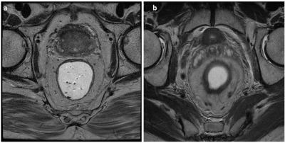 |
9 | Rectal Cancer MRI Motion Quality Assessment using a Convolutional Neural Network
Avishkar Sharma1, Ke Lei2, Shreyas Vasanawala1, and Vipul Sheth1
1Radiology, Stanford University, Stanford, CA, United States, 2Electric Engineering, Stanford University, Stanford, CA, United States
Magnetic resonance (MR) imaging plays a pivotal role in the staging and treatment planning of rectal cancer. Accurate staging depends on good-quality high-resolution axial T2-weighted images orthogonal to the rectal tumor. Rectal MRI is often confounded by motion artifacts secondary to bowel peristalsis and patient movement. We propose a CNN model that automatically assesses image quality instantaneously after a scan is finished to reduce the frequency of patient recalls and non-diagnostic images. Our model achieves high accuracy in identifying motion degradation on an individual slice basis and perfect accuracy when classifying the entire sequence.
|
||
3414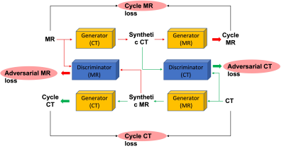 |
10 | Deep Learning Approaches Using Convolutional Neural Networks to Generate Synthetic CT from Spinal MRI for Radiotherapy Planning
Yang Zhang1,2, Farouk Nouizi1, Ning Lang3, Xiaoying Xing3, Yongye Chen3, Qizheng Wang3, Enlong Zhang3, Huishu Yuan3, Ke Nie2, and Min-Ying Su1
1Department of Radiological Sciences, University of California, Irvine, CA, United States, 2Department of Radiation Oncology, Rutgers-Cancer Institute of New Jersey, Rutgers-Robert Wood Johnson Medical School, New Brunswick, NJ, United States, 3Department of Radiology, Peking University Third Hospital, Beijing, China
A total of 27 patients receiving both spinal CT and MR for evaluation of back pain were identified for analysis. MR images and CT image were co-registered first, and the CT was used as ground truth for training a deep learning algorithm using MR images to generate synthetic CT. In this study, we implemented cycleGAN to generate these synthetic CT images from their corresponding MR slices. Five-fold cross validation was used to evaluate the performance of the trained model. Compared to the original images, the Mean Average Error was 27.63±11.51, and the Peak Signal-to Noise Ratio was 19.44±5.72.
|
||
3415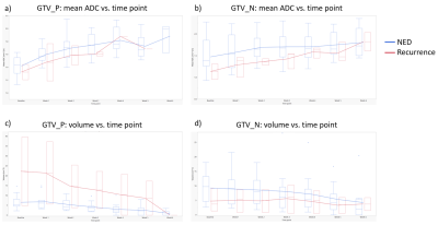 |
11 | Weekly Apparent Diffusion Coefficient Kinetics in HPV-positive Oropharyngeal Cancer Patients Undergoing Definitive Radiation Therapy
Brigid McDonald1, Renjie He1, Yao Ding2, Travis Salzillo2, Jihong Wang2, Clifton Fuller2, and Abdallah Mohamed2
1Radiation Oncology, UT MD Anderson Cancer Center, Houston, TX, United States, 2UT MD Anderson Cancer Center, Houston, TX, United States
In this study, we investigate weekly changes in mean and median apparent diffusion coefficient (ADC) values from diffusion-weighted MRI for HPV+ oropharyngeal cancer patients undergoing definitive radiation therapy (RT). Trends of increasing ADC values in GTV_P and GTV_N were observed with clear separation at each time point between patients with no evidence of disease vs. recurrence at 2 months post-RT. Because of our small sample size (15), a larger cohort is needed for more robust outcome prediction analysis.
|
||
3416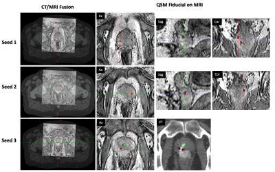 |
12 | Quantitative Susceptibility Mapping for Gold Fiducial Marker Localization in Prostate for MRI-Only Radiation Therapy Treatment Planning
Jie Deng1, Chenyang Shen1, Junjie Wu1, and Steve B Jiang1
1Radiation Oncology, UT Southwestern Medical Center, Dallas, TX, United States
MRI-only simulation permits superior soft tissue contrast for tumor tissue characterization and target margin delineation. One bottleneck preventing MRI-only simulation to be widely applied in prostate cancer EBRT is the tedious marker localization process on MR images as compared to that on a CT. The fiducial and calcification on MRI also appear to be similar, which reduces the confidence level of the clinician in fiducial localization, and further increases the time needed. In this study, we developed a framework using quantitative susceptibility mapping to automatically identify the fiducial markers based on phase information of gradient-echo MRI.
|
||
3417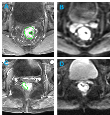 |
13 | Comparison between machine learning and radiologists’ readings for prediction of chemoradiation therapy response in rectal cancer using MRI
Yang Zhang1,2, Liming Shi3, Weiwen Zhou3, Xiaonan Sun3, Salma Jabbour1, Ning Yue1, Min-Ying Su2, and Ke Nie1
1Department of Radiation Oncology, Rutgers-Cancer Institute of New Jersey, Rutgers-Robert Wood Johnson Medical School, New Brunswick, NJ, United States, 2Department of Radiological Sciences, University of California, Irvine, CA, United States, 3Department of Radiation Oncology, Sir Run Run Shaw Hospital, Zhejiang University School of Medicine, Hangzhou, China
A radiomics model with pre-treatment MRI for pCR prediction and compared with two oncologists’ readings. Their performance with and without the model assistances was cross-compared to see the potential role of radiomics model in assisting clinical decision. A total of 203 patients receiving neoadjuvant CRT followed by total mesorectal excision (TME) were enrolled. For training set, AUC from radiomics model is 0.85 and AUC from clinical model is 0.74. For testing set, AUC from radiomics model is 0.82 and AUC from clinical model is 0.69. For oncologist’s reading, with the assistance of radiomics model, positive prediction value (PPV) and specificity increased significantly for both experienced and inexperienced oncologist.
|
||
3418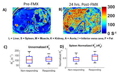 |
14 | Pre-Therapy Ferumoxytol-Enhanced Magnetic Resonance Imaging (MRI) Predicts Response of Metastatic Breast Cancer to Liposomal Irinotecan
Harshan Ravi1, Andres M. Arias Lorza1, James R. Costello2, Hyo Sook Han3, Daniel K. Jeong2, Stephan G. Klinz Klinz4, Jasgit C. Sachdev5, Ronald L. Korn6, and Natarajan Raghunand1,7
1Department of Cancer Physiology, Moffitt Cancer Center and Research Institute, Tampa,, FL, United States, 2Department of Radiology, Moffitt Cancer Center and Research Institute, Tampa,, FL, United States, 3Department of Breast Oncology, Moffitt Cancer Center and Research Institute, Tampa,, FL, United States, 4Ipsen Bioscience, Cambridge, MA, United States, 5HonorHealth Research Institute, Scottsdale, AZ, United States, 6Imaging Endpoints Core Lab, Scottsdale, AZ, United States, 7Department of Oncologic Sciences, University of South Florida, Tampa,, FL, United States
R*2 and the apparent concentration of FMX in the tumor (FMXC) are considered biomarkers of liposomal irinotecan (nal-IRI) drug uptake into that tumor lesion, which in turn would determine response of that tumor to nal-IRI. Historically, quantification of R*2 and FMXC had been implemented by using a calibration phantom and acquiring patient scans both pre-FMX and post-FMX. Here we have demonstrated a pre-treatment FMX-enhanced MRI companion biomarker of response to nal-IRI in mBC patients that can be computed from a single16-24 h post-FMX MRI scan without the need for calibration phantoms or pre-FMX scans.
|
||
3419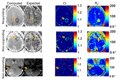 |
15 | R1 and R2* Mismatch on Ferumoxytol-Enhanced MRI Predicts Response of Breast Cancer Brain Metastases to Liposomal Irinotecan
Harshan Ravi1, Andres M. Arias Lorza1, James R. Costello2, Hyo Sook Han3, Daniel K. Jeong2, Stephan G. Klinz4, Jasgit C. Sachdev5, Ronald L. Korn6, and Natarajan Raghunand1,7
1Department of Cancer Physiology, Moffitti Cancer Center and Research Institute, Tampa,, FL, United States, 2Department of Radiology, Moffitti Cancer Center and Research Institute, Tampa, FL, United States, 3Department of Breast Oncology, Moffitti Cancer Center and Research Institute, Tampa, FL, United States, 4Ipsen Bioscience,, Cambridge,, MA, United States, 5Honor Health Research Institute, Scottsdale,, AZ, United States, 6Imaging Endpoints Core Lab, Scottsdale,, AZ, United States, 7Department of Oncologic Sciences, University of South Florida, Tampa, FL, United States
Phagocytosis of ferumoxytol (FMX) by tumor-associated macrophages (TAMs) results in intracellular compartmentation, which has opposing effects on longitudinal (R1) and transverse (R2*) relaxivity of FMX. Pixelwise mismatch between apparent ferumoxytol concentration (FMXc) computed from post-FMX R1 and R2* maps can be exploited to identify pixels with high concentrations of phagocytosed (compartmentalized) FMX. Here we show mismatch on MRI, acquired 24 h post-FMX, measured in terms of compartmentation index (CI) greater than 1 and hypothesized to be indicative of FMX intracellular compartmentation, was predictive of the response of breast cancer brain metastases to liposomal irinotecan (nal-IRI) at the individual tumor level.
|
||
3420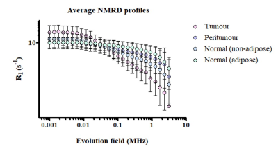 |
16 | Pre-clinical Fast Field-Cycling NMR for the detection and classification of breast cancer Video Permission Withheld
Katie Hanna1, Ehab Husain2, Yazan Masannat3, Rasha Abu-Eid4, David Lurie5, Valerie Speirs1, and Lionel Broche5
1Institute of Medical Sciences, School of Medicine, Medical Sciences and Nutrition, University of Aberdeen, Aberdeen, Scotland, 2Department of Pathology, Aberdeen Royal Infirmary, Aberdeen, Scotland, 3Breast Unit, Aberdeen Royal Infirmary, Aberdeen, Scotland, 4Institute of Dentistry, School of Medicine, Medical Sciences and Nutrition, University of Aberdeen, Aberdeen, Scotland, 5Aberdeen Biomedical Imaging Centre, University of Aberdeen, Aberdeen, Scotland
This study aims to explore novel biomarkers of breast cancer using FFC-NMR. NMR dispersion profiles were generated by analysing patient-matched tumour, peritumoral zone and distant normal fixed tissue samples from twenty breast cancer patients. These profiles were analysed using piecewise power models and the numerical parameters derived from these models could significantly classify the different regions of breast tissue and distinguish patients from different prognostic categories. The numerical parameters investigated in this study may have potential as quantitative biomarkers for breast cancer detection and risk stratification.
|
||
3421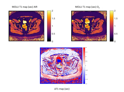 |
17 | Feasibility and repeatability of oxygen-enhanced T1 measurements in primary colorectal cancer: a prospective study in 22 patients. Video Not Available
Davide Prezzi1, Radhouene Neji1,2, Isabel Dregely1, Sami Jeljeli1, Paul Bassett3, Gary Cook1, and Vicky Goh1
1King's College London, London, United Kingdom, 2Siemens Healthineers, Frimley, United Kingdom, 3Statsconsultancy Ltd, Amersham, United Kingdom Oxygen-enhanced ΔT1 tumor measurements were feasible in 12/22 colorectal cancer patients by means of single-slice MOLLI. A small but statistically significant negative tumor ΔT1 was found overall, in line with the expected response in well perfused and oxygenated tissue [mean (95% CI) ΔT1 = -33 (-41, -25) msec; mean tumor T1 = 1545 ± 163 msec]. A significant negative MOLLI ΔT1 was found in muscle. Repeatability and interobserver agreement were acceptable. Multi-slice variable-flip-angle VIBE tumor T1 measurements were degraded by motion or susceptibility artefact. Technical developments aimed at mitigating artefact and amplifying ΔT1 are needed for future clinical translation. |
||
 Back to Meeting Home
 Back to the Program-at-a-Glance
The International Society for Magnetic Resonance in Medicine is accredited by the Accreditation Council for Continuing Medical Education to provide continuing medical education for physicians.
 Back to Meeting Home
 Back to the Program-at-a-Glance
 View the Presentation