Online Gather.town Pitches
Advances in Data Acquisition II
Joint Annual Meeting ISMRM-ESMRMB & ISMRT 31st Annual Meeting • 07-12 May 2022 • London, UK

Online Gather.town Pitches
Advances in Data Acquisition II
| Booth # | ||||
|---|---|---|---|---|
4316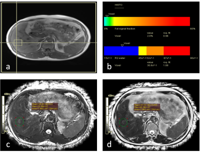 |
1 | Comparison of multi-echo MRS and radial TSE sequences in patients with HCC
Chenhui li1, Liling Long1, Huiting Zhang2, and Fei Han3
1The First Affiliated Hospital of Guangxi Medical University, Nanning, China, 2MR Scientific Marketing, Siemens Healthineers, Wuhan, China, 3Siemens Medical Solutions, Los Angeles, CA, United States
Multi-echo MRS (HISTO) and a novel radial TSE (RadTSE) sequence were compared in patients with hepatic cellular cancer (HCC). Results showed that T2 from RadTSE with fat saturation had a good consistency with that from HISTO. The difference in T2 between RadTSE without and with FS showed excellent correlation with fat fraction from HISTO. Radial TSE and multi-echo MRS sequences may have similar value in terms of quantitative imaging of the liver.
|
||
4317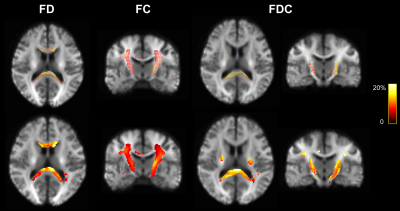 |
2 | 3D diffusion MRI with Twin-navigator-based GRASE for comparison of fiber-tracking using 2D and 3D sequences in human whole brain
Haotian Li1, Yi-Cheng Hsu2, Tao Zu1, Yi Sun2, Yi Zhang1, and Dan Wu1
1Key Laboratory for Biomedical Engineering of Ministry of Education, Department of Biomedical Engineering, College of Biomedical Engineering & Instrument Science, Zhejiang University, Hangzhou, China, 2MR Collaboration, Siemens Healthineers Ltd., Shanghai, China
3D pulse sequences enable high-resolution acquisition with high SNR and ideal slice profiles, which however, is particularly difficult for diffusion MRI (dMRI) due to the additional phase errors from diffusion encoding. Here we proposed a twin-navigator based 3D diffusion-weighted gradient spin-echo (DW-GRASE) sequence to correct the phase errors between shots for human whole-brain acquisition. Moreover, we tested whether acquisitions may impact the fiber-tracking results by comparing the 3D-GRASE with 2D-EPI using the fixel-based analysis, which indicated a significant difference between the 3D and 2D sequences in several microstructural parameters of the long cerebrospinal tract and splenium of corpus callous.
|
||
4318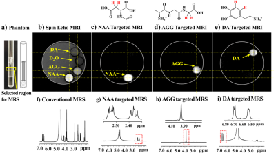 |
3 | Molecularly Targeted Magnetic Resonance Imaging and Spectroscopy
Ye-Feng Yao1, Jia-Xiang Xin1, Guang Yang1, Huojun Zhang2, Jiaqi Li1, Caixia Fu3, Jiachen Wang1, Rui Tong1, and Daxiu Wei1
1Shanghai Key Laboratory of Magnetic Resonance, East China Normal University, Shanghai, China, 2Shanghai Changhai Hospital, Shanghai, China, 3Siemens Shenzhen Magnetic Resonance Ltd., Shenzhen, China
This study developed a molecularly targeted magnetic resonance imaging/magnetic resonance spectroscopy (MRI/MRS) method to selectively probe a specific metabolite molecule of tissues/organs in vivo. Several biomolecules have been used for molecularly targeted MRI and MRS. We used N-acetyl aspartate (NAA)- and glutamate (GLU)-targeted 1H MRS spectra of the human brain and reported a novel approach to measure the local pH values of tissue in vivo.
|
||
4319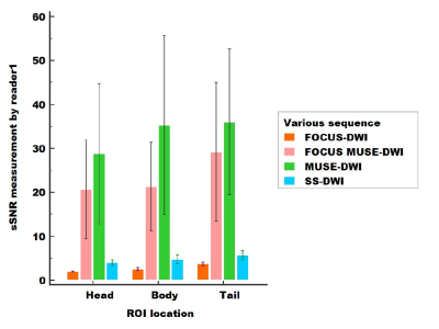 |
4 | A comparison study of novel and conventional diffusion weighted imaging techniques in subjective and objective evaluation of the pancreas
Yigang Pei1, Yu Bai1, Wenzheng Li1, Wenguang Liu1, Weiyin Vivian Liu2, Zhangxuan Hu2, and Lingling Peng2
1Radiology, Xiangya Hospital Central South University, Changsha, China, 2GE heathcare, Beijing, China
Based on field-of-view optimized and constrained undistorted single shot (FOCUS) combined with multiplexed sensitivity-encoding (MUSE) techinique, a new diffusion-weighted imaging (DWI) sequence was developed in our study (named FOCUS MUSE-DWI). Our aim is to assess FOCUS MUSE-DWI’ reliability with comparison to single-shot DWI (ss-DWI), FOCUS-DWI and MUSE-DWI with the evaluation of apparent diffusion coefficient (ADC) repeatability, surrogate signal-to-noise ratio (sSNR) and image quality. FOCUS MUSE-DWI can provide sufficient sSNR and excellent image quality, best ADC repeatability in above four DWIs. It suggests that FOCUS MUSE-DWI is a reliability technique and should be recommended for the clinical application of pancrentic DWI.
|
||
4320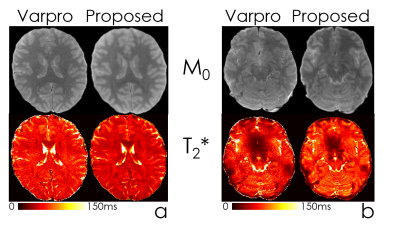 |
5 | Accurate parameter estimation using scan-specific unsupervised deep learning for relaxometry and MR fingerprinting
Mengze Gao1, Huihui Ye2, Tae Hyung Kim3,4, Zijing Zhang2, Seohee So5, and Berkin Bilgic3,4
1Department of Precision Instrument, Tsinghua University, Beijing, China, 2State Key Laboratory of Modern Optical Instrumentation, College of Optical Science and Engineering, Zhejiang University, Hangzhou, China, 3Harvard Medical School, Boston, MA, United States, 4Athinoula A. Martinos Center for Biomedical Imaging, Charlestown, MA, United States, 5School of Electrical Engineering, Korea Advanced Institute of Science and Technology, Daejeon, Korea, Republic of
We propose an unsupervised convolutional neural network (CNN) for relaxation parameter estimation. This network incorporates signal relaxation and Bloch simulations while taking advantage of residual learning and spatial relations across neighboring voxels. Quantification accuracy and robustness to noise is shown to be significantly improved compared to standard parameter estimation methods in numerical simulations and in vivo data for multi-echo T2 and T2* mapping. The combination of the proposed network with subspace modeling and MR fingerprinting (MRF) from highly undersampled data permits high quality T1 and T2 mapping.
|
||
4321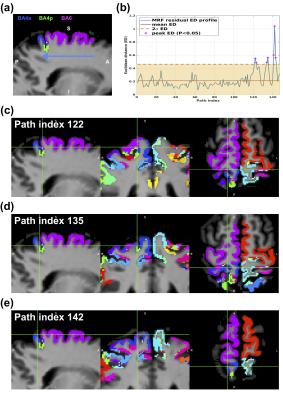 |
6 | In vivo microstructural border delineation between areas of the human cerebral cortex using magnetic resonance fingerprinting (MRF) residuals
Shahrzad Moinian1,2, Viktor Vegh1,2, and David Reutens1,2
1ARC training Centre for Innovation in Biomedical Imaging Technology, The University of Queensland, Brisbane, Australia, 2Centre for Advanced Imaging, The University of Queensland, Brisbane, Australia
We previously showed that magnetic resonance fingerprinting (MRF) residual signals can be used for in vivo voxel-wise parcellation of the human cerebral cortex, using supervised machine learning classification algorithms. However, previous work relied on brain atlases to provide probabilistic masks of cortical region to label samples to train a classification model. Here, we investigate the feasibility of developing automated atlas-free cortical border delineation in individuals. We demonstrate that 90% of the cortical border voxels identified by the proposed framework are co-localised with the borders between two cortical areas on the Juelich maximum probability map of cerebral cortex in six participants.
|
||
4322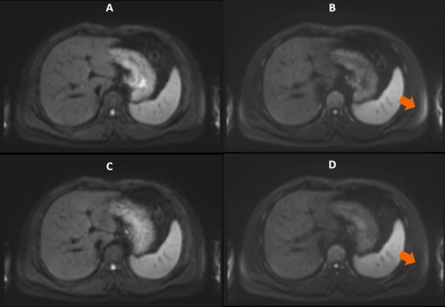 |
7 | Improved fat suppression in diffusion MRI using eddy current compensation gradient
Wei Liu1, Michael Koehler2, Flavio Carinci2, Adam Kettinger2, Thorsten Feiweier2, Kun Zhou1, and Mario Zeller2
1Siemens Shenzhen Magnetic Resonance Ltd, Shenzhzen, China, 2Siemens Healthcare GmbH, Erlangen, Germany
In this work, we propose to compensate the residual eddy current field caused by diffusion gradients at the time when chemically selective fat suppression gets applied with an additional gradient applied after the EPI readout. Using an analytic solution, the amplitude of the eddy currents can be cancelled under certain assumptions. The experimental results based on a volunteer scan demonstrate improved fat suppression in diffusion MRI with the proposed method.
|
||
4323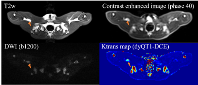 |
8 | Dynamic quantitative T1 mapping (dyQT1) series for quantitative DCE imaging using variable flip angle method
Qing Li1, Xinyu Song2, Jienan Wang2, Caixia Fu3, Yi Sun1, and Yuehua Li2
1MR Collaborations, Siemens Healthineers Ltd., Shanghai, China, 2Institute of Diagnostic and Interventional Radiology, Shanghai Jiao Tong University Affiliated Sixth People's Hospital, Shanghai, China, 3MR Application Development, Siemens Shenzhen Magnetic Resonance Ltd., Shenzhen, China
In this work, a new acquisition scheme is proposed for directly measuring the continuous quantitative T1 changes during the contrast enhancement. By alternatively changing the flip angles from phase to phase, the temporal resolution of T1 mapping could be the same as conventional DCE (conv-DCE) imaging, i.e., 4.36 s in our experiments. The dynamic quantitative T1 mapping (dyQT1) DCE method were validated on animal study (rabbit with VX2 tumor), and compared with conv-DCE. The preliminary results showed dyQT1-DCE was more time efficient without native T1 scans and higher sensitivity in the detection of tumor than conv-DCE.
|
||
4324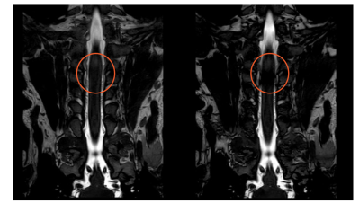 |
9 | Banding artifacts reduction for brachial plexus (BP) imaging Using RF phase cycling balanced FFE technique combined with compressed SENSE Video Not Available
Qiaoling Wu1 and Geli Hu2
1Department of Radiology, Peking Union Medical College Hospital,Chinese Academy of Medical Sciences, Beijing, China, 2Philips Healthcare, Beijing, China
Banding artifacts are frequently observed in brachial plexus imaging, particularly within the spinal canal, with the balanced FFE sequence (b-FFE) at 3.0T due to increased influence of higher magnetic field inhomogeneity compared with clinical 1.5T. In this work we propose using a RF phase cycling technique with b-FFE (namely b-FFE-XD) to reduce the banding artifacts in clinical. This design RF phase limits the intravoxel dephasing of scan object voxels, therefore the artifacts caused by field inhomogeneity decrease. Meanwhile, the compressed SENSE technique was adopted to reduce the scan time. Results demonstrated successful removal of banding artifacts with the proposed technique.
|
||
4325 |
10 | An accelerated magnetic resonance imaging pulse sequence for 3D T1 mapping based on magnetization-prepared rapid gradient-echo (MPRAGE)
Xinpei Wang1, Jichang Zhang1, and Chengbo Wang1
1Faculty of Science and Engineering, University of Nottingham Ningbo China, Ningbo, China
Plenty of studies have reported correlations between the tissue pathological changes and abnormal T1 value. However, T1 mapping pulse sequences often suffer from the B1 inhomogeneity and long scan time. To improve the time efficiency without leading to the worse robustness to B1 inhomogeneity, we develop an accelerated MPRAGE-based 3D T1 mapping method by using SPGR steady state. Our proposed method is demonstrated in phantom experiments and the preliminary in-vivo brain scan.
|
||
4326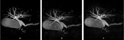 |
11 | Feasibility of 3D breath hold MRCP: Prospective Comparison With parallel imaging technique and compressed sensing method
Zhiyong Chen1, Yunjing Xue1, Bin Sun1, and Yang Song2
1Radiology, Fujian Medical University Union Hospital, Fuzhou, China, 2MR Scientific Marketing, Siemens Healthineers Ltd, Shanghai, China, Shanghai, China
The modified 3D-BH-PI-MRCP technique allowing direct exciting the area of interest, could not only decrease the slice number but also could eliminate folding artifacts.
|
||
4327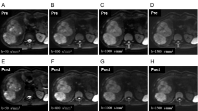 |
12 | Comparison of the image quality and ADC values on diffusion weighted imaging at both low and high b-values with gadoxetic acid-enhanced MR imaging
Shang Wan1, Yi Wei1, Hehan Tang1, Lisha Nie2, Xiaocheng Wei2, and Bin Song3
1Radiology, West China Hospital, Sichuan University, Cheng Du, China, 2GE Healthcare Beijing China, Beijing, China, 3West China Hospital, Sichuan University, Cheng Du, China
Gd-EOB-DTPA has been widely used in liver MR imaging for the evaluation of hepatic lesion, whereas, the long intervals between the dynamic enhanced imaging and hepatobiliary phase imaging usually influence patient throughput. Notably, the adjustment of diffusion weighted imaging (DWI) sequence’s scanning order from pre-contrast to post-contrast is crucial to overcome this limitation. However, few studies have investigated the apparent diffusion coefficient (ADC) values that at low and high b values should be affected, respectively, thus, we aimed to prospectively determine whether DWI at both low and high b-values should be affected before and after gadoxetic acid-enhanced MR imaging.
|
||
4328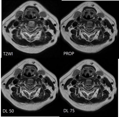 |
13 | Axial T2-weighted MR imaging of the cervical spine using PROPELLER with deep learning reconstruction to improve image quality
You Seon Song1, In Sook Lee1, Moon jung Hwang2, Kyungeun Jang2, Maggie Fung3, and Xinzeng Wang4
1Pusan National University Hospital, Busan, Korea, Republic of, 2GE Healthcare Korea, Seoul, Korea, Republic of, 3GE Healthcare, New York, NY, United States, 4GE Healthcare, Houston, TX, United States
We evaluated the utility of PROPELLER T2 FSE with deep-learning (DL) reconstruction in the cervical spine MRI, with the goal of overcoming respiratory and swallowing motion, improving image sharpness, and achieving high-resolution imaging within a comparable scan time to the conventional T2 FSE sequences. With utilization of DL reconstruction, the axial PROPELLER T2 was able to obtain images with high resolution and reduced noise.
|
||
 Back to Meeting Home
 Back to the Program-at-a-Glance
The International Society for Magnetic Resonance in Medicine is accredited by the Accreditation Council for Continuing Medical Education to provide continuing medical education for physicians.
 Back to Meeting Home
 Back to the Program-at-a-Glance
 View the Presentation