Online Gather.town Pitches
Diffusion I
Joint Annual Meeting ISMRM-ESMRMB & ISMRT 31st Annual Meeting • 07-12 May 2022 • London, UK

| Booth # | ||||
|---|---|---|---|---|
 |
3544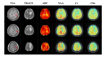 |
1 | Simultaneous Mapping of Water Diffusion Coefficients and Metabolite Distributions of the Brain Using MRSI without Water Suppression
Rong Guo1,2, Yudu Li1,2, Yibo Zhao1,2, Tianyao Wang3, Yao Li4, Brad Sutton1,2,5, and Zhi-Pei Liang1,2
1Department of Electrical and Computer Engineering, University of Illinois at Urbana-Champaign, Urbana, IL, United States, 2Beckman Institute for Advanced Science and Technology, University of Illinois at Urbana-Champaign, Urbana, IL, United States, 3Radiology Department, Fifth People's Hospital of Shanghai, Fudan University, Shanghai, China, 4School of Biomedical Engineering, Shanghai Jiao Tong University, Shanghai, China, 5Department of Bioengineering, University of Illinois at Urbana-Champaign, Urbana, IL, United States
The feasibility of high-resolution metabolic imaging using water-unsuppressed MRSI has been recently demonstrated in clinical settings using SPICE. This work utilizes the unsuppressed water spectroscopic signals for distortion-free mapping of water diffusion coefficients, thus adding a new feature to SPICE for multi-modal brain mapping. Experimental results demonstrated that simultaneous distortion-free diffusion imaging at 1.0×1.0×2.0 mm3 resolution and metabolic imaging at 2.0×3.0×3.0 mm3 nominal resolution were successfully obtained in a total 8-min scan.
|
|
3545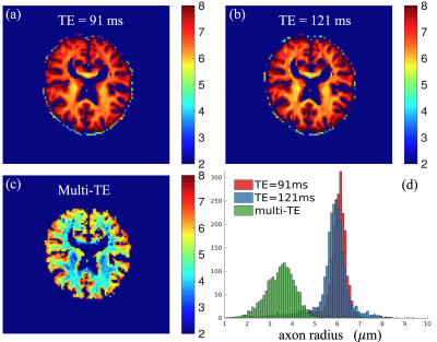 |
2 | Axon radius mapping using multi-TE diffusion MRI from clinical scanners
Lipeng Ning1 and Yogesh Rathi1
1Harvard Medical School, BWH, Boston, MA, United States
Diffusion MRI is sensitive to heterogeneity tissue microstructure. Recent methods for axon diameter estimation rely on diffusion MRI data acquired using advanced scanners with b-values higher than 6000 s/mm2 so that signals from extra-axonal components vanish because of the underlying relative high diffusivity. In this work, we introduce a method for axon diameter estimation that uses diffusion MRI with two TEs and relative lower b-values acquired using clinical scanners. Our method separates signals from intra- and extra-axonal components by exploiting the b-value-dependent relaxation coefficients. The performance of the proposed method is demonstrated using in vivo data acquired from a clinical scanner.
|
||
3546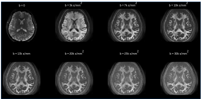 |
3 | Brain Microstructure Imaging with Ultrahigh B-Encoding using MAGNUS High Performance Gradients
Nastaren Abad1, Radhika Madhavan1, Tim Sprenger2, Chitresh Bhushan1, Ante Zhu1, Luca Marinelli1, Eric Fiveland1, Seung-Kyun Lee1, J. Kevin DeMarco3, Robert Shih3, Maureen Hood3,4, Gail Kohls3, H Doug Morris3, Kimbra Kenny4,
Vincent B Ho3,4, and Thomas KF Foo1
1GE Global Research Center, Niskayuna, NY, United States, 2MR Applied Science Laboratory Europe, GE Healthcare, Stockholm, Sweden, 3Walter Reed National Military Medical Center, Bethesda, MD, United States, 4Uniformed Services University of the Health Sciences, Bethesda, MD, United States An ultra-high diffusion b-value protocol (b=7,000-30,000s/mm2) was acquired in a high-performance 3.0 T MAGNUS head gradient system (max gradient amplitude=200 mT/m and max slew rate=500 T/m/s) for white matter microstructure imaging.With shorter TE enabled by MAGNUS, measurements of intra-axonal diffusivities are demonstrated at ultra-high b-values. At these ultra-high b-values, signal from extra-axonal water is effectively suppressed but SNR is also reduced. We show that real-valued image reconstruction maintains Gaussian noise properties and reduces Rician bias, allowing better quantification of intra-axonal diffusivities and effective axonal radii in healthy volunteers and in ongoing human subject research studies of mild traumatic brain injury. |
||
3547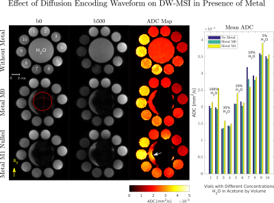 |
4 | Effect of Static Background Gradients on Diffusion Contrast Near Large Metallic Implants
Philip Kenneth Lee1,2, Daehyun Yoon2, and Brian Andrew Hargreaves1,2,3
1Electrical Engineering, Stanford University, Stanford, CA, United States, 2Radiology, Stanford University, Stanford, CA, United States, 3Bioengineering, Stanford University, Stanford, CA, United States Metallic implants create severe off-resonance adjacent to the implant, in the range of 2-20 kHz at 3T. The off-resonance pattern creates a static, “always on” background gradient with unknown amplitude and polarity. Static non-linear background gradients are a known source of local encoding errors, but their effect on diffusion contrast near implants has not yet been evaluated. Static background gradients are distinct from diffusion gradient non-linearities, which affect the achieved b-value if sufficiently large. We apply theoretical results and phantom experiments to evaluate two different diffusion encoding schemes and evaluate the effect of metal-induced off-resonance on ADC quantification. |
||
3548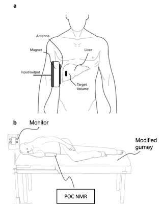 |
5 | NOVEL POINT-OF-CARE MAGNETIC RESONANCE TECHNOLOGY TO MEASURE LIVER FAT: PHANTOM AND FIRST-IN-HUMAN PILOT STUDY Video Permission Withheld
Mark Barahman1, Eduardo Grunvald2, Pablo J Prado3, Alejandro Bussandri3, Walter C Henderson1, Tanya Wolfson1, Kathryn J Fowler1, and Claude B Sirlin1
1Radiology, UCSD, San Diego, CA, United States, 2Internal Medicine, UCSD, San Diego, CA, United States, 3Livivos Inc, San Diego, CA, United States LiverScope® is a novel point-of-care (POC) NMR technology for liver fat quantification. · POC NMR PDFF agreed closely with ground-truth PDFF values in commercial PDFF phantoms (R2 = 0.99) · POC NMR was feasible in an outpatient clinic, had no demonstrated adverse events, and agreed closely with contemporaneous MRI-PDFF reference standard values (R2 = 0.94) in human adults with obesity and at risk for NAFLD: |
||
3549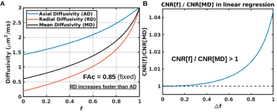 |
6 | Sensitivity advantage of a two- over one-compartment diffusion model for studying aging: An analytic demonstration
Jordan A. Chad1,2, Nir Sochen3,4, J. Jean Chen1,2, and Ofer Pasternak5,6
1Rotman Research Institute, Baycrest Health Sciences, Toronto, ON, Canada, 2Department of Medical Biophysics, University of Toronto, Toronto, ON, Canada, 3School of Mathematical Sciences, Tel Aviv University, Tel Aviv, Israel, 4School of Neuroscience, Tel Aviv University, Tel Aviv, Israel, 5Department of Radiology, Brigham and Women's Hospital, Harvard Medical School, Boston, MA, United States, 6Department of Psychiatry, Brigham and Women's Hospital, Harvard Medical School, Boston, MA, United States
Recent diffusion modeling studies aim to distinguish age-related degenerative processes specific to white matter fibers, such as axonal myelination and dispersion, from age-related elevated isotropic diffusivity, resulting from, e.g., enlargement of the extracellular space. Here we analytically investigate the relationship between two single-shell approaches for distinguishing these effects, respectively based on a one- and two-compartment model. We derive a nonlinear relationship between the models that renders a sensitivity advantage to the two-compartment model, and find that a linear elevation of an isotropic compartment with age explains the well-documented phenomenon of accelerated MD elevations (faster radially than axially) in aging.
|
||
3550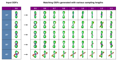 |
7 | Optimization of sampling lengths in Radial DSI empowers ODF-Fingerprinting: A phantom study on fibers crossing at narrow angles
Patryk Filipiak1, Lee Basler2, Benjamin Rodack2, John Dzikiy2, Anthony Zuccolotto2, Ying-Chia Lin1, Dimitris G. Placantonakis3, Timothy Shepherd1, Walter Schneider4, Fernando E. Boada1, and Steven H. Baete1
1Center for Advanced Imaging Innovation and Research (CAI2R), Department of Radiology, NYU Langone Health, New York, NY, United States, 2Psychology Software Tools, Inc., Pittsburgh, PA, United States, 3Department of Neurosurgery, Perlmutter Cancer Center, Neuroscience Institute, Kimmel Center for Stem Cell Biology, NYU Langone Health, New York, NY, United States, 4University of Pittsburgh, Pittsburgh, PA, United States We optimize the sampling length parameter of Radial Diffusion Spectrum Imaging (RDSI) to improve fiber reconstruction accuracy of ODF-Fingerprinting (ODF-FP). As the ground truth, we use anisotropic diffusion phantoms containing taxons (textile water-filled tubes) with inside diameter of 0.8µm, approaching the anatomical scale of axons found in the human brain. Thanks to our optimized approach, ODF-FP reconstructs fibers crossing at angles as narrow as 10, 15, and 20 degrees with the average accuracy of approximately 10 degrees. We generalize our observations by proposing a criterion for selecting near-optimal sampling lengths to accelerate and simplify the application of ODF-FP with RDSI. |
||
3551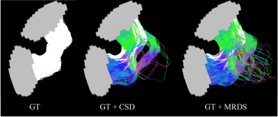 |
8 | Tractography from Gaussian multi-compartmental ODFs
Erick Hernandez-Gutierrez1, Alonso Ramirez-Manzanares2, Antoine Théberge1, Maxime Descoteaux1, Luis Concha3, and Ricardo Coronado-Leija4
1Université de Sherbrooke, Sherbrooke, QC, Canada, 2Centro de Investigación en Matemáticas A.C., Guanajuato, Mexico, 3Universidad Nacional Autónoma de México, Querétaro, Mexico, 4New York University School of Medicine, New York, NY, United States
In this study, we propose a pipeline to compute the structural connectome from diffusion-weighted magnetic resonance imaging (dMRI). The pipeline is based on a novel method called MRDS, which computes independent Gaussian profiles per intravoxel diffusion compartments. The effectiveness of our pipeline is shown on the DiSCo challenge synthetic dataset. Our findings show that the pipeline is competitive and outperforms a pipeline based on constrained spherical deconvolution (CSD).
|
||
3552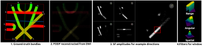 |
9 | Intuitive Angle-Aware Bilateral Filtering Revealing Asymmetric Fiber ODF for Improved Fiber Tractography
Charles Poirier1 and Maxime Descoteaux1
1Computer Sciences, Université de Sherbrooke, Sherbrooke, QC, Canada Although the voxel-wise signal acquired by diffusion MRI is symmetric, it is not always the case of the underlying fiber configurations. We present angle-aware bilateral filtering, an intuitive method for denoising fiber ODF fields revealing asymmetric fiber ODFs. To evaluate the effect of the filtering, tractography is performed on the resulting asymmetric fiber ODF field. Compared to the tractogram obtained from the original fiber ODF field, we show that using asymmetric filtered fiber ODF field as input to a fiber tracking algorithm reduces the ratio of incomplete streamlines and false connections and increases the proportion of true connections. |
||
3553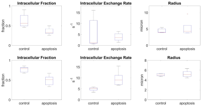 |
10 | Two-pool model with exchange is more sensitive to exchange increases during apoptosis than the partially absorbing wall model
Daniel Djayakarsana1, Greg Stanisz1, and Colleen Bailey1
1Sunnybrook Research Institute, Toronto, ON, Canada
Modeling diffusion that incorporates the microscopic partially absorbing wall is a more realistic description compared to the standard Bloch equation based two-site model with exchange. The resulting signal equations are more complex and involve working in the Laplace domain. Here, we demonstrate that the increased spatial relevance of the partially absorbing wall model does not improve fitting in our AML model treated with cisplatin for cell death. In fact, the partially absorbing wall model fits and parameters are poorer. Therefore, extracting diffusion parameters from the Bloch equation based two-site exchange model is sufficient for our diffusion MRI datasets.
|
||
3554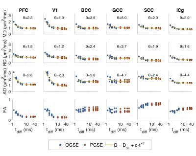 |
11 | Diffusion time dependence in the non-human primate brain
Sean P Devan1,2, Xiaoyu Jiang1,3, Kurt G Schilling1,3,4, Feng Wang1,3, Li Min Chen1,3, John C Gore1,3,4,5, and Junzhong Xu1,3,4,5
1Vanderbilt University Institute of Imaging Science, Vanderbilt University Medical Center, Nashville, TN, United States, 2Chemical and Physical Biology Program, Vanderbilt University, Nashville, TN, United States, 3Department of Radiology and Radiological Sciences, Vanderbilt University Medical Center, Nashville, TN, United States, 4Department of Biomedical Engineering, Vanderbilt University, Nashville, TN, United States, 5Department of Physics and Astronomy, Vanderbilt University, Nashville, TN, United States Time-dependent diffusivity is sensitive to microstructural geometry and has previously been described by a power-law scaling. However, the limits of validity of this relationship across different ranges of diffusion time are unclear. We therefore acquired brain images of nonhuman primates in vivo with diffusion times from 1.9 to 40 ms to assess how well a power-law fits diffusivity in this range. We assess fitting in the time and frequency domains, whether pulsed (PGSE) and oscillating (OGSE) gradient spin echo can be combined for better results, and the sensitivity of diffusivity estimates to varying power-law exponents. |
||
3555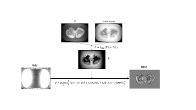 |
12 | Quantitative Susceptibility Mapping Artifact Reduction in Ex Vivo Tissue Specimens Using Adaptive Multi-Contrast Informed Regularization Video Permission Withheld
Priya S Balasubramanian1,2, Lingfei Guo2,3, Weiyuan Huang2,4, Ilhami Kovanlikaya2, Alisa Ramineni5, Nector Garcia5, Benjamin Liechty5, Thanh Nguyen2, Pascal Spincemaille2, David Pisapia5, and Yi Wang2
1Electrical and Computer Engineering, CORNELL UNIVERSITY, New York City, NY, United States, 2Department of Radiology, Weill Cornell Medicine, New York City, NY, United States, 3Department of Radiology, Shandong Provincial Hospital Affiliated to Shandong First Medical University, Jinan, Shandong, Jinan, Shandong, China, 4Department of Radiology, Hainan General Hospital, Hainan Hospital Affiliated to Hainan Medical College, Hainan, China, 5Department of Pathology and Laboratory Medicine, Weill Cornell Medicine and New York Presbyterian Hospital, New York City, NY, United States
Quantitative susceptibility mapping (QSM) solves an ill-posed dipole field inversion problem and is prone to streaking and shadowing artifacts. This report presents a novel QSM reconstruction algorithm that effectively suppresses these artifacts using prior information from traditional MRI contrasts. The proposed algorithm was evaluated using ex vivo high resolution QSM of cerebellum and brainstem specimens using COSMOS as the reference method, showing improved QSM accuracy and artifact reduction and better agreement with myelin sensitive Luxol Fast Blue staining compared to conventional MEDI and MEDI-SMV methods.
|
||
3556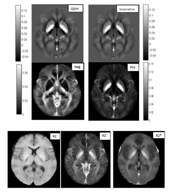 |
13 | Repeatability of Susceptibility Separation in Brain
Javad Hamidi Esfahani1, Nashwan Naji1, Peter Seres1, Christian Beaulieu1, and Alan H Wilman1
1Biomedical Engineering, University of Alberta, Edmonton, AB, Canada |
||
3557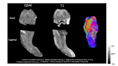 |
14 | Quantitative Susceptibility Mapping of the Ex Vivo Human Brainstem with Luxol Fast Blue Staining Correlation Video Permission Withheld
Priya S Balasubramanian1,2, Lingfei Guo2,3, Weiyuan Huang2,4, Ilhami Kovanlikaya2, Alisa Ramineni5, Nector Garcia5, Benjamin Liechty5, Jonathan Dyke2, Henning Voss2, Eric Aronowitz2, Pascal Spincemaille2, David Pisapia5, and Yi Wang2
1Electrical and Computer Engineering, CORNELL UNIVERSITY, New York City, NY, United States, 2Department of Radiology, Weill Cornell Medicine, New York City, NY, United States, 3Department of Radiology, Shandong Provincial Hospital Affiliated to Shandong First Medical University, Jinan, Shandong, Jinan, Shandong, China, 4Department of Radiology, Hainan General Hospital, Hainan Hospital Affiliated to Hainan Medical College, Hainan, China, 5Department of Pathology and Laboratory Medicine, Weill Cornell Medicine and New York Presbyterian Hospital, New York City, NY, United States
This study utilized sub-millimeter 3T gradient echo MRI of the brainstem ex vivo to obtain a magnetic susceptibility map (QSM). ROIs of both deep brain nuclei and myelinated fiber tracts were chosen and quantified from QSM obtained from MRI field. The ex vivo specimens were then stained with Luxol Fast Blue and the resultant absorbance of the ROIs were compared to QSM. A strong linear correlation was observed (R2 = 0.897, p = 0.0041).
|
||
3558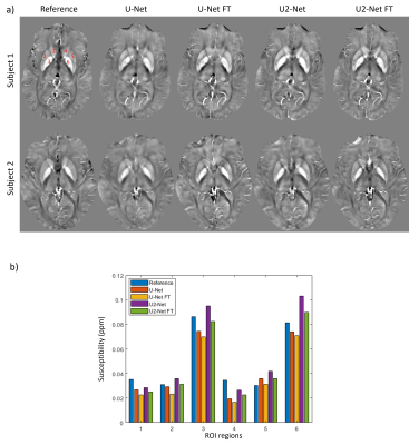 |
15 | Physics-based network fine-tuning for robust susceptibility mapping from high-pass filtered phase
Jinwei Zhang1, Alexey Dimov2, Hang Zhang1, Chao Li1, Thanh Nguyen2, Pascal Spincemaille2, and Yi Wang2
1Cornell University, New York, NY, United States, 2Weill Cornell Medicine, New York, NY, United States
A network fine-tuning step based on signal physics is proposed for deep learning based quantitative susceptibility mapping using high-pass filtered phase only to susceptibility. The proposed method showed better robustness compared to the pre-trained networks without fine-tuning when the test dataset deviated from the training dataset, such as a change in voxel size or high-pass filter cutoff frequency.
|
||
3559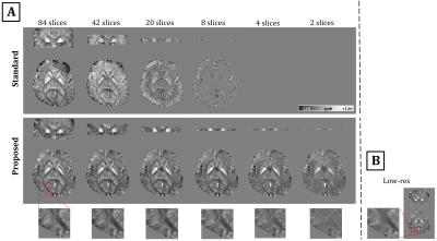 |
16 | Towards fast single-slice QSM: Challenges and possible solutions
Nashwan Naji1 and Alan H. Wilman1
1Department of Biomedical Engineering, University of Alberta, Edmonton, AB, Canada Susceptibility maps reconstructed from small-axial slabs suffer underestimation due to background-field removal imperfections near slab boundaries and the increased difficulty of solving a 3D-inversion problem with reduced-support in the direction of B0. Reliable QSM reconstruction from small slabs would enable higher resolution imaging within a reasonable time-frame and further accelerate clinical adoption. This work proposed using additional rapid low-resolution data, that serves as prior information, to improve background-field estimation and regularize the inversion-to-susceptibility process. Consistent high-resolution QSM at 3T was obtained from slabs of width as small as 6.8mm, aided by a lower-resolution dataset of 16 times coarser voxels. |
||
 Back to Meeting Home
 Back to the Program-at-a-Glance
The International Society for Magnetic Resonance in Medicine is accredited by the Accreditation Council for Continuing Medical Education to provide continuing medical education for physicians.
 Back to Meeting Home
 Back to the Program-at-a-Glance
 View the Presentation