Online Gather.town Pitches
Genitourinary & Women's Imaging I
Joint Annual Meeting ISMRM-ESMRMB & ISMRT 31st Annual Meeting • 07-12 May 2022 • London, UK

Online Gather.town Pitches
Genitourinary & Women's Imaging I
| Booth # | ||||
|---|---|---|---|---|
3686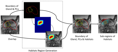 |
1 | Radiomics Analysis of the Prostate Cancer Habitats in Diagnosing Extracapsular Extension on Multi-Parametric MRI
Yihong Zhang1, Ying Hou2, Jie Bao3, Yang Song4, Dongmei Wu1, Yu-dong Zhang2, Xu Yan4, and Guang Yang1
1Shanghai Key Laboratory of Magnetic Resonance, East China Normal University, Shanghai, China, 2Department of Radiology, the First Affiliated Hospital with Nanjing Medical University, Nanjing, China, 3Department of Radiology, the First Affiliated Hospital with Soochow University, Soochow, China, 4MR Scientific Marketing, Siemens Healthineers Ltd, Shanghai, China We extracted radiomics features from subregions of the area where the lesion was close to the capsular boundary to diagnose the extracapsular extension (ECE). The model was trained with 574 cases, using 5-fold cross-validation and evaluated with an independent internal test cohort of 144 cases and an external test cohort of 146 cases. The model built with subregion features achieved AUC (area under receiver operating characteristic curve) values of 0.792 and 0.713 on the internal and external test cohort, respectively, outperforming the model built with whole lesion features whose internal and external test AUCs are 0.724 and 0.581, respectively. |
||
3687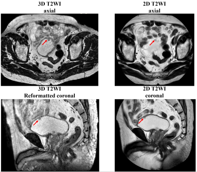 |
2 | Comparison of detection rate using compressed SENSE accelerated 3D T2-weighted FSE acquisitions and standard 2D acquisitions in bladder cancer Video Permission Withheld
Lei Ye1, Xiaoyong Zhang2, Yuntian Chen1, and Hui Xu1
1Department of Radiology, West China Hospital, Sichuan University, Chengdu, Sichuan, China, 2Clinical Science, Philips Healthcare, Chengdu, Sichuan, China
Conventional T2-weighted images using two-dimensional (2D) fast-spin-echo (FSE) are commonly used in clinical scanning of bladder, with a drawback of high missing rate. This study compared the BCa detection rate between compressed SENSE accelerated 3D and conventional 2D FSE T2WI within the same patients. The results showed that the accelerated 3D FSE T2-weighted acquisitions had excellent performance at detecting lesions smaller than 3cm and on the uncommon site of bladder wall.
|
||
3688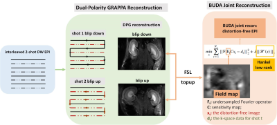 |
3 | DPG-BUDA: Distortion-free diffusion-weighted renal MRI in small rodents
Qiang Liu1,2,3, Yingjie Mei1,2,3, Quan Tao1,2,3, Qiqi Lu1,2,3, Xinyuan Zhang1,2,3, and Yanqiu Feng1,2,3
1School of Biomedical Engineering, Southern Medical University, Guangzhou, China, 2Guangdong Provincial Key Laboratory of Medical Image Processing & Guangdong Province Engineering Laboratory for Medical Imaging and Diagnostic Technology, Southern Medical University, Guangzhou, China, 3Guangdong-Hong Kong-Macao Greater Bay Area Center for Brain Science and Brain-Inspired Intelligence & Key Laboratory of Mental Health of the Ministry of Education, Southern Medical University, Guangzhou, China
We introduced blip-up/down acquisition (BUDA) strategy for interleaved 2-shot EPI to achieve motion robust, distortion-free rat renal diffusion-weighted imaging (DWI). Dual-polarity GRAPPA (DPG) method was implemented to correct Nyquist ghost artifact while simultaneously reconstructing the undersampled 2-shot data, then the field map was estimated from the opposing-distorted images. The field map was incorporated into a joint reconstruction with a structure low-rank constraint across the two shots to eliminate distortion and phase inconsistencies. DPG-BUDA permits high anatomical integrity while holding high scan efficiency for renal DWI in small rodents, it has the potential to benefit the characterization of the renal microstructure.
|
||
3689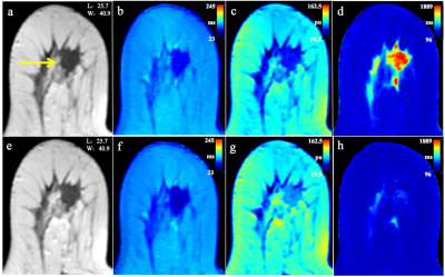 |
4 | Synthetic MRI with quantitative mappings for identifying HER2-enriched breast cancers and differentiating them from other molecular subtypes Video Not Available
Weibo Gao1, Quanxin Yang1, and Xiaocheng Wei2
1Second Affiliated Hospital of Xi’an Jiaotong University, Xi'an, China, 2GE Healthcare, MR Research China, Beijing, China
In this study, we aim to investigate whether quantitative parameters of synthetic MRI can be used to differentiate among molecular subtypes and prognostic factors in patients with breast cancer. It was concluded that SD of PD-Pre was significantly different between HER2-enriched and non-HER2-enriched breast cancers, which displayed optimal performance than others. Quantitative T2-Pre, T2-Gd, SD of PD-Pre, and SD of PD-Gd were significantly different in the molecular subtypes and can further differentiate luminal breast cancers from non-luminal subtypes. T1-Pre, T2-Pre, and SD of PD-Gd were significantly different between triple negative (TN) and non-TN breast cancers.
|
||
3690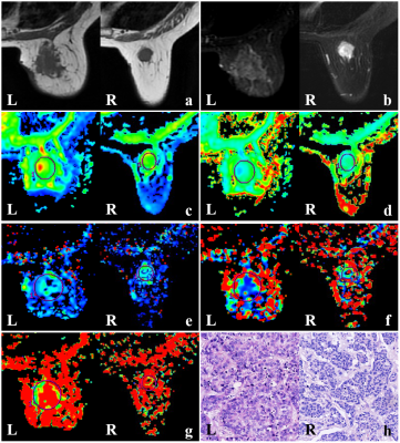 |
5 | Predictive value of IVIM combined with DKI for breast cancer axillary lymph node metastasis: A retrospective study Video Not Available
Zhe Zhou1, Yueqin Chen1, Fan Zhao1, Zhanguo Sun1, Hao Yu1, Weiqiang Dou2, and Weiwei Wang1
1the affiliated hospital of Jining medical university, Jining, China, 2GE Healthcare, MR Research China, Beijing, China An accurate preoperative prediction of axillary lymph node (ALN) metastasis is essential in clinic treatment. Intravoxel incoherent motion (IVIM) and diffusion kurtosis imaging (DKI) have been reported as effective non-invasive imaging methods in reflecting internal complexity of tumors. Few studies have used DKI combined with IVIM in diagnosing ALN metastasis of breast cancer. Therefore, in this study we aimed to explore if IVIM and/or DKI was useful in evaluating the ALN metastasis. |
||
3691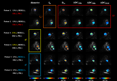 |
6 | Effects of oscillating frequency and SNR on quantifying microstructural properties in breast tumor using oscillating gradient diffusion MRI Video Not Available
Ruicheng Ba1, Xiaoxia Wang2, Zelin Zhang1, Hsu Yi-Cheng3, Yi Sun3, Jiuquan Zhang2, and Dan Wu1
1Key Laboratory for Biomedical Engineering of Ministry of Education, Department of Biomedical Engineering, College of Biomedical Engineering & Instrument Science, Zhejiang University, Hangzhou, China, 2Department of Radiology, Chongqing University Cancer Hospital, Chongqing, China, 3MR Collaboration, Siemens Healthineers Ltd., Shanghai, China
The oscillating frequency and SNR are important for accurate microstructural mapping in diffusion-time-dependent diffusion MRI, which however, are limited on clinical scanners. This study demonstrated that the dMRI acquisition protocol with frequency up to 50 Hz achieved significant higher accuracy than that with 33Hz, based on 1) Monte-carlo simulation, 2) clinical data on 37 breast tumor patients, and 3) correlation with histological data. Also, for the first time, we demonstrated the feasibility of oscillating gradient dMRI-based microstructural mapping in distinguishing breast tumor status clinically.
|
||
3692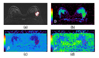 |
7 | Differentiation of benign and malignant breast lesions using diffusion-weighted imaging with a fractional order calculus model Video Permission Withheld
Guanying Wang1, Xingjun Gao1, Jianhong Li1, Dan Yang1, Dongmei Wu2, Yunfei Zhang3, Yongming Dai3, and Chunhong Wang3
1Department of Radiology, Xinyang Central Hospital, Xinyang, China, 2East China Normal University, Shanghai, China, 3Central Research Institute, United Imaging Healthcare, Shanghai, China
Diffusion-weighted imaging (DWI) is a powerful tool for cancer imaging. Many advanced DWI techniques have been developed and applied during clinical practice. Since fractional order calculus (FROC) model was proposed, it has shown huge clinical potential for cancer diagnosis, prognostic prediction and so on. However, scarcely has the FROC-DWI been utilized for breast imaging. The purpose of this study is to demonstrate that a new set of parameters (D, β and μ) from a fractional order calculus (FROC) diffusion model can be used to improve the accuracy of differentiating among benign and malignant breast lesions. Results suggested it’s feasible to use FROC diffusion model to improve the differentiation between benign and malignant breast lesions.
|
||
3693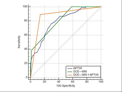 |
8 | Differentiation between benign and malignant breast masses using 3D amide proton transfer weighted imaging and dynamic contrast-enhanced MRI
Ruhua Wang1, Yan Zhang1, Liangjie Lin2, and Jingliang Cheng1
1Department of Magnetic Resonance Imaging, The First Affiliated Hospital of Zhengzhou University, Zhengzhou, China, 2Philips Healthcare, Beijing, China
This study evaluated the diagnostic value of dynamic contrast-enhanced MRI (DCE-MRI) combined with 3D amide proton transfer weighted imaging (APTWI) for differentiation between benign and malignant breast masses. Results indicated the quantitative MTRasym(3.5 ppm) value calculated by APTWI was significantly different between benign and malignant lesions, and the combination of APTWI and DCE-MRI improves the diagnostic performance for discriminating benign and malignant breast masses, especially category 4 masses.
|
||
3694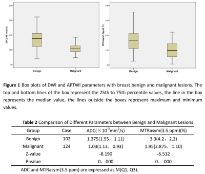 |
9 | 3D Amide Proton Transfer-weighted Imaging In Evaluation Of Breast Lesions: Comparison With Diffusion-weighted Imaging
Ruhua Wang1, Yan Zhang1, Liangjie Lin2, and Jingliang Cheng1
1Department of Magnetic Resonance Imaging, The First Affiliated Hospital of Zhengzhou University, Zhengzhou, China, 2Philips Healthcare, Beijing, China
This purpose of this study is to investigate the value of 3D APTWI and evaluate the diagnostic efficacy of its combination with DWI for discriminating breast benign from malignant lesions. Results showed the MTRasym(3.5 ppm) value calculated by APTWI for malignant breast lesions were significantly lower than those of benign lesions, and APTWI combined with DWI obtained a better diagnostic performance than the single method. Therefore, APTWI is a novel technique, which can be used to differentiate breast benign from malignant lesions noninvasively.
|
||
3695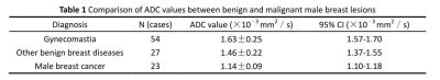 |
10 | The value of diffusion-weighted imaging and dynamic contrast-enhanced imaging in the differential diagnosis of male breast diseases
Ruhua Wang1, Yan Zhang1, and Jingliang Cheng1
1Department of Magnetic Resonance Imaging, The First Affiliated Hospital of Zhengzhou University, Zhengzhou, China
The male breast is often ignored because of its rudimentary and nonfunctional nature. However, the male breast can develop many benign and neoplastic diseases, including breast cancer.Much less is known regarding the appearance of benign and malignant male breast conditions. Therefore, in this retrospective study, we explored the value of diffusion-weighted imaging (DWI) and dynamic contrast-enhanced imaging (DCE-MRI) in the differential diagnosis of male breast diseases and confirmed the importance of DWI and DCE-MRI in the end.
|
||
3696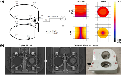 |
11 | Fabrication and evaluation of bilateral Helmholtz radiofrequency coil for thermo-stable breast image with reduced artifacts Video Not Available
Young Han Lee1, Kyu-Ho Song2, Jaemoon Yang1, Won Jun Kang3, Keum Sil Lee4, Min Jung Kim3, Eun-Kyung Kim1, Dan Heo1, Bo-Young Choe5, and Jin-Suck Suh1
1Department of Radiology, Research Institute of Radiological Science, Severance Hospital, Yonsei University College of Medicine, Seoul, Korea, Republic of, 2Department of Radiology, Washington University School of Medicine, St. Louis, MO, United States, 3Department of Nuclear Medicine, Severance Hospital, Yonsei University College of Medicine, Seoul, Korea, Republic of, 4Department of Radiology, Stanford University School of Medicine, Stanford, CA, United States, 5Department of Biomedical Engineering, and Research Institute of Biomedical Engineering, College of Medicine, The Catholic University of Korea, Seoul, Korea, Republic of
The positron emission tomography (PET)-magnetic resonance (MR) system is a newly emerging technique that yields hybrid images with high-resolution anatomical and metabolic information. With PET-MR imaging, a definitive diagnosis of breast abnormalities will be possible with high spatial accuracy and images will be acquired for the optimal fusion of anatomic locations. Therefore, we propose a PET-compatible 2-channel breast MR coil with minimal disturbance to image acquisition which can be used for simultaneous PET-MR imaging in patients with breast cancer.
|
||
3697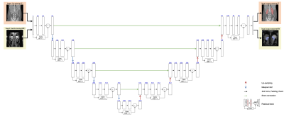 |
12 | Kidney segmentation in MR images using CT-trained ResUNet and transfer learning
Chang Ni1, Zhe Wang2, Mengkang Lu3, and Jeff L. Zhang1
1Vascular and Physiologic Imaging Research (VPIR) Lab, School of Biomedical Engineering, ShanghaiTech University, Shanghai, China, 2X-ray Systems Lab, School of Biomedical Engineering, ShanghaiTech University, Shanghai, China, 3SAIIP, School of Computer Science and Technology, Northwestern Polytechnical University, Xi'an, China Kidney segmentation is often necessary for analyzing renal MRI data. Deep learning approach shows much promise, but typically requires large numbers of images for model training. In this study, we explored the feasibility of segmenting MRI images using ResUNet pre-trained with CT images and fine-tuning with transfer learning. The fine-tuning step used 60 MRI images (from 5 subjects) only. The trained model performed excellently in segmenting an independent set of MRI images, with DICE similarity of 0.94 and volume error of 13%±9%. This study demonstrates the power of transfer learning in utilizing images of a different modality in kidney segmentation. |
||
3698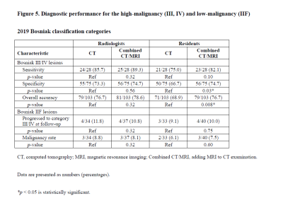 |
13 | Diagnostic value of adding MRI to CT examination for evaluating cystic renal masses using the 2019 Bosniak classification.
Yuki Arita1, Soichiro Yoshida2, Thomas C Kwee3, Hirotaka Akita1, Hiromi Edo4, Ryo Takeshita1, Haruka Okamura1, Misa Nagasaka1, Ryo Ueda5, Shigeo Okuda1, and Yasuhisa Fujii2
1Radiology, Keio University School of Medicine, Tokyo, Japan, 2Urology, Tokyo Medical and Dental University Graduate School, Tokyo, Japan, 3Radiology, University Medical Center Groningen, Groningen, Netherlands, 4Radiology, National Defence Medical College, Saitama, Japan, 5Radiation Technology, Keio University Hospital, Tokyo, Japan
We compared the interobserver agreement and diagnostic performance of CT alone and combined CT/MRI among two groups of readers (radiologists and residents) using the 2019 Bosniak classification (BC2019) to determine the malignancy of cystic renal masses (CRMs). Our study demonstrated that combined CT/MRI resulted in substantially high interobserver agreement between radiologists and residents. In addition, the diagnostic performance for category III/IV malignancy improved significantly for residents with combined CT/MRI when compared with evaluations based on CT alone. Thus, combined CT/MRI may be useful for diagnosing malignancy in CRMs, especially for non-expert readers, using the (BC2019).
|
||
3699 |
14 | A new radiomic tool based on apparent diffusion coefficient to predict the pathological grade of Bladder Cancer
Danyan Li1, Cheng Wang1, Chuanqi Sun2, Jie Meng1, Jilei Zhang3, Chengyu Ding3, Zijian Bian3, and Bing Zhang1
1department of radiology, Nanjing Drum Tower Hospital, The Affiliated Hospital of Nanjing University Medical School, Nanjing, China, 2Department of Biomedical Engineering, Guangzhou Medical University, Guangzhou, China, 3Philips Healthcare, Shanghai, China
The present study tried to develop a new radiomics tool based on apparent diffusion coefficient to predict the pathological grade of Bladder cancer (BCa). The results demonstrate that the radient boosting classifier (GBC) achieved the best effect in distinguishing pathological low or high grade of bladder cancer. And GBC also shows the best accuracy when we added the arch bridge sign on MRI as a new feature. This classification performance may suggest that the proposed method is a promising approach for preoperatively evaluating pathological grade in bladder cancer, which is very important for the choice of clinical treatment options.
|
||
3700 |
15 | Evaluation of isotropic high-resolution IRIS diffusion-weighted MRI in bladder cancer: clinical experience
Hui Xu1, Lei Ye2, Yuntian Chen2, and Xiaoyong Zhang3
1radiology, westchina hospital, Chengdu, China, 2westchina hospital, Chengdu, China, 3Philips Healthcare, Chengdu, China
Conventional single-shot echo planar imaging is widely used in bladder DWI for evaluating the malignant degree of lesions, however, it usually suffers from magnetic susceptibility artifacts and image blurring, which have limitations to identify the small lesions or lesions presented as slightly thickened bladder wall. This study this study was to investigate the feasibility and effectiveness of isotropic high-resolution IRIS DWI for the evaluation bladder lesions at 3T. The results suggested that the IRIS DWI with isotropic high resolution can effectively improve image quality and diagnostic confidence for bladder lesions.
|
||
 Back to Meeting Home
 Back to the Program-at-a-Glance
The International Society for Magnetic Resonance in Medicine is accredited by the Accreditation Council for Continuing Medical Education to provide continuing medical education for physicians.
 Back to Meeting Home
 Back to the Program-at-a-Glance
 View the Presentation