Online Gather.town Pitches
Preclinical Imaging II
Joint Annual Meeting ISMRM-ESMRMB & ISMRT 31st Annual Meeting • 07-12 May 2022 • London, UK

Online Gather.town Pitches
Preclinical Imaging II
| Booth # | ||||
|---|---|---|---|---|
3422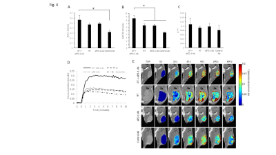 |
1 | MRI assessment of abscopal effect induced by radiation/immune checkpoint blockade combination therapy Video Permission Withheld
Kota Yamashita1, Yasunori Otowa1, Kazutoshi Yamamoto1, Jeffery R. Brender1, Nallathamby Devasahayam1, Murali C. Krishna1, and Shun Kishimoto1
1Radiation Biology branch, National Cancer Institute, Bethesda, MD, United States
Radiation therapy (RT) occasionally induces regression of non-irradiated metastatic lesions, which is called abscopal effect. Immune checkpoint inhibitors enhance abscopal effects. In this study we examined the physiological changes induced by abscopal effect and identify using MRI-based imaging biomarkers which predict the successful abscopal effect. Hypoxic fraction < 10 mmHg (HF10), permeability, perfusion, and CD8+ T cell infiltration in unirradiated tumor increased after the combination of RT and PD-1 blockade therapy. Interestingly, higher permeability/perfusion and lower HF10 in irradiated tumor before treatment is associated with slower growth of the unirradiated tumor after treatment of the tumor on the contralateral side.
|
||
3423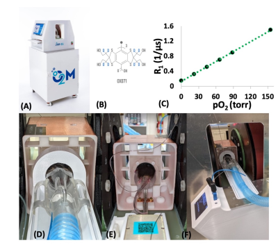 |
2 | In Vivo Oxygen Imaging of Implanted Islet Encapsulation Devices
Mrignayani Kotecha1, Navin Viswakarma1, Eliyas Siddiqui1, Sonny Patel1, and Boris Epel2
1O2M Technologies, LLC, Chicago, IL, United States, 2Radiation and Cellular Oncology, The University of Chicago, Chicago, IL, United States
The lack of oxygen supply to the highly metabolic pancreatic islet cells is one of the major factors contributing to the failure of islet transplantation devices designed to cure type I diabetes (T1D). Several approaches to improve oxygenation in these devices have been developed. However, the lack of available technologies to provide reliable pO2 assessment in and around devices hinders the progress severely. We performed in vivo oxygen imaging of cell loaded encapsulation devices using pulse electron paramagnetic resonance oxygen imaging (EPROI) technique. For the first time, whole-body EPROI is used to monitor pO2 of implanted devices in this work.
|
||
3424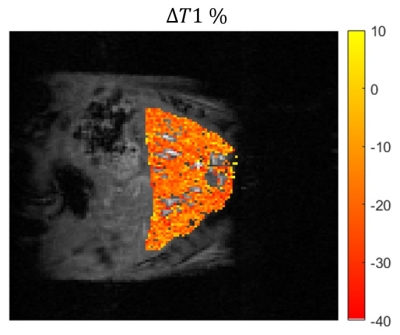 |
3 | Free-breathing liver T1 mapping for quantitative oxygen consumption
Janaka Wansapura1
1Advanced Imaging Research Center, University of Texas Southwestern, Dallas, TX, United States
We introduce a dual flip angle T1 method based on respiratory triggered cine spoiled gradient echo acquisition. The method is used to assess the change in T1 of liver tissue between breathing medical air and 100% O2 in an animal model. Our results show small but significant decrease in T1 of liver due to hypoxia, indicating increased oxygen partial pressure in tissue.
|
||
3425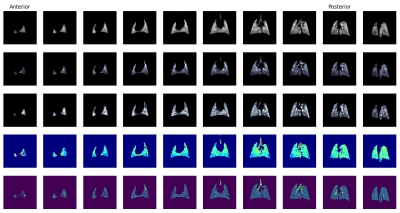 |
4 | Dynamic Spiral Imaging of Rodent Pulmonary Ventilation using Natural-Abundance Hyperpolarized 129Xe Imaging Video Not Available
Luis Loza1, Tahmina Achekzai1, Stephen Kadlecek1, Hooman Hamedani1, Faraz Amzajerdian1, Mostafa Ismail1, Ian Duncan1, Kai Ruppert1, and Rahim Rizi1
1University of Pennsylvania, Philadelphia, PA, United States
While a hyperpolarized 129Xe MRI-derived fractional ventilation is a valuable metric for assessing lung function, currently implemented schemes use enriched xenon and rely on static images acquired over breath holds to derive FV. In order to probe pulmonary ventilation under more physiologically natural conditions while keeping costs low, we acquired images during continuous controlled breathing in a mechanically ventilated rat using natural-abundance xenon. In addition to FV, we were able to derive the additional physiologically relevant metrics of tidal volume, functional residual capacity, and phase.
|
||
3426 |
5 | The relationship of arterial input function between abdominal aorta and hepatic artery
Jie Huang1, Yuancheng Liu2, Xianchun Zeng2, Chunqi Qian1, and Erik M. Shapiro1
1Michigan State University, East Lansing, MI, United States, 2Guizhou Provincial People's Hospital, Guiyang, China
The hepatic artery (HA) input is required in the dual-input, two-compartment model of studying liver function with dynamic gadoxetate-enhanced MRI. The dynamic contrast agent concentration in abdominal aorta (AA) is usually measured and used as the arterial input function instead of that measured in the HA because its small size. In this study, we determined the relationship between these two concentrations. We found that the measured arterial input function in AA can be reliably used as the HA input function with a suppression scaling factor taken into account.
|
||
3427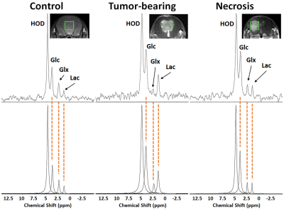 |
6 | Deuterium (2H) MR Spectroscopy Distinguishes Tumor (Glioblastoma) from Radiation Necrosis in Mouse Brain
Kyu-Ho Song1, John A. Engelbach1, James D. Quirk1, Keith M. Rich2,3, Joseph J.H. Ackerman1,4,5,6, and Joel R. Garbow1,6
1Department of Radiology, Washington University, St. Louis, MO, United States, 2Department of Neurosurgery, Washington University, St. Louis, MO, United States, 3Department of Radiation Oncology, Washington University, St. Louis, MO, United States, 4Department of Chemistry, Washington University, St. Louis, MO, United States, 5Department of Internal Medicine, Washington University, St. Louis, MO, United States, 6Alvin J. Siteman Cancer Center, Washington University School of Medicine, St. Louis, MO, United States
Employing mouse models of radiation necrosis (one that displays the histologic hallmarks of the clinical condition) and glioblastoma (GL261), deuterium (2H) MR spectroscopy, in concert with infusion of 2H-labelled glucose, was employed to ascertain whether the 2H MRS signatures could differentiate the two lesions, an unmet clinical need. The 2H MR metabolic profiles of the two lesions were markedly different. In the tumor, the Warburg effect (aerobic glycolysis, fermentation) converted glucose nearly exclusively to lactate. In radiation necrosis, oxidative phosphorylation (respiration) dominated, with glucose converting to TCA cycle intermediates glutamate and glutamine. Thus, 2H MR distinguishes glioblastoma vs. radiation necrosis.
|
||
3428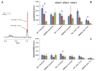 |
7 | Detection of Metabolic Biomarkers of Response to BRAF and MEK Inhibitor Therapy of Melanoma
Pradeep Kumar Gupta1, Stepan Orlovskiy1, David S. Nelson1, Stephen Pickup1, Alexander A. Shestov1, Fernando Arias-Mendoza1, Mary E. Putt2, Dennis B. Leeper3, Jerry D. Glickson1, and Kavindra Nath1
1Department of Radiology, Perelman School of Medicine, University of Pennsylvania, Philadelphia, PA, United States, 2Department of Biostatistics and Epidemiology, Perelman School of Medicine, University of Pennsylvania, Philadelphia, PA, United States, 3Department of Radiation Oncology, Thomas Jefferson University, Philadelphia, PA, United States
In vivo 1H and 31P MRS were used to monitor the effects of BRAF and MEK inhibitors therapy in two metabolically different melanoma xenograft models. Our approach is to combine BRAF inhibitor with MEK inhibitor in melanoma xenograft models. Combination of BRAF and MEK inhibitors has been shown to be more effective than the use of either drug alone. Differences in relative levels of glycolysis between two melanoma xenograft models may produce differential therapeutic responses to BRAF and MEK inhibitors . In melanoma, metabolic changes in response to targeted kinase inhibitor therapy occur rapidly and are related to subsequent tumor response.
|
||
3429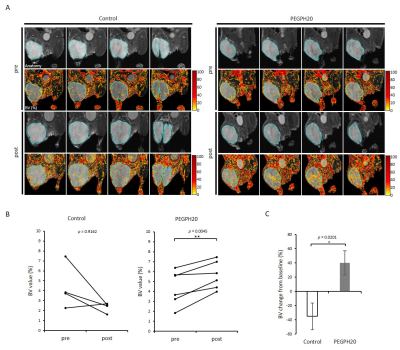 |
8 | Multimodal molecular imaging assessment of tumor microenvironment. Hyaluronan depletion induces tumor reoxygenation and radiosensitization
Shun C Kishimoto1, Tomohiro Seki2, Yu Saida3, Yasunori Otowa1, Kota Yamashita1, Kazutoshi Yamamoto1, Nallathamby Devasahayam1, Jeffrey R Brender1, and Murali C Krishna1
1NCI, Bethesda, MD, United States, 2Josai University, Sakado, Japan, 3Medical and Dental Hospital, Niigata University, Niigata, Japan
PEGPH20 is a PEGylated form of recombinant human hyaluronidase PH20. The purpose of this study is to investigate physiologic changes in pancreatic adenocarcinoma xenograft after PEGPH20 treatment using multi-modal imaging and further determine the utility of PEGPH20 as radiosensitizer. PEGPH20 treatment significantly increased intratumor pO2, blood perfusion and volume, and decreased glycolytic flux assessed by EPR, MRI with USPIO or Gd, and 13C DNP MRI, respectively. PEGPH20 enhanced treatment effect of radiotherapy. The results validated the utility of the imaging methods to monitor the changes in the tumor microenvironment after hyaluronan depletion and to predict the radio-sensitizing effect of PEGPH20.
|
||
3430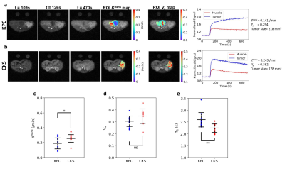 |
9 | DWI Metrics Differentiating Intraductal Papillary Mucinous Neoplasms from Pancreatic Cancer: A study in GEM Models
Miguel Romanello Giroud Joaquim1, Emma E Furth2, Yong Fan2, Hee Kwon Song2, Stephen Pickup2, Jianbo Cao3, Hoon Choi2, Mamta Gupta2, Cynthia Clendenin2, Thomas Karasic2, Jeffrey Duda2, James Gee2, Peter O'Dwyer2, Mark Rosen2,
and Rong Zhou1
1Department of Radiology, University of Pennsylvania, Philadelphia, PA, United States, 2University of Pennsylvania, Philadelphia, PA, United States, 3University of Cambridge, Cambridge, United Kingdom
Intraductal Papillary Mucinous Neoplasms (IPMN) are recognized as important precursors to invasive pancreatic ductal adenocarcinoma (PDAC). While IPMN requires surveillance without treatment, a clinical marker is lacking which can identify those undergoing malignant transformation. In two genetic engineered mouse models (KPC and CKS), which resemble human PDAC and IPMN, respectively, we tested the hypothesis that differences in cellular architecture and stromal features between PDAC and IPMN present themselves in DW-MRI and /or DCE-MRI metrics. Our data revealed an almost complete separation of ADC values between CKS (benign) vs. KPC (malignant) tumors and identified histopathological features corroborating the imaging metrics.
|
||
3431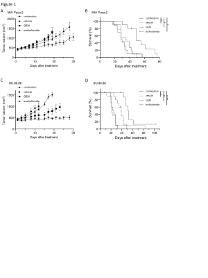 |
10 | EPR and MRI studies of microenvironment in pancreatic cancers. Mechanistic studies of combination therapy with gemcitabine and evofosfamide.
Yasunori Otowa1, Kota Yamashita1, Kazutoshi Yamamoto1, Jeffery R Brender1, Nallathamby Devasahayam1, Shun Kishimoto1, and Murali C Krishna1
1National Cancer Institute, Bethesda, MD, United States
Although gemcitabine (GEM) and evofosfamide as monotherapy has a weak effect on MIA Paca-2 and SU 86.86 pancreatic ductal adenocarcinomas respectively, yet the combination therapy shows synergistic effect on both tumor models. The purpose of this study is to understand the synergistic effects of combination therapy using multi-modal imaging methods. In MIA Paca-2 tumors, treatment with GEM induced hypoxia in tumor microenvironment by decreasing the perfusion, which can potentiate evofosfamide. On the other hand, in SU.86.86 tumors, evofosfamide increased blood volume and pO2 in the tumor microenvironment, to potentiate efficacy of anti-proliferatives such as GEM.
|
||
3432 |
11 | Assessment of anti-mitotic treatment response in breast cancer using temporal diffusion spectroscopy
xiaoyu jiang1,2, Sean P p Devan1, Jingping Xie1, John C. Gore1,3, and junzhong xu1,4
1Vanderbilt University Institute of Imaging Science, nashville, TN, United States, 2Department of Radiology and Radiological Sciences, Vanderbilt University Institute of Imaging Science, nashville, TN, United States, 3Department of Radiology and Radiological Sciences, Vanderbilt University Medical Center, nashville, TN, United States, 4Department of Radiology and Radiological Sciences, Vanderbilt University Medical Center, Nashville, TN, United States
Reliable and sensitive methods for assessing the response of breast cancer to treatment are critical for rapid selection of the most appropriate therapy for individual patients, and development of novel therapies. Paclitaxel has been reported to slow/block mitosis at the metaphase-anaphase transition and induce apoptosis, leading to cell shrinkage and membrane breakdown. Using a biophysical model that allows derivation of microstructural parameters (e.g., cell size d) and an indicator of the intracellular water lifetime (τin ) from the tdiff dependence of diffusion MRI signals, we monitored the treatment response of breast cancer in both cell cultures and solid tumors in vivo.
|
||
3433 |
12 | Optimization of a highly sensitivity 19F MRI probe for NK cell immunotherapy tracking
Gary Martinez1, Dmitry Nevozhay2, Juan C Bournat3, Donghang Cheng3, Ajay Sharma3, Tyler King4, Emily Que5, Vidya Gopalakrishnan6, Konstantin Sokolov1, and James A Bankson1
1Imaging Physics, The University of Texas MD Anderson Cancer Center, Houston, TX, United States, 2Dmitry Nevozhay, The University of Texas MD Anderson Cancer Center, Houston, TX, United States, 3Pediatrics, The University of Texas MD Anderson Cancer Center, Houston, TX, United States, 4Chemistry, The University of Texas at Austin, Austin, TX, United States, 5The University of Texas at Austin, Austin, TX, United States, 6The University of Texas MD Anderson Cancer Center, Houston, TX, United States
NK cell immunotherapy has great potential as a safe and effective anticancer therapy yet there are significant challenges for immunotherapy in brain tumors. We have developed a 19F probe with enhanced sensitivity for the purposes of 19F labeling of NK cells to monitor NK cell biodistribution and immunotherapy. In this paper, we report on the relaxometric characteristics of the probe, acquisition optimization, and initial ex vivo imaging tests in murine models. The agent displayed improved sensitivity relative to Celsense and was clearly visible in the cerebral cortex in approximately equivalent amounts that will be used for cell tracking experiments.
|
||
3434 |
13 | Fluorine-19 MRI of Stem Cell-Derived Alveolar-Like Macrophages Tagged with Perfluoropolyether
Janny Yeyoung Kim1,2, Michael Litvack1, Daniel Li1, Brandon Zanette1, Martin Post1, and Giles Santyr1,2
1Translational Medicine, The Hospital for Sick Children, Toronto, ON, Canada, 2Medical Biophysics, University of Toronto, Toronto, ON, Canada
When stem cell-derived alveolar-like macrophages (ALMs) are labeled with perfluoropolyether (PFPE), they can potentially be monitored and tracked longitudinally in vivo using 19F MRI. In this work, different concentrations of PFPE and 5 million PFPE-labeled ALMs were imaged in vitro using 19F MRI at 3T. The results demonstrated the feasibility of detecting a small number of PFPE-labeled ALMs. This work will provide guidance for the upcoming in vivo instillation experiments in rat lungs. Longitudinal in vivo studies will further aid ALMs to be clinically translated as a stem cell therapy to treat chronic lung diseases.
|
||
3435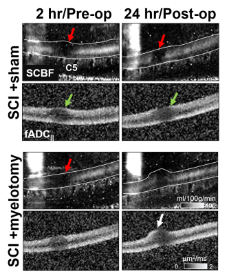 |
14 | Multimodal MRI of cervical spinal cord injury before and after surgical myelotomy
Seung-Yi Lee1, Briana P Meyer2, Shekar N Kurpad3, and Matthew D Budde3
1Medical College of Wisconsin, Milwaukee, WI, United States, 2Biophysics, Medical College of Wisconsin, Milwaukee, WI, United States, 3Neurosurgery, Medical College of Wisconsin, Milwaukee, WI, United States
Surgical myelotomy, a procedure intended to relieve pressure on an acutely-injured spinal cord, improves long-term functional and pathologic outcomes in animal models. However, the direct consequences of myelotomy on the cord pathophysiology have not been evaluated in the acute setting. In this work, we used multimodal MRI tailored for the spinal cord to monitor changes in edema, hemorrhage, axonal injury, and perfusion immediately before and 24 hours after surgical myelotomy. The results demonstrate spatial and temporal changes and provide unique insight into the pathophysiology of acute injury and its intervention.
|
||
3436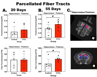 |
15 | Brain Connectomics in a Mouse Model of Early and Late Stage Pruritus
Talaignair N Venkatraman1, Ouyang Chen2, Allen W Song3, Ru-Rong Ji2, and Chris D Lascola3
1Radiology, Duke University Medical Center, Durham, NC, United States, 2Anesthesiology, Duke University Medical Center, Durham, NC, United States, 3BIAC and Radiology, Duke University Medical Center, Durham, NC, United States
We performed high-resolution ex-vivo DTI in mice with cutaneous T-Cell lymphoma (CTCL) in a mouse model of pruritus. Our results show significant network changes within and between specific brain regions, namely thalamus and hippocampus, by the later stage of disease. At the chronic stage (55 days), micro-structural changes and enhanced connectivity are established between the hippocampus and thalamus.
|
||
3437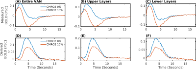 |
16 | Investigating the origin of the BOLD initial dip with biophysical simulations based on realistic microvascular networks: flow or metabolism?
Joerg Peter Pfannmoeller1, Grant Adison Hartung1, Avery Berman1, and Jonathan Rizzo Polimeni1
1Martinos Center for Biomedical Imaging, Massachusetts General Hospital, Charlestown, MA, United States
We used simulations to achieve insights about the nature of the initial dip and found that it is indeed possible that retrograde propagation causes a local decoupling between hemodynamics and neural activity localized to the upper layers of the cortex. The simulations allowed to assess the contributions of the different simultaneously occurring contributions and point towards a high specificity of the initial dip to the capillaries or the veins. A methodological difficulty was posed by the limited availability of arterial dilation data. Therefore, we derived dilation traces from the measurements and determine a range of possible propagation velocities.
|
||
3438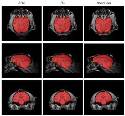 |
17 | Automated Brain Extraction from T1-weighted MRI of Rhesus Macaques using a U-Net Deep Learning Framework
Anqi Zhang1, Donghoon Kim1,2, Jeong-chul Kim3, Brad A. Hobson1, Sonny R. Elizaldi4, John Morrison5, Smita Iyer4, Abhijit J. Chaudhari2,4, and Youngkyoo Jung1,2,3
1Biomedical Engineering, University of California, Davis, CA, United States, 2Radiology, University of California, Davis, CA, United States, 3Radiology, Wake Forest School of Medicine, Winston-Salem, NC, United States, 4California National Primate Research Center, University of California, Davis, CA, United States, 5Neurology, University of California, Davis, CA, United States Transfer learning of an advanced deep learning framework, utilizing 3D U-Net pre-trained model, and trained on human brain MRI scans is proposed for brain extraction from MRI of other species. The proposed network architecture successfully performed whole brain extraction from rhesus macaque brain MRIs automatically with high accuracy, reduced errors, and lower computational cost. Options for data augmentation and different learning rates were also tested. Successful implementation of automated brain extraction would offer the potential to apply the same strategy on other animal models. |
||
3439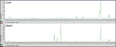 |
18 | Investigating time-restricted feeding and metabolic sexual dimorphism in Nile grass rats by time-domain NMR lipid profiling
Hayden Johnson1, Thomas Yates1, Chidambaram Ramanathan2, Melissa Puppa2, Marie van der Merwe2, and Aaryani Tipirneni-Sajja1
1Biomedical Engineering, University of Memphis, Memphis, TN, United States, 2College of Health Sciences, University of Memphis, Memphis, TN, United States
NMR-based metabolomics is a powerful technique for elucidating the metabolic effects of diet through the identification and quantification of molecules present in tissues. This study utilizes time-domain processing of NMR data for robust, reproducible resonance characterization in the profiling of lipid species in heart and liver tissues of male and female animals subject to high fat diets with and without time-restriction. Results show triglyceride and fatty acid accumulation was associated with eating a high fat diet, but these effects were attenuated by time-restricted feeding. Hepatic lipids expressed a sexually dimorphic response, with higher concentrations of many lipids in female subjects.
|
||
3440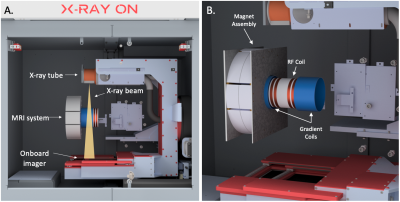 |
19 | Integrating a Unilateral MRI Scanner with a Small Animal Irradiator for Preclinical MR-Guided Radiation Experiments
Jace Grandinetti1, Yuncheng Zhong1, Yuting Peng1, Chenyang Shen1, Shu Zhang1, and Xun Jia1
1Radiation Oncology, UT Southwestern Medical Center, Dallas, TX, United States
Magnetic Resonance-guided Radiation Therapy (MRgRT) has recently become available in human RT, however, this technology does not currently exist for preclinical systems. We propose a novel approach to develop and integrate an MR system with an existing small animal irradiator and report our ongoing progress constructing such a system. The MR system is comprised of a unilateral Halbach cylinder with permanent magnets and split gradient coils to prevent obstruction of the radiation beam while targeting a 4cm region of interest. Further development is ongoing for a split broadband RF coil and custom pulse sequences to maximize signal-to-noise.
|
||
 Back to Meeting Home
 Back to the Program-at-a-Glance
The International Society for Magnetic Resonance in Medicine is accredited by the Accreditation Council for Continuing Medical Education to provide continuing medical education for physicians.
 Back to Meeting Home
 Back to the Program-at-a-Glance
 View the Presentation