Online Gather.town Pitches
MR Contrasts III
Joint Annual Meeting ISMRM-ESMRMB & ISMRT 31st Annual Meeting • 07-12 May 2022 • London, UK

| Booth # | ||||
|---|---|---|---|---|
4084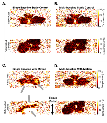 |
1 | Dynamic 3D Stack-of-Radial PRF MR Thermometry to Monitor High-Intensity Focused Ultrasound Heating: Validation in a Tissue Motion Phantom
Qing Dai1,2, Le Zhang1,3, Xinzhou Li1,2, Pengkang Yu4, Tsu-Chin Tsao4, and Holden H. Wu1,2
1Department of Radiological Sciences, University of California, Los Angeles, Los Angeles, CA, United States, 2Department of Bioengineering, University of California, Los Angeles, Los Angeles, CA, United States, 3Department of Radiology, Mayo Clinics, Rochester, MN, United States, 4Department of Mechanical and Aerospace Engineering, University of California, Los Angeles, Los Angeles, CA, United States
Multi-baseline proton resonant frequency (PRF) shift MR thermometry has been proposed to address the potential mismatch between baseline images and images during thermal therapy in moving organs. Previous methods had to compromise the spatial coverage to increase temporal resolution for resolving motion. This work developed a dynamic 3D stack-of-radial multi-baseline PRF MR thermometry method to monitor high-intensity focused ultrasound heating in moving tissue. The proposed method achieved stable and accurate thermometry with volumetric coverage and high spatiotemporal resolution, validated in a tissue motion phantom with temperature probe measurements.
|
||
4085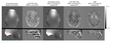 |
2 | Simultaneous Time-of-Flight (TOF) MR Angiography, SWI and QSM without compromising TOF features
Ashmita De1, Justin Grenier1, and Alan Wilman1
1Biomedical Engineering, University of Alberta, Edmonton, AB, Canada
Multiple-echo gradient echo sequences developed to simultaneously compute TOF-MRA, SWI and QSM in previous studies have either compromised TOF-MRA features or have not computed QSM. In this abstract, images were acquired in healthy volunteers at 3T with different parameter variations of the proposed 3D triple-echo gradient echo sequence with all TOF-MRA features. The effect of TOF-MRA features on phase and QSM were explored and corrected to obtain artifact-free QSM. The parameters of the sequence have been optimized to obtain TOF-MRA, SWI, QSM and R2* maps simultaneously.
|
||
4086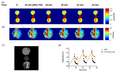 |
3 | MR thermometry using fingerprinting: application to lead heating
Enlin Qian1, Pavan Poojar1, David H. Gultekin1, J. Thomas Vaughan1, Devashish Shrivastava1, Zhezhen Jin2, Brett E. Youngerman3, and Sairam Geethanath1
1Columbia Magnetic Resonance Research Center, Columbia University, New York, NY, United States, 2Department of Biostatistics, Columbia University, New York, NY, United States, 3Columbia University Irving Medical Center, Columbia University, New York, NY, United States
We explore the feasibility of noninvasive temperature measurement around deep brain stimulation (DBS) leads using magnetic resonance fingerprinting (MRF) by leveraging 1) the dependency of T1 on temperature; 2) MRF’s efficient T1 and B0 mapping. We conducted a T1 and temperature calibration experiment and a lead calorimetry experiment on bovine muscle. The effects of B0 inhomogeneity and temperature profiles of different regions of interest (ROI) were investigated. The calibration showed a strong correlation between temperature and T1 (R2>0.98). We observed a maximum change of 10.2 oC near leads (~1mm away) during calorimetry experiments, validated by fluoroptic probes.
|
||
4087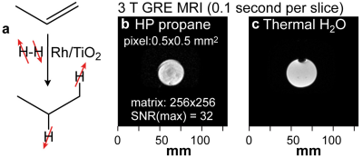 |
4 | Disposable, Handheld Clinical-Scale HP Propane Hyperpolarizer for Pulmonary MRI
Eduard Y Chekmenev1, Anna Samoilenko2, Jonathan R. Birchall2, Kirill V. Kovtunov3, Larisa M Kovtunova3, Valeriy I. Bukhtiyarov4, Igor V. Koptyug3, Chunqi Qian5, Boyd M Goodson6, and Nuwandi M. Ariyasingha2
1Chemistry and Oncology, Wayne State University, Detroit, MI, United States, 2Chemistry, Wayne State University, Detroit, MI, United States, 3International Tomography Center RAS, Novosibirsk, Russian Federation, 4Boreskov Institute of Catalysis RAS, Novosibirsk, Russian Federation, 5Radiology, Michigan State University, Lansing, MI, United States, 6Southern Illinois University Carbondale, Carbondale, IL, United States
We demonstrate a disposable and handheld hyperpolarizer for clinical-scale production of hyperpolarized propane gas for application as an inhalable contrast agent for pulmonary MRI. The device consists of an aluminum commercial spray can, which contains a mixture of pressurized propylene and parahydrogen gases. This filled container can be easily transported and stored for weeks. The hyperpolarized propane production is initialized by valve actuation, which releases the propylene-parahydrogen mixture into a mini-catalytic reactor producing a stream of pure (without catalyst) hyperpolarized propane gas on demand. The feasibility of 3 T EPI, FIESTA, and GRE MRI in hyperpolarized phantoms is demonstrated.
|
||
4088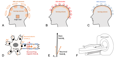 |
5 | Quantum Sensing of Local Neuronal Firings (qsLNF) in Human Brain via Proton (1H) MRI: Proof of Concept
Yongxian Qian1, Liz Calderon1, Xingye Chen1, Anli Liu2, Yvonne W. Lui1, and Fernando E. Boada1
1Radiology, New York University, New York, NY, United States, 2Neurology, New York University, New York, NY, United States
Neuronal firing generates fast action potentials along axon and slow postsynaptic currents at dendrites, which are difficult to detect and locate by scalp EEG and MEG placed above the skull. Here we propose local quantum sensing via endogenous proton (1H) nuclear spins inside firing neurons and detect magnetic fields indued by both action potentials and postsynaptic currents. Computer simulations and human studies showed that the proposed technique has the potential to non-invasively detect and locate neuronal firings in the brain through the acquisitions of FID signal on a 3T MRI scanner with multi-channel array coil.
|
||
4089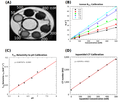 |
6 | In vivo pH Measurement by T2ex Relaxometry of Iodinated Contrast Agent and Comparison with CEST pH Imaging
Pietro Irrera1, Bruna Victorasso Jardim-Perassi1, Epifanio Ruiz2, Mikalai Budzevich2, Justin YC Lau2, and Robert J Gillies1
1Cancer Physiology, Moffitt Cancer Center, Tampa, FL, United States, 2Small Animal Imaging Laboratory, Moffitt Cancer Center, Tampa, FL, United States
This work is based on a well-known MRI property, the transverse relaxation changes of bulk water due to the presence of a solute with water-exchangeable protons. Many molecules can exhibit this ability to change water T2, such as glucose, urea, ammonium chloride, metals and paramagnetic complexes. Here we used the FDA-approved iodinated agent iopamidol (Isovue), widely used for CEST applications, as a T2ex agent to quantify pH in vitro and in vivo, since its –NH groups exhibit pH-dependent exchange rates with water protons. Computed tomography provided iopamidol concentrations to complement the MR relaxometry measurements, allowing determination of pH in vivo.
|
||
4090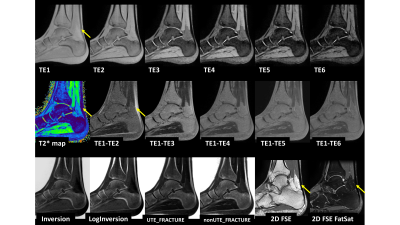 |
7 | Visualizing and generating a quantitative map of short T2* tissues and producing CT-like imaging of bony structures using multi-echo UTE
Hung Phi Do1, Dawn Berkeley1, Brian Tymkiw1, and Mo Kadbi1
1Canon Medical Systems USA, Inc., Tustin, CA, United States
Two-dimensional Fast Spin Echo is a workhorse of clinical MRI. However, its minimum TE is in the order of milliseconds or tens of milliseconds, which limits its ability to image tissues with short T2*. This work aims to optimize the multi-echo ultra-short echo time (UTE) sequence to (i) visualize, (ii) generate an associated quantitative map of tissues with short T2*, and (ii) to produce CT-like images of bony structures, which often are not visible in clinical images routinely acquired using FSE sequence.
|
||
4091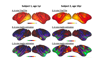 |
8 | Multivariate mapping of cortical myeloarchitecture in the macaque cortex
Erika P Raven1, Jeffrey Bennett2, Jelle Veraart1, Claude Lepage3, Joey Charbonneau2, Eliza Bliss-Moreau2, and Jiangyang Zhang1
1Radiology, NYU School of Medicine, New York, NY, United States, 2Psychology, UC Davis, Davis, CA, United States, 3Montreal Neurological Institute, McGill University, Montreal, QC, Canada
Mapping cortical myelination has been a long-standing goal for accurate parcellation and classification of cytoarchitectonic boundaries. We investigated a multivariate myelin mapping approach for the accurate parcellation of small cortical subregions of the insula by exploiting the co-localization of iron with dense bands of myelin in cortical gray matter. We assessed the performance of multivariate myelin mapping in the rhesus macaque, both in vivo and ex vivo, and compared its performance with T1w/T2w ratio imaging. We demonstrated that multivariate myelin mapping is a translatable myelin sensitive technique for the differentiation of insula subregions.
|
||
4092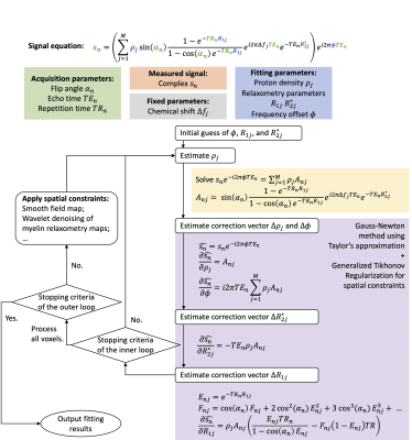 |
9 | Iterative decomposition of multi-compartment relaxometry with least square estimations (IDMCR) for UTE relaxometry in brain
Jingwen Yao1, Nikhil Deveshwar1,2, and Peder E. Z. Larson1
1Department of Radiology and Biomedical Imaging, University of California, San Francisco, San Francisco, CA, United States, 2Graduate Program in Bioengineering, UCSF/UC Berkeley, San Francisco, CA, United States
To improve the fitting performance of a UTE relaxometry model, we proposed an iterative decomposition method of multi-compartment relaxometry with least square estimations (IDMCR), based on Gauss-Newton estimations and incorporating spatial constraints. Monte Carlo simulation showed that IDMCR provided robust and unbiased estimation of ultra-short T2* myelin fraction, which was further improved by adding spatial constraints. We examined the fitting algorithm in one healthy volunteer UTE relaxometry data and showed clear contrast of gray and white matter on the fitted myelin fraction map. The framework of IDMCR could also be easily adapted for other nonlinear parameter fitting problems.
|
||
4093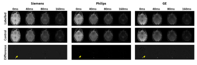 |
10 | Measuring cerebral venous oxygenation: multi-site multi-vendor standardization of TRUST MRI and association with end-tidal CO2
Abubakr Eldirdiri1, Jiachen Zhuo1, Zixuan Lin2, Hanzhang Lu2,3,4, Rao Gullapalli1, and Dengrong Jiang2
1Department of Diagnostic Radiology and Nuclear Medicine, University of Maryland School of Medicine, Baltimore, MD, United States, 2The Russell H. Morgan Department of Radiology & Radiological Science, Johns Hopkins University School of Medicine, Baltimore, MD, United States, 3Department of Biomedical Engineering, Johns Hopkins University School of Medicine, Baltimore, MD, United States, 4F.M. Kirby Research Center for Functional Brain Imaging, Kennedy Krieger Research Institute, Baltimore, MD, United States This work presents a multi vendor multi-site MRI study in which a TRUST sequence was harmonized across three MRI platforms from GE, Siemens, and Philips to measure the cerebral venous oxygenation Yv. We carried out intra-scanner and inter-scanner analysis on the variability of the venous oxygenation measurements and demonstrated high measurements reproducibility across the three platforms. Moreover, we examined the relationship between the fluctuations in end-tidal CO2 and the Yv measurements and showed that end-tidal CO2 can reduce the variability in Yv measurements in multi-site setting. |
||
4094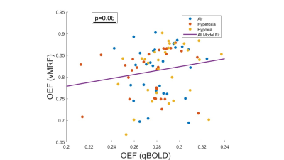 |
11 | Comparison of Quantitative BOLD and Vascular MRF for Mapping Brain Oxygenation
Linh N N Le1, Gregory J Wheeler1, Thomas Christen2, Greg Zaharchuk3, and Audrey P Fan 1,4
1Biomedical Engineering, University of California, Davis, Davis, CA, United States, 2Grenoble Institute of Neuroscience, Grenoble, France, 3Radiology, Stanford University, Palo Alto, CA, United States, 4Neurology, University of California, Davis, Davis, CA, United States
We compared two approaches of quantitative Blood-Oxygenation Level-Dependent (qBOLD) and vascular MR Fingerprinting (vMRF) to map brain oxygenation from Gradient-Echo Sampling of Free Induction Decay and Echo (GESFIDE) scans acquired during normoxic, hyperoxic, and hypoxic gas breathing conditions. In 12 healthy subjects, OEF calculated from qBOLD (qOEF) and OEF generated from vMRF (vOEF) were inversely correlated to pulse oxygenation (SaO2) (p=0.032 and p=0.014, respectively), indicating that qBOLD and vMRF are reliable methods for brain oxygenation mapping.
|
||
4095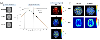 |
12 | Oxygen Extraction Fraction using Quantitative BOLD and Cerebral Blood Flow during Vasodilation
Linh N N Le1, Gregory J Wheeler2, Alique M Momjian2, Corinne A Donnay3, Nicholas P Blockley4, and Audrey P Fan2,3
1Biomedical Engineering, University of California, Davis, San Diego, CA, United States, 2Biomedical Engineering, University of California, Davis, Davis, CA, United States, 3Neurology, University of California, Davis, Davis, CA, United States, 4School of Life Sciences, University Of Nottingham, Nottingham, United Kingdom
We quantified brain oxygen extraction fraction (OEF) using quantitative Blood-Oxygenation Level-Dependent (qBOLD) modeling of asymmetric spin echo (ASE) scans acquired at baseline and after pharmacological vasodilation with acetazolamide. In 9 healthy subjects, 60.51±42.00% CBF elevation and -8.45±9.71% OEF decrease in response to vasodilation was observed in multiple gray matter regions in the cortex. The correlation between cerebral blood flow (CBF) and OEF was inversely proportional (p=0.02), indicating complementary vascular responses to the acetazolamide stimulus.
|
||
4096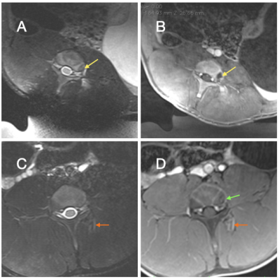 |
13 | MR-guided Focused Ultrasound of the Dorsal Root Ganglion for the Treatment of Low Back Pain: Preclinical Study in a Peripheral Nerve Injury Model
Viola Rieke1, Candace Floyd1, Allison Payne1, Henrik Odeen1, Matt Zabriskie1, Michelle Kline1, Rock Hadley1, Robb Merrill1, and Lubdha Shah1
1University of Utah, Salt Lake City, UT, United States
Many of the currently available low back pain (LBP) treatments are invasive with associated risks and complications. MR-guided Focused Ultrasound (MRgFUS) is a lower risk, completely non-invasive alternative modality. We developed a large animal chronic LBP model, induced by peripheral nerve injury, and evaluated the response with quantitative sensory testing (QST). Here we investigate the ability to decrease neuropathic LBP with FUS neuromodulation of the spinal dorsal root ganglion with MRgFUS. Our data suggest that the QST techniques are sensitive indicators of pain and that effects on pain reduction can be detected using this clinically relevant evaluation method.
|
||
4097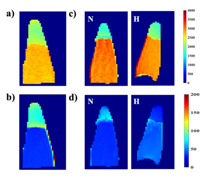 |
14 | An iron-based hypoxia targeting contrast agent Video Permission Withheld
Babak Moghadas1, C. Chad Quarles2, and Vikram D. Kodibagkar3
1Arizona State University, Tempe, AZ, United States, 2Barrow Neurological Institute, Phoenix, AZ, United States, 3School of Biological and Systems Engineering, Arizona State University, Tempe, AZ, United States
We developed and tested a novel iron based contrast agent for imaging hypoxia using magnetic resonance imaging (MRI). After synthesis, the cytotoxicity data and the magnetic properties were studied to measure the relaxivity of the contrast agent. The ability of the contrast agent in targeting hypoxic was demonstrated in vitro by comparing cell cultures under hypoxia by T1 and T2 mapping. The cytotoxicity assay showed no significant changes to the cells’ viability over the period of 8h exposure to the contrast agent. The retention in the hypoxic condition is an indication of its performance under the proposed mechanism.
|
||
4098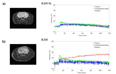 |
15 | Imaging hypoxia in two brain tumor models using GdDO3NI
Babak Moghadas1, Jonathan Scirone1, Matthew Scarpelli2, Alberto Fuentes2, C. Chad Quarles2, and Vikram D. Kodibagkar3
1Arizona State University, Tempe, AZ, United States, 2Barrow Neurological Institute, Phoenix, AZ, United States, 3School of Biological and Systems Engineering, Arizona State University, Tempe, AZ, United States
In this study we have used the hypoxia-targeting MRI contrast agent GdDO3NI, (a nitroimidazole-based T1 contrast agent) to image the development of hypoxia in two types of rodent brain tumor models. Our results indicate a range of signal enhancements from 0-17% over baseline in the C6 and 9L tumors using GdDO3NI with clearance from contralateral brain and muscle tissue. This study further demonstrates the utility of GdDO3NI in non-invasive imaging of tissue hypoxia with high resolution.
|
||
4099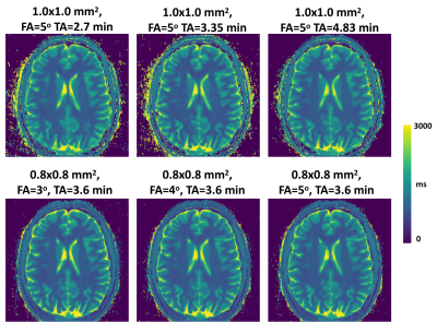 |
16 | Repeatability and Robustness of MP-GRASP T1 Mapping
Zhitao Li1, Yang Yang2, and Li Feng2
1Department of Radiology, Stanford University, Palo Alto, CA, United States, 2Biomedical Engineering and Imaging Institute and Department of Radiology, Icahn School of Medicine at Mount Sinai, New York, NY, United States The repeatability of fast 3D T1 mapping using MP-GRASP (Magnetization-Prepared Golden-angle RAdial Sparse Parallel) MRI and its robustness to variation of imaging parameters including flip angle in phantom and brain were demonstrated. The experiments showed that MP-GRASP has a good robustness to B1 inhomogeneity, with intra-slice repeatability below 1% in the single tube phantom experiment. The longitudinal experiments also yielded good repeatability both in phantom (<2.5%) and in the brain (<2%) under various imaging conditions. The T1 values estimated are accurate relative to inversion recovery spin echo (IR-SE) gold standard (R2=0.997, Lin’s CCC=0.996). |
||
4100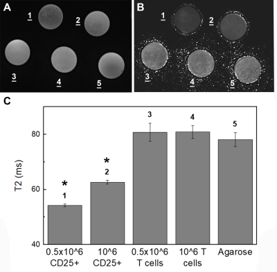 |
17 | Cell sorting microbeads as novel contrast agent for magnetic resonance imaging
Aman Khurana1, Francesc Marti2, Roberto Gedaly2, and Fanny Chapelin3
1Radiology, University of Kentucky, Lexington, KY, United States, 2Surgery, University of Kentucky, Lexington, KY, United States, 3Biomedical Engineering, University of Kentucky, Lexington, KY, United States
Due to their low cytoplasmic capacity and phagocytic activity, T cells have consistently been challenging to label for MR tracking applications. We developed a one-stop shop approach where the T cell sorting agent also labels the cells which can subsequently be imaged using non-invasive MRI. We demonstrate positive intracellular localization of the beads in Treg cells by means of histopathology analyses and MRI signal effects in phantoms containing different amounts of labeled cells.
|
||
 Back to Meeting Home
 Back to the Program-at-a-Glance
The International Society for Magnetic Resonance in Medicine is accredited by the Accreditation Council for Continuing Medical Education to provide continuing medical education for physicians.
 Back to Meeting Home
 Back to the Program-at-a-Glance
 View the Presentation