Online Gather.town Pitches
fMRI I
Joint Annual Meeting ISMRM-ESMRMB & ISMRT 31st Annual Meeting • 07-12 May 2022 • London, UK

| Booth # | ||||
|---|---|---|---|---|
3025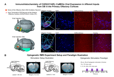 |
1 | Unravel the Role of Primary Olfactory Cortices in Olfactory Processing via Optogenetic fMRI
Teng Ma1,2,3, Xunda Wang1,2, Linshan Xie1,2, Pit Shan Chong4, Peng Cao3, Pek-Lan Khong3, Lee Wei Lim4, Ed. X Wu1,2,4, and Alex T. L. Leong1,2
1Laboratory of Biomedical Imaging and Signal Processing, The University of Hong Kong, Hong Kong SAR, China, 2Department of Electrical and Electronic Engineering, The University of Hong Kong, Hong Kong SAR, China, 3Department of Diagnostic Radiology, Li Ka Shing Faculty of Medicine, The University of Hong Kong, Hong Kong SAR, China, 4School of Biomedical Sciences, Li Ka Shing Faculty of Medicine, The University of Hong Kong, Hong Kong SAR, China
The anterior olfactory nucleus (AON) and the piriform cortex (Pir) are the two primary sensory cortices critical for olfaction. Although it is well documented that both cortices overlap significantly in their functions in olfactory processing, molecular and anatomical tracing studies have indicated otherwise. Consequently, our present understanding of the functions of AON and Pir in olfactory processing at the systems level remains incomplete. In this study, we employed optogenetic fMRI to interrogate the role of AON and Pir in processing olfactory inputs and beyond, and the associated long-range olfactory pathways and their spatiotemporal response properties.
|
||
3026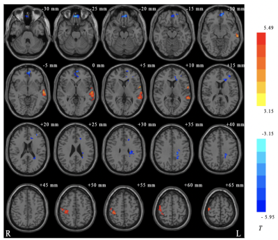 |
2 | Effects of Dobutamine on Neural Activity in Normal Subjects: A Exploratory Resting-State Functional MRI Study. Video Not Available
Yawen Liu1, Pengling Ren2, TingTing Zhang1,2, Linkun Cai1, Hao Wang2, Hongxia Yin2, Wei Zheng3, Yishi Wang4, Haijun Niu1, Zhenghan Yang2, Dehong Luo5, and Zhenchang Wang2
1School of Biological Science and Medical Engineering, Beihang University, Beijing, China, 2Department of Radiology, Beijing Friendship Hospital, Capital Medical University, Beijing, China, 3National Space Science Center, Chinese Academy of Sciences, Beijing, China, 4Philips Heathcare, Beijing, China, 5National Cancer Center/Cancer Hospital and Shenzhen Hospital, Shenzhen, China
To explore the alternations in spontaneous brain activities in normal subjects after dobutamine infusion, the regional homogeneity (ReHo) method was used to analyze the interregional synchronized activity of all participants. The results showed that compared with normal resting state, significantly decreased ReHo was found after dobutamine infusion in bilateral frontal lobe, while increased in left temporal lobe and right parietal lobe, suggesting that dobutamine may have an effect on functional brain activity.
|
||
3027 |
3 | Thalamic Reticular Nucleus Exerts Long-range Cross-modal Modulation on Sensory Processing Depending on Inputs?
Xunda Wang1,2, Linshan Xie1,2, Teng Ma1,2,3, Pit Shan Chong4, Lee-Wei Lim4, Peng Cao3, Pek-Lan Khong3, Alex T. L. Leong1,2, and Ed X. Wu1,2
1Laboratory of Biomedical Imaging and Signal Processing, The University of Hong Kong, Hong Kong SAR, China, 2Department of Electrical and Electronic Engineering, The University of Hong Kong, Hong Kong SAR, China, 3Department of Diagnostic Radiology, Li Ka Shing Faculty of Medicine, The University of Hong Kong, Hong Kong SAR, China, 4School of Biomedical Sciences, Li Ka Shing Faculty of Medicine, The University of Hong Kong, Hong Kong SAR, China
Thalamic reticular nucleus (TRN) has been shown to gate sensory thalamo-cortical interactions and selectively modulate thalamic sensory information processing according to behavioral demands. However, whether TRN can exert long-range, i.e., beyond thalamus, cross-modal modulation of sensory processing remains unclear. In this fMRI study, we demonstrate that optogenetic excitation of somatosensory-specific TRN enhances cross-modal excitatory sensory inputs but suppresses cross-modal competing inputs at somatosensory cortices. Our work provides insight into how TRN differentially gate the processing of distinct cross-modal sensory information at large-scale, which may be critical for ensuring the balance between various task demands.
|
||
3028 |
4 | The effect of total sleep deprivation under workload on spontaneous brain activity in medical staff: a resting-state functional MR imaging study Video Permission Withheld
Cong Peng1, Ding bo Guo1, lisha Nie2, Liuheng Liu 1, Dongling Xiao 1, and Hua Yang1
1Chongqing Hospital of Traditional Chinese Medicine, Chongqing, China, 2GE Healthcare, MR Research China, Beijing, Chongqing, China
The current study aims to assess the effect of total sleep deprivation (TSD) under workload on spontaneous brain activity in medical staff and to further investigate its latent associations with clinical markers. It was concluded that spontaneous brain activity abnormalities occurred during TSD under workload, which might explain the reduced performance of these participants on neurocognitive tests.
|
||
3029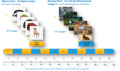 |
5 | fMRI response to individualized cue-reactivity paradigm of males with gaming behavior
Pavel Tikhonov1, Alexander Efimtcev2, Dmitriy Iskhakov2, and Mikhail Zubkov1
1School of Physics and Engineering, ITMO University, Saint-Petersburg, Russian Federation, 2Department of Radiology, Federal Almazov North‐West Medical Research Center, Saint-Petersburg, Russian Federation
Task-related fMRI studies are providing increasing amount of information on the neurobiological aspects of the Gaming Disorder. This study aims to Explore both the activation patterns and the functional connectivities present in the gaming disorder-like subject brains via task-based fMRI study using individualized visual stimuli. 24 male test participants and 25 male control participants took part in the fMRI scanning with gaming-related and neutral visual stimuli. Data analysis showed altered nucleus accumbens connectivity and amygdala resembling that in cases of substance addiction1,2.
|
||
3030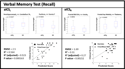 |
6 | Association Between the Recall Verbal Memory and Side of Asymptomatic Carotid Artery Stenosis
Jyun-Ru Chen1, Chun-Jen Lin2,3, I-Hui Lee2,3,4, and Chia-Feng Lu1
1Department of Biomedical Imaging and Radiological Sciences, National Yang Ming Chiao Tung University, Taipei, Taiwan, 2School of Medicine, National Yang-Ming University, Taipei, Taiwan, 3Neurological Institute, Taipei Veterans General Hospital, Taipei, Taiwan, 4Institute of Brain Science, National Yang Ming Chiao Tung University, Taipei, Taiwan
Previous studies reported that the patients with asymptomatic internal carotid stenosis (aICS) had a higher risk of stroke and recall verbal memory impairment. However, whether the side of aICS could cause different verbal memory outcomes was less explored. In this study, significant difference of functional connectivity (FC) was found between the left and right aICS groups. The correlation analysis estimating the association between FC and recall verbal memory also presented different profiles between two aICS groups.
|
||
3031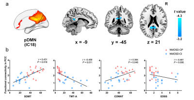 |
7 | Altered functional connectivity associated with cognitive impairment in neuromyelitis optica spectrum disorder
Yang Yang1, Qiuyu Yu1, Peng Wu2, Shuting Han1, Xiaojuan Wu1, Hui Dai1, and Yonggang Li1
1The First Affiliated Hospital of Soochow University, Suzhou, China, 2Philips Healthcare, Shanghai, China Patients with neuromyelitis optica spectrum disorder (NMOSD) develop cognitive symptoms during the course of disease. We observed decreased intra- and internetwork connectivity in the main cognitive networks by Independent Component Analysis in cognitively impaired patients compared to cognitively preserved patients and healthy controls. The decreased intra- and internetwork connectivity was significantly correlated with cognitive performance in the whole NMOSD group. Our findings indicated that aberrant connectivity patterns across the main cognitive networks might suggest the neural substrates underlying cognitive decline in NMOSD. |
||
3032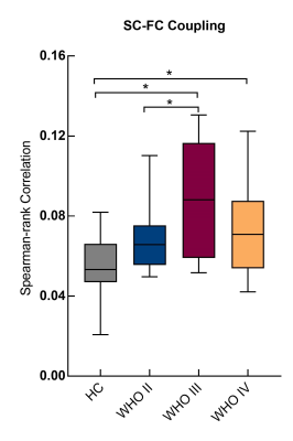 |
8 | Malignancy-specific structural-functional dyscoupling in brains with glioma
Siqi Cai1,2, Chunxiang Jiang1,2, Zhifeng Shi3, Shihui Zhou1,2, and Lijuan Zhang*1
1Shenzhen Institutes of Advanced Technology, Chinese Academy of Sciences, Shenzhen, China, 2University of Chinese Academy of Sciences, Beijing, China, 3Huashan Hospital of Fudan University, Shanghai, China
The structural-functional coupling in the connectivity of the contralesional hemisphere was enhanced and specific to the malignancy grades of glioma. Structural and functional networks in brains with low grade glioma featured with increased global and local efficiency, whereas the plasticity of the functional networks was limited in brians with high grade glioma. These findings provide new knowledge toward innovating the characterization of glioma.
|
||
3033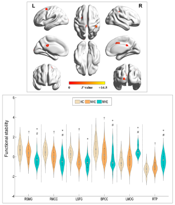 |
9 | Abnormal functional stability in patients with minimal hepatic encephalopathy. Video Not Available
LiMin Cai1 and HuaJun Chen1
1Fujian Medical University Union Hospital, Fuzhou, China
We investigated the stability of brain functional architectures and its relationship with cognitive impairment in cirrhotic patients. From healthy controls, to NHE, and to minimal hepatic encephalopathy(MHE) group, a stepwise reduction of FS was found in right supramarginal gyrus , right middle cingulate cortex, left superior frontal gyrus, and BPCC; whereas, a progressive increment of FS was observed in left middle occipital gyrus and right temporal pole. Mean FS was correlated with Psychometric Hepatic Encephalopathy Score among cirrhotic patients. FS index showed moderate discrimination potential between NHE and MHE. FS alteration could be potential biomarkers serving for diagnosis about MHE.
|
||
3034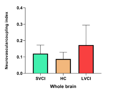 |
10 | Altered macroscale neurovascular coupling in vascular cognitive impairment Video Not Available
Zhao Ruan1 and Weiyin Vivian Liu2
1Department of Radiology, Zhongnan Hospital of Wuhan University, Wuhan, China, 2GE Healthcare, Beijing, China This study aims to integrate psychological behavior, clinical biochemistry and other indicators, and use multi-mode MRI technology to explore specific imaging indicators of VCIND neurovascular coupling (NVC). NVC analysis and CBF/ ALFF ratio calculation were performed to investigate the neurovascular coupling changes and clinical significance of LVCI, SVCI and HC. Regardless of the level of the whole brain or brain regions, we have found neurovascular uncoupling in VCIND patients compared with healthy controls. These findings will be helpful in screening patients with VCIND, and has important theoretical and clinical significance in preventing and delaying the development of dementia. |
||
3035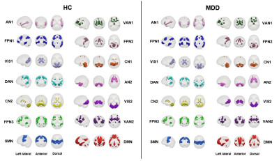 |
11 | Stressful life events associate with adolescent depression through dysfunction of frontoparietal network
Yingxue Gao1, Ruohan Feng2, Lihua Zhuo2, Kaili Liang1, Weijie Bao1, Hui Qiu1, Zilin Zhou1, Guoping Huang3, and Xiaoqi Huang1
1Huaxi MR Research Center (HMRRC), Department of Radiology, West China Hospital, Sichuan University, Chengdu, China, 2Department of Radiology, the Third Hospital of Mianyang, Sichuan Mental Health Center, Mianyang, China, Mianyang, China, 3Department of Psychiatry, the Third Hospital of Mianyang, Sichuan Mental Health Center, China, Mianyang, China
The current study investigate functional network alterations in adolescent MDD using the clustering analysis. We found that adolescents with MDD showed decreased connectivity between the frontoparietal network (FPN) and other networks relative to HC. The global connectivity strength of FPN was negatively correlated with the punishment and academic pressure factors, and mediated the association between these two stressors and depression. Our findings suggest that punishment and academic pressure may contribute to the development of depression in adolescence through dysfunction of the FPN, which may promote future prevention of adolescent MDD.
|
||
3036 |
12 | Altered spontaneous brain activity in patients with comitant exotropia before and after surgery: a resting-state fMRI study
Qian Wu1, Hao Hu1, Wen Chen1, Lu Chen1, Jiang Zhou1, Xiaoquan Xu1, and Feiyun Wu1
1Department of Radiology, The First Affiliated Hospital of Nanjing Medical University, Nanjing, China
We investigate the brain functional alterations in patients with comitant exotropia (CE) before and after surgery, using resting-state functional magnetic resonance imaging with amplitude of low-frequency fluctuation. Our study suggested that CE may lead to decreased brain functional activities in visual-associated areas and the brain function could partially restore along with recovery of exodeviation after strabismus surgery. Besides, our results provided an explanation to the postoperative remnant of stereopsis impairment, which would be helpful to improve the clinical interventions for patients with CE.
|
||
3037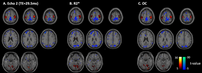 |
13 | Improved reliability of functional connectivity with multi-echo task fMRI
Amod S Jog1, Radhika Madhavan2, Nastaren Abad3, Eric Fiveland3, Brice Fernandez4, Chitresh Bhushan3, Luca Marinelli3, H Doug Morris5, and Thomas K Foo3
1GE Global Research, Bangalore, India, 2GE Healthcare, Niskayuna, NY, United States, 3GE Global Research, Niskayuna, NY, United States, 4GE Healthcare, Buc, France, 5Walter Reed National Military Medical Center, Bethesda, MD, United States Signal from conventional single-echo fMRI depends on multiple factors including cerebral metabolic rate for oxygen (CMRO2), blood flow (CBF) and blood volume (CBV). Interpretation of functional connectivity analyses derived from conventional single-echo fMRI is complicated due to coupling of various factors in the fMRI signal. In this work, we use multi-echo task-fMRI to estimate functional connectivity from fluctuations in R2* (transverse relaxation rate) assumed to be surrogates for fluctuations in CMRO2. We also investigate the similarities and differences between single-echo and R2* multi-echo fMRI derived functional activation in a visuo-motor decision task paradigm. |
||
3038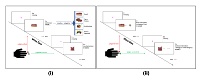 |
14 | Food pictures versus corresponding calorie information stimuli: an event-related fMRI study at 7 T of brain response differences in adult males Video Not Available
Jiawei Han1,2, Qiangfeng Wang1, Ying-Hua Chu3, Ying Liu1, Junqi Xu1, and He Wang1,2
1Institute of Science and Technology for Brain-Inspired Intelligence, Fudan University, Shanghai, China, 2Human Phenome Institute, Fudan University, Shanghai, China, 3MR Collaboration, Siemens Healthineers Ltd., Shanghai, China
Balancing food calories and nutrients has become a great concern for contemporary young people. In this study, we found that the calorie information of food activates the response of left middle occipital gyrus, which is related to the current sense of individual satiety. Therefore, it is likely that a sensory hub of the digestive system, corresponding to the visual stimulation of food, is existing.
|
||
3039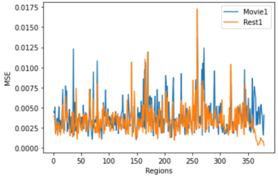 |
15 | Modelling the Brain Functional Difference of Movie Watching and Resting State with Autoencoder
Yifei Sun1, Fernando Calamante1,2,3, and Jinglei Lv1,2
1School of Biomedical Engineering, University of Sydney, Sydney, Australia, 2Brain and Mind Centre, University of Sydney, Sydney, Australia, 3Sydney Imaging, University of Sydney, Sydney, Australia
The human brain is a complex dynamic system. Understanding the functional difference of the brain between the resting state and performing tasks helps to decipher the functional brain architecture. In this work, we trained an autoencoder to optimally reconstruct the resting state fMRI (rs-fMRI) data of 50 subjects, and this model was subsequently used to reconstruct the movie watching fMRI (mw-fMRI) data of the same subjects. We have compared the reconstruction error between movie watching and resting state, and significant regional functional differences were found.
|
||
 Back to Meeting Home
 Back to the Program-at-a-Glance
The International Society for Magnetic Resonance in Medicine is accredited by the Accreditation Council for Continuing Medical Education to provide continuing medical education for physicians.
 Back to Meeting Home
 Back to the Program-at-a-Glance
 View the Presentation