Online Gather.town Pitches
Molecular Imaging IV
Joint Annual Meeting ISMRM-ESMRMB & ISMRT 31st Annual Meeting • 07-12 May 2022 • London, UK

Online Gather.town Pitches
Molecular Imaging IV
| Booth # | ||||
|---|---|---|---|---|
4451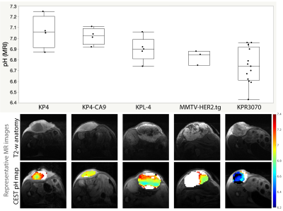 |
1 | Determining the conditions for robust acidoCEST MRI to measure extracellular pH in tumors in vivo
Rohan S. Virgincar1, Kai H. Barck1, Mary Ann Go2, Geoffrey Del Rosario2, Shang-Fan Yu2, ManKin Choy1, Alvin Gogineni1, Justin Elstrott1, Herman S. Gill1, Jan Marik1, Genee Lee2, Mark D. Pagel3, Alex de Crespigny4, Teemu T.
Junttila5, Christoph Spiess6, Robby M. Weimer1, and Luke Xie1
1Biomedical Imaging, Genentech, South San Francisco, CA, United States, 2Translational Oncology, Genentech, South San Francisco, CA, United States, 3Department of Cancer Systems Imaging, University of Texas MD Anderson Cancer Center, Houston, TX, United States, 4Early Clinical Development, Genentech, South San Francisco, CA, United States, 5In Vivo Pharmacology, Genentech, South San Francisco, CA, United States, 6Antibody Engineering, Genentech, South San Francisco, CA, United States
AcidoCEST MRI is a promising technique to directly measure extracellular pH in vivo and is well-suited to imaging tumors, which can exhibit acidosis. However, few studies have demonstrated the conditions and the minimum contrast agent concentrations needed for robust acidoCEST MRI. In this study, we employed micro-CT to determine the uptake of iopamidol in tumors with intratumoral and intravenous delivery of isovue-370, estimated the accuracy of pH measured by MRI vs. a pH meter, demonstrated the minimum iopamidol concentration required for acidoCEST MRI, and reported pH measurements in different tumor xenograft models.
|
||
4452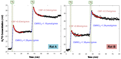 |
2 | Simultaneously Assessing Cerebral Glucose and Oxygen Metabolism and Blood Flow in Rat Brain at Ultrahigh Field of 16.4T
Guangle Zhang1, Wei Zhu1, Wei Chen1, and Xiao-Hong Zhu1
1University of Minnesota, Minneapolis, MN, United States In this study, we used a newly developed tri-frequency RF surface coil that enables 1H MRI and interleaved 2H/17O MRS measurements to simultaneously assess cerebral glucose and oxygen metabolism, cerebral blood flow and oxygen extraction fraction in rat brain at 16.4T with administration of 2H-labeled glucose and 17O-labeled oxygen gas. Our results demonstrate that this approach can capture the activities of major pathways relevant to glucose and oxygen metabolism and perfusion in the same brain at the same time, thereby provide comprehensive information crucial for understanding the metabolic and vascular coupling or uncoupling relationship in healthy and diseased brains. |
||
4453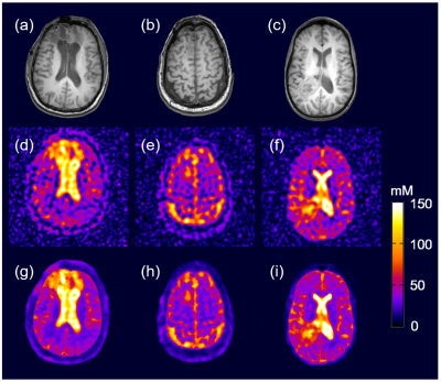 |
3 | Reconstructing High-Quality Sodium MR Images from Limited Noisy k-Space Data with Model-Assisted Deep Learning
Yibo Zhao1,2, Yudu Li1,2, Rong Guo1,2, Keith R. Thulborn3, and Zhi-Pei Liang1,2
1Beckman Institute for Advanced Science and Technology, University of Illinois, Urbana-Champaign, Urbana, IL, United States, 2Department of Electrical and Computer Engineering, University of Illinois, Urbana-Champaign, Urbana, IL, United States, 3Center for Magnetic Resonance Research, University of Illinois at Chicago, Chicago, IL, United States
Sodium MRI can acquire important biological information about cell integrity and tissue viability, but its clinical application has been limited by low SNR and poor spatial resolution. We propose a novel method to reconstruct high-quality sodium images from limited and noisy k-space data. The new method synergistically integrates model-based reconstruction with deep learning. Simulation and experimental results show that the proposed method can reconstruct high-SNR and high-resolution sodium images, which clearly delineate lesions such as brain tumors.
|
||
4454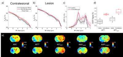 |
4 | MR imaging of stroke rats using CEST with an Average Saturation Efficiency Filter (ASEF)
Julius Juhyun Chung1 and Tao Jin1
1University of Pittsburgh, Pittsburgh, PA, United States
Average Saturation Efficiency Filter (ASEF) is a fast and intuitive technique for achieving CEST imaging with improved sensitivity removing fast exchanges and semi-solid MT background with minimal loss to sensitivity. Our results in MCAO rodents showed that it can detect the ischemic lesion from CEST signal contrasts at 3.6, 2.6, and 2 ppm which may provide different metabolic-related information with higher sensitivity to 3-point measurement at 3.6 ppm and comparable contrast. Its low requirement on number of imaged signals also opens up possibilities for dynamic imaging or signal averaging.
|
||
4455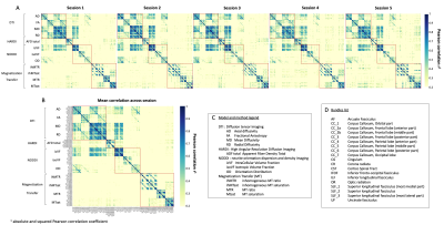 |
5 | Measures of reliability in high frequency longitudinal white matter multi-shell diffusion and inhomogeneous magnetization transfer database
Manon Edde1,2, Guillaume Theaud1,2, Matthieu Dumont2, Antoine Théberge3, Alex Valcourt-Caron1,2, Stefano Magon4, and Maxime Descoteaux1,2
11Sherbrooke Connectivity Imaging Lab (SCIL), University of Sherbrooke, SCIL, Sherbrooke, QC, Canada, 2Imeka Solutions, Inc., Sherbrooke, QC, Canada, 3Videos & Images Theory and Analytics Laboratory (VITAL), University of Sherbrooke,, Sherbrooke, QC, Canada, 4Pharma Research and Early Development, Roche Innovation Center Basel, F. Hoffmann-La Roche Ltd., Basel, Switzerland
Longitudinal imaging studies are widely used to capture and characterize progressive brain changes. However, evaluation of the reproducibility and the variation of measures in MRI is crucial to reliably detect or predict brain changes with quantitative MRI measures. Using the coefficient of variation and intraclass correlation, we evaluated the reproducibility of diffusion and myelin-based MRI measures from a multi-shell diffusion and inhomogeneous magnetization transfer MRI dataset of twenty healthy adults, each with five MRI acquisitions over six months. Moderate to high reproducibility of diffusion and myelin measures is observed within white matter tracks of interests.
|
||
4456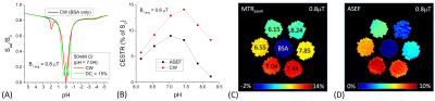 |
6 | Average Saturation Efficiency Filter (ASEF) for CEST Imaging
Tao Jin1 and Julius Juhyun Chung1
1University of Pittsburgh, Pittsburgh, PA, United States
Endogenous CEST signal usually has low specificity due to contamination from the magnetization transfer effect and from fast exchanging labile protons with close Larmor frequencies. We propose to improve CEST signal specificity with an ASEF which measures the difference between CEST signals acquired with similar average saturation power but largely different duty cycles (DC), e.g., a continuous wave or a high DC pulse train versus a low DC one. Simulation and creatine phantom studies showed that ASEF can improve the specificity of slow to intermediate exchanging CEST signal with a relatively small loss of sensitivity.
|
||
4457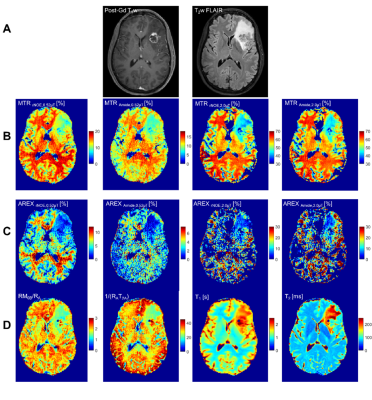 |
7 | Saturation Transfer MRI in a Clinical Setting for Differentiating Radiation Necrosis from Tumour Progression in Brain Metastases
Rachel W. Chan1, Hatef Mehrabian1, Arjun Sahgal2, Hanbo Chen2, Aimee Theriault2, Wilfred W. Lam1, Sten Myrehaug2, Chia-Lin Tseng2, Zain Husain2, Jay Detsky2, Hany Soliman2, and Greg J. Stanisz1,3,4
1Physical Sciences Platform, Sunnybrook Research Institute, Toronto, ON, Canada, 2Department of Radiation Oncology, Sunnybrook Health Sciences Centre, Toronto, ON, Canada, 3Department of Medical Biophysics, University of Toronto, Toronto, ON, Canada, 4Department of Neurosurgery and Pediatric Neurosurgery, Medical University of Lublin, Lublin, Poland
Stereotactic radiosurgery for brain metastases delivers a focal dose of radiation and has excellent local tumour control but leads to radiation necrosis (RN) in up to 22% of patients. This study used saturation transfer MRI for distinguishing between RN and tumour progression (TP), extending our previous work to a larger cohort of 70 patients (75 lesions). Eleven out of 14 metrics (including quantitative MT and CEST) showed statistically significant differences between the RN and TP cohorts, including magnetization transfer ratio (MTR) metrics showing the best separation. Univariable logistic regression resulted in the high-power MTR having the highest AUC=0.88 (with AIC=67.3).
|
||
4458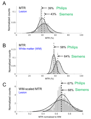 |
8 | Differentiating Radiation Necrosis from Tumour Progression in Brain Metastases using CEST: A Cross-Vendor Comparison
Rachel W. Chan1, Wilfred W. Lam1, Patrick Liebig2, Leedan Murray1, Hatef Mehrabian1, Aimee Theriault3, Ruby Endre1, Garry Detzler1, Sten Myrehaug3, Chia-Lin Tseng3, Jay Detsky3, Pejman J. Maralani4, Arjun Sahgal3, Hany
Soliman3, and Greg J. Stanisz1,5,6
1Physical Sciences Platform, Sunnybrook Research Institute, Toronto, ON, Canada, 2Siemens Healthineers, Erlangen, Germany, 3Department of Radiation Oncology, Sunnybrook Health Sciences Centre, Toronto, ON, Canada, 4Department of Medical Imaging, Sunnybrook Health Sciences Centre, University of Toronto, Toronto, ON, Canada, 5Department of Medical Biophysics, University of Toronto, Toronto, ON, Canada, 6Department of Neurosurgery and Pediatric Neurosurgery, Medical University of Lublin, Lublin, Poland
Stereotactic radiosurgery for the treatment of brain metastases delivers a high dose of radiation with excellent local control, but increases the likelihood of radiation necrosis. CEST is a promising technique for distinguishing radiation necrosis from tumour progression in brain metastases, but its application has been limited to a single MRI system and CEST sequence. This study explores the use of scaling of the magnetization transfer ratio (MTR) by the white matter (WM) of each patient for comparison across vendors/sequences. It was found that the WM-scaled MTR showed improved correspondence across the MR systems, across two CEST sequences.
|
||
4459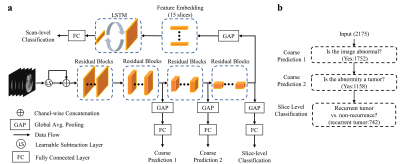 |
9 | Predicting tumor recurrence in patients with gliomas via deep learning-based analysis of structural and amide proton transfer weighted MRI
Pengfei Guo1,2, Mathias Unberath2, Jinyuan Zhou1, Hye-Young Heo1, Charles G. Eberhart3, Michael Lim4, Jaishri O. Blakeley5, Peter van Zijl1,6, and Shanshan Jiang1
1Department of Radiology, Johns Hopkins University, Baltimore, MD, United States, 2Whiting School of Engineering, Johns Hopkins University, Baltimore, MD, United States, 3Department of Pathology, Johns Hopkins University, Baltimore, MD, United States, 4Department of Neurosurgery, Johns Hopkins University, Baltimore, MD, United States, 5Department of Neurology, Johns Hopkins University, Baltimore, MD, United States, 6F.M. Kirby Research Center for Functional Brain Imaging, Kennedy Krieger Institute, Baltimore, MD, United States
Amide protein transfer weighted (APTw) MRI has been validated to accurately detect recurrent malignant gliomas across different studies. However, APTw image interpretation is time consuming and requires professional knowledge. Therefore, reliable, automated imaging diagnostic tools to assess malignant glioma response to therapies are urgently needed. Here, we develop and verify a CNN-based deep-learning algorithm to identify tumor progression versus response by adding APTw MRI data to structural MR images as the proposed model input. Our results suggest that the use of APTw images can increase the diagnostic accuracy to structural MRI for the treatment response assessment.
|
||
4460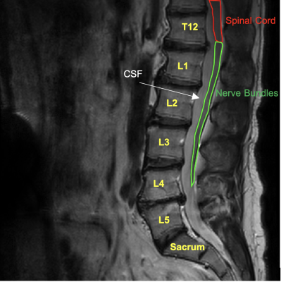 |
10 | R1rho Dispersion in Spinal Cord and Nerve
Alan E. Rivera-Garcia1, Juan Uribe2, Jay D. Turner2, John C. Gore3, and Ping Wang1
1Translational Neuroscience, Barrow Neurological Institute, Phoenix, AZ, United States, 2Barrow Neurological Institute, Phoenix, AZ, United States, 3Radiology and Radiological Sciences, Vanderbilt University Medical Center, Nashville, TN, United States
Previous studies have demonstrated that at high fields (3T and beyond), dispersion of R1rho with different locking fields may reflect chemical exchange processes in biological tissues. This study aimed to investigate if R1rho dispersion is measurable in the spinal cord and nerve bundles. The results show that the dispersion is significant and measurable in both tissues, suggesting that R1rho dispersion has potential to characterize changes in composition or other physicochemical properties in nerve injuries and/or neurological disorders.
|
||
4461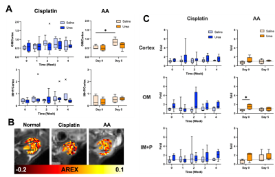 |
11 | Differentiating mild and severe nephropathies via multiparametric urea CEST, NOE and quantitative MT imaging
Soo Hyun Shin1, Michael Wendland2, and Moriel Vandsburger1
1Department of Bioengineering, University of California, Berkeley, Berkeley, CA, United States, 2Berkeley Preclinical Imaging Core (BPIC), University of California, Berkeley, Berkeley, CA, United States
Standardized blood tests lack adequate sensitivity to renal function and its underlying pathophysiology. We examined whether urea CEST, along with NOE CEST and quantitative MT imaging, can be used to quantitatively differentiate cisplatin and aristolochic acid (AA) models of mild and severe nephropathy, respectively. The increase of T1 relaxation time and decrease of qMT contrast were parallel in two models, while the decreased intrarenal gradient of urea CEST and NOE contrast were only observed in AA models. These results indicate that our integrated approach has the potential to noninvasively quantitate renal injuries and characterize different types of renal pathophysiology.
|
||
 Back to Meeting Home
 Back to the Program-at-a-Glance
The International Society for Magnetic Resonance in Medicine is accredited by the Accreditation Council for Continuing Medical Education to provide continuing medical education for physicians.
 Back to Meeting Home
 Back to the Program-at-a-Glance
 View the Presentation