Online Gather.town Pitches
Advances in Data Acquisition I
Joint Annual Meeting ISMRM-ESMRMB & ISMRT 31st Annual Meeting • 07-12 May 2022 • London, UK

Online Gather.town Pitches
Advances in Data Acquisition I
| Booth # | ||||
|---|---|---|---|---|
 |
3488 |
1 | SuperRes-EPTI: in-vivo mesoscale distortion-free dMRI at 500μm-isotropic resolution using short-TE EPTI with rotating-view super resolution
Zijing Dong1,2, Jonathan R. Polimeni1,2, Lawrence L. Wald1,2, and Fuyixue Wang1,2
1Athinoula A. Martinos Center for Biomedical Imaging, Massachusetts General Hospital, Charlestown, MA, United States, 2Department of Radiology, Harvard Medical School, Boston, MA, United States
To achieve high spatial resolution in-vivo dMRI (mesoscale at isotropic 500μm), we propose a novel SuperRes-EPTI technique that combines our recently developed EPTI technique with a rotating-view super-resolution acquisition and reconstruction. The SuperRes-EPTI can achieve an overall 3.5× SNR gain (SuperRes×EPTI = 2.7×1.3), while providing images completely-free from distortions that allow for improved super-resolution reconstruction and high effective resolution. The SuperRes acquisition scheme was designed to avoid spin-history artifacts, and incorporates rigid motion correction between thick-slice volumes for high motion-robustness. In-vivo distortion-free dMRI data were acquired at 500μm-isotropic resolution at 3T that reveals detailed fibers in gray matter.
|
|
3489 |
2 | MAP-MRI diffusion estimates are biased by simultaneous multi-slice acceleration Video Not Available
L. Tugan Muftuler1, Andrew S. Nencka2, Volkan E. Arpinar2, Jaemin Shin3, Baolian Yang4, Graeme McKinnon4, Suchandrima Banerjee5, and Kevin M. Koch2
1Neurosurgery, Medical College of Wisconsin, Milwaukee, WI, United States, 2Radiology, Medical College of Wisconsin, Milwaukee, WI, United States, 3GE Healthcare, New York, NY, United States, 4GE Healthcare, Waukesha, WI, United States, 5GE Healthcare, Menlo Park, CA, United States
Advanced diffusion MRI models are being explored to study the complex microstructure of the brain with higher accuracy. However, these techniques require long acquisition times. Simultaneous multi-slice (SMS) accelerates data acquisition by exciting multiple image slices simultaneously and separating the overlapping slices using a mathematical model. However, SMS acceleration introduces increased noise in reconstructed images and crosstalk between simultaneously excited slices. These compounded effects from SMS acceleration could affect tissue microstructure parameters derived from advanced diffusion MRI models.
|
||
3490 |
3 | How Many Diffusion Directions Are Needed to Suppress Spherical Harmonic Aliasing in Fiber Orientation Density Functions?
Hunter Moss1 and Jens Jensen1
1MUSC, CHARLESTON, SC, United States
A fiber orientation density function (fODF) gives the angular density of axonal fibers within a white matter voxel. Estimated fODFs obtained with diffusion MRI are prone to aliasing errors, depending on the number of diffusion directions used in the data acquisition. Here these errors are quantified for several typical fODFs represented with spherical harmonic expansions truncated at a specified degree. Choosing the number of directions equal to 2 to 3 times the number of adjustable parameters in the spherical harmonic expansion is found to be sufficient to reduce aliasing errors to a low level.
|
||
3491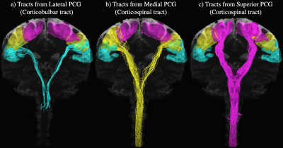 |
4 | Characterizing Corticobulbar and Corticospinal Tracts into the Cervical Spinal Cord with Sub-Millimeter Isotropic DTI
Bruce Iain1, Yixin Ma1, and Allen Song1
1Brain Imaging and Analysis Center, Duke University, Durham, NC, United States
An ability to characterize the corticobulbar and corticospinal tracts from the motor cortex into the spinal cord with diffusion tensor imaging remains a challenge due to limiting factors such as achievable spatial resolutions, spatial coverages, and signal-to-noise ratio in the spinal cord. By extending the field-of-view through a combination of sub-millimeter isotropic axial and sagittal acquisitions, this study presents a technique to delineate the complete pyramidal tracts with data acquired at a sufficient spatial resolution to resolve intricate structural details such as the pyramidal decussation. Such a delineation can facilitate placement of spinal cord stimulation electrodes for movement disorder treatments.
|
||
3492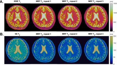 |
5 | Evaluation of MR Fingerprinting at 0.55T
Zhibo S. Zhu1, Nam Gyun Lee2, Ye Tian1, Ahsan Javed3, Mark Griswold4, Adrienne Campbell-Washburn3, and Krishna S. Nayak1
1Department Of Electrical and Computer Engineering, University of Southern California, Los Angeles, CA, United States, 2Department Of Biomedical Engineering, University of Southern California, Los Angeles, CA, United States, 3Division of Intramural Research, National Heart, Lung, and Blood Institute, National Institutes of Health, Bethesda, MD, United States, 4Department of Radiology, Case Western Reserve University, Cleveland, OH, United States
We perform a thorough comparison of MRF with reference relaxometry approaches on a NIST/ISMRM phantom and a healthy volunteer. In the NIST phantom, relative bias from -4.7% to 27.7% for the NiCl2 vials and from 5.01% to 28% for the MnCl2 vials within a biological relevant T1 and T2 range. In-vivo, MRF has good repeatability (variations <2%) but substantial bias in WM and GM against the reference approach.
|
||
3493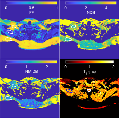 |
6 | Simultaneous triglyceride characterization and water T1 estimation in a breath-hold applied to brown adipose tissue using MR fingerprinting
Jason Ostenson1,2, Bruce M. Damon3, and Evan L. Brittain4
1Vanderbilt University Institute of Imaging Science, Vanderbilt University Medical Center, Nashville, TN, United States, 2Radiology and Radiological Sciences, Vanderbilt University Medical Center, Nashville, TN, United States, 3Stephens Family Clinical Research Institute Carle Foundation Hospital, Urbana, IL, United States, 4Division of Cardiovascular Medicine, Vanderbilt University Medical Center, Nashville, TN, United States
Multi-parametric fat-water imaging of brown adipose tissue (BAT) is an important tool to study BAT’s composition and metabolism. We propose a single breath-hold MR fingerprinting method to simultaneously estimate the triglyceride number of double bonds (NDB) and number of methylene-interrupted double bonds (NMIDB), as well as water T1. We provide results of simulation, phantom, and example human BAT studies. The proposed method’s NDB and NMIDB estimates strongly correlate with MRS estimates (ρ = 0.998), and the T1 estimates are similar to those from MRS. The NDB, NMIDB, and T1 estimates in/around BAT are similar to those from the literature.
|
||
3494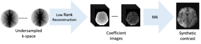 |
7 | MRI contrast synthesis from low-rank coefficient images
Xiaoxia Zhang1, Sebastian Flassbeck1, and Jakob Assländer1
1Center for Biomedical Imaging, Department of Radiology, New York University Grossman School of Medicine, New York University, New York, NY, United States
Synthetic contrasts are commonly derived from parameter maps via Bloch simulation.Typically, model imperfections, in particular partial volume effects, cause artifacts in those images. Recently, it has been proposed to overcome this problem by mapping directly from MR-Fingerprinting data to synthetic contrasts with neural networks. Those methods, however, face the MRF-typical undersampling artifacts, as well as the computational burden of hundreds of input images. We propose to first reconstruct images in a low-rank sub-space, which maintains the correct partial volume contrast, but allows for removal of undersampling artifacts, and to map from this space to synthetic contrasts with a neural network.
|
||
3495 |
8 | Simultaneous T1, T2 and T2* Quantification with MR Fingerprinting Using Dual-Echo Echo Planar Imaging
Di Cui1, Xiaoxi Liu1, Peng Cao2, Angela Jakary1, Jing Liu1, Janine Lupo1, Yan Li1, and Duan Xu1
1University of California, San Francisco, San Francisco, CA, United States, 2The University of Hong Kong, Hong Kong, China
An MR fingerprinting method using dual echo EPI was developed in this study for simultaneous quantification of T1, T2, T2*, proton density and off resonance. In vivo evaluations performed in patients with lower grade glioma tumor demonstrated the ability of the technique to acquire multiple reflexivity maps within a clinically manageable scan time with each slice using 16 seconds and achieving total brain coverage in approximately four minutes.
|
||
3496 |
9 | Towards Whole Brain Diffusion Tensor Spectroscopic Imaging
Bruno Sa de la Rocque Guimaraes1,2, Khaled Talaat1,2, Michael Mullen3, Essa Yacoub3, and Stefan Posse1,4
1Neurology Department, University of New Mexico, Albuquerque, NM, United States, 2Nuclear Engineering Department, University of New Mexico, Albuquerque, NM, United States, 3Center for Magnetic Resonance Research, University of Minnesota, Minneapolis, MN, United States, 4Physics and Astronomy Department, University of New Mexico, Albuquerque, NM, United States
Here we describe the development of a high-spatial resolution multi-slice single-shot Diffusion-Tensor proton-echo-planar-spectroscopic-imaging (PEPSI) technique using a binomial spatial-spectral refocusing RF pulse for mapping the diffusion tensor of water, Cho, Cr and NAA. Analysis of NAA and water diffusion in white matter obtained in a 4-slice acquisition with 1cc voxel size in 6 minutes (bmax = 2430.7 mm2/s) showed a mean ADC value of 0.15*10-3 and 0.66*10-3 mm2/s, which is consistent with values reported in the literature. Single-shot high spatial resolution diffusion sensitive MR spectroscopic imaging has the potential to probe metabolite diffusion across extended brain regions.
|
||
3497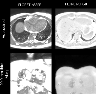 |
10 | High-quality Lung imaging with FLORET UTE and Fibonacci interleaved trajectory ordering
Guruprasad Krishnamoorthy1, Matthew M Willmering2, Jason C Woods2,3, and James G Pipe4
1MR R&D, Philips Healthcare, Rochester, MN, United States, 2Center for Pulmonary Imaging Research, Divisions of Pulmonary Medicine and Radiology, Cincinnati Children’s Hospital Medical Center, Cincinnati, OH, United States, 3Departments of Pediatrics, Radiology, and Physics, University of Cincinnati College of Medicine, Cincinnati, OH, United States, 4Department of Radiology, Mayo Clinic, Rochester, MN, United States
FLORET (Fermat looped, orthogonally encoded trajectories) is an efficient center-out 3D spiral trajectory, and it supports ultra-short echo times. In this work, a new trajectory ordering scheme and FLORET balanced steady-state free precession (FLORET-bSSFP) were developed at 1.5T with inline reconstruction. Simulations, phantom, and human lung imaging were performed to compare the performance of the proposed trajectory ordering with standard ordering. The proposed trajectory ordering scheme minimized artifacts caused by the changing gradient moments while improving temporal stability and motion robustness. High-quality free-breathing lung images were obtained using FLORET-bSFFP and spoiled gradient echo-based UTE FLORET.
|
||
3498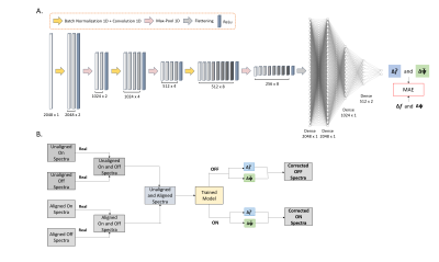 |
11 | Magnetic Resonance Spectroscopy Spectral Registration with Deep Learning
David Ma1, Hortense Le1, Scott Small2,3,4, and Jia Guo2,5
1Department of Biomedical Engineering, Columbia University, New York, NY, United States, 2Department of Psychiatry, Columbia University, New York, NY, United States, 3Department of Neurology, Columbia University, New York, NY, United States, 4Taub Institute Research on Alzheimer's Disease and the Aging Brain, Columbia University, New York, NY, United States, 5Mortimer B. Zuckerman Mind Brain Behavior Institute, Columbia University, New York, NY, United States
Deep learning is an effective image processing approach that has been enthusiastically adopted in Magnetic Resonance Spectroscopy (MRS). Methods such as multilayer perceptrons (MLP) and convolutional neural networks (CNN) have been applied to frequency and phase correction (FPC) to help resolve frequency and phase shifts that arise in MRS. However, both methods need to be trained separately with frequency and phase offsets to perform FPC. In this study, we aim to introduce a spectrum registration technique using CNNs that perform simultaneous correction of both frequency and phase shifts of single voxel MEGA-PRESS MRS simulated data.
|
||
 Back to Meeting Home
 Back to the Program-at-a-Glance
The International Society for Magnetic Resonance in Medicine is accredited by the Accreditation Council for Continuing Medical Education to provide continuing medical education for physicians.
 Back to Meeting Home
 Back to the Program-at-a-Glance
 View the Presentation