Online Gather.town Pitches
Advances in Data Acquisition I
Joint Annual Meeting ISMRM-ESMRMB & ISMRT 31st Annual Meeting • 07-12 May 2022 • London, UK

Online Gather.town Pitches
Advances in Data Acquisition I
| Booth # | ||||
|---|---|---|---|---|
3288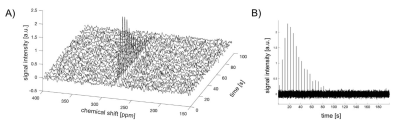 |
1 | Hyperpolarized 15N-BBCP as a novel probe of H2O2 Video Permission Withheld
Hyejin Park1, Jun Chen2, Ivan Dimitrov2,3, Jae Mo Park2,4, and Qiu Wang1
1Chemistry, Duke University, Durham, NC, United States, 2Advanced Imaging Research Center, University of Texas Southwestern Medical Center, Dallas, TX, United States, 3Philips Healthcare, Dallas, TX, United States, 4Department of Radiology, University of Texas Southwestern Medical Center, Dallas, TX, United States
In this work, we have developed a novel de novo 15N-molecular probe, 15N-boronobenzyl-4-cyanopyridinium (15N-BBCP), for in vivo detection of H2O2. The long spin-lattice relaxation time (T1), high H2O2 sensitivity
and selectivity, low cytotoxicity, and large chemical shift changes upon reaction suggest that the probe is suited for in vivo hyperpolarized 15N MRI.
|
||
3289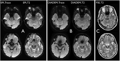 |
2 | Clinical DIADEM: Distortion-Free High-Resolution Diffusion-Weighted Imaging of the Brain.
Myung-Ho In1, Norbert G Campeau1, Joshua D Trzasko1, Daehun Kang1, Kirk M Welker1, John Huston III1, Yunhong Shu1, and Matt A Bernstein1
1Department of Radiology, Mayo Clinic, Rochester, MN, United States
Echo-planar based diffusion-weighted imaging (DWI) of the brain is prone to local image distortion which can lead to misdiagnosis or nondiagnostic image quality in areas prone to susceptibility effects. A novel distortion-free imaging scheme, termed DIADEM (Distortion-free Imaging: A Double Encoding Method) was recently introduced and optimized for brain imaging on a high-performance experimental Compact 3T system. In this study, we fully implement the DIADEM DWI technique on two 510-k cleared, whole-body 3T scanners and explore the clinical feasibility for its use in routine clinical practice.
|
||
3290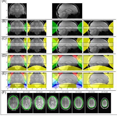 |
3 | Multi-slab 3D ECLIPSE for whole brain lipid suppressed MRSI
Chathura Kumaragamage1, Peter B Brown1, Scott McIntyre1, Terence W Nixon1, Henk M De Feyter1, and Robin A de Graaf1
1Radiology and Biomedical Imaging, Yale University, New Haven, CT, United States
The utility of ECLIPSE, a pulsed second order gradient insert allowing elliptical localization, was previously shown to provide high axial coverage for single slab MRSI acquisitions with robust lipid suppression. In this work we extend the ECLIPSE method with multi-slab localization that provides improved coverage, for applications in 3D MRSI acquisitions. A two-slab ECLIPSE-OVS method providing > 100-fold in mean lipid suppression was developed, with improved superior-inferior coverage. Simulation results show that a four-slab ECLIPSE method, based on OVS and IVS, can provide high-quality lipid suppression with whole-brain coverage.
|
||
3291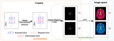 |
4 | Holistic Acceleration from Acquisition to Reconstruction for Submilimeter 3D MR Fingerprinting via Deep Learning
Yilin Liu1, Feng Cheng1, Yong Chen2, and Pew-Thian Yap3
1Computer Science, UNC-Chapel Hill, Chapel Hill, NC, United States, 2Radiology, Case Western Reserve University, Cleveland, OH, United States, 3Radiology, UNC-Chapel Hill, Chapel Hill, NC, United States
We developed a novel deep-learning empowered pipeline for both rapid acquisition and reconstruction of high-resolution 3D MRF with 16X acceleration, thereby significantly improving the clinical feasibility of MRF for quantitative imaging.
|
||
3292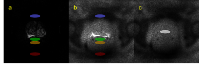 |
5 | Spatial properties of noise in prostate DWI acquired with an endo-rectal coil as critical information for clinical QA
Milica Medved1,2, Aritrick Chatterjee1,2, Ajit Devaraj3, Ambereen Yousuf1,2, Aytekin Oto1,2, and Gregory S Karczmar1,2
1Department of Radiology, The University of Chicago, Chicago, IL, United States, 2Sanford J. Grossman Center of Excellence in Prostate Imaging and Image Guided Therapy, The University of Chicago, Chicago, IL, United States, 3Philips Research NA, Cambridge, MA, United States
Development of quantitative QC methods for prostate DWI is important in clinical practice, necessitating reliable estimates of spatially heterogenous noise. In data obtained at 3T with the use of an endo-rectal coil, we use raw noise measurements to simulate pure-noise k-space datasets that are post-processed to yield noise maps. Good agreement is found between noise levels measured from noise maps in the central prostate and the estimates of noise produced from the anterior rectal ROIs in high TE / high b-value DWI. Thus, noise can be estimated from an anterior rectum region when the endo-rectal coil is used.
|
||
3293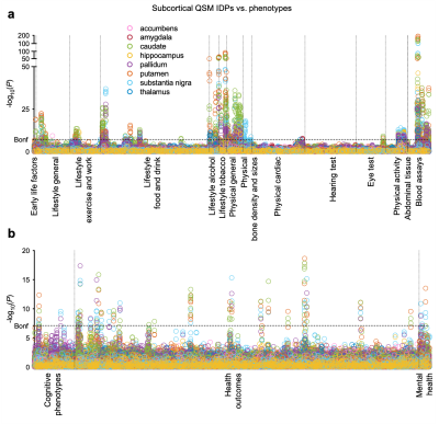 |
6 | Phenotypic associations of magnetic susceptibility in the brain
Chaoyue Wang1, Stephen M. Smith1, Fidel Alfaro-Almagro1, Gwenaëlle Douaud1, Johannes C. Klein1,2, Alberto Llera3, Aurea B. Martins-Bach1, Cristiana Fiscone4,5, Richard Bowtell4, Lloyd T. Elliott6, Benjamin C. Tendler1, and Karla L. Miller1
1Wellcome Centre for Integrative Neuroimaging, FMRIB, Nuffield Department of Clinical Neurosciences, University of Oxford, Oxford, United Kingdom, 2Oxford Parkinson’s Disease Centre, University of Oxford, Oxford, United Kingdom, 3Donders Institute for Brain, Cognition and Behaviour, Radboud University, Nijmegen, Netherlands, 4Sir Peter Mansfield Imaging Centre, School of Physics and Astronomy, University of Nottingham, Nottingham, United Kingdom, 5Department of Biomedical and Neuromotor Sciences, University of Bologna, Bologna, Italy, 6Department of Statistics and Actuarial Science, Simon Fraser University, Vancouver, BC, Canada In this abstract, we describe our findings from the phenotypic association analyses (univariate Pearson correlations) between 17,485 non-imaging phenotypes and QSM (and T2*) measures using data from 35,885 subjects in UK Biobank. In total, we identify statistically-significant associations of 251 phenotypes with QSM IDPs. Here we describe example associations in blood assays, health outcomes, food and drink, and alcohol consumption categories in detail. Our results demonstrate that QSM and T2* contribute complementary information. This new QSM resource provides an opportunity to investigate susceptibility contrast in previously unexplored territory, which might lead to advances in application of QSM in neuroscience. |
||
3294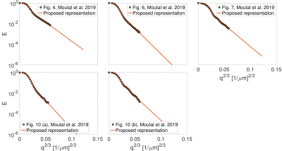 |
7 | Cumulant expansion with localization
Maryam Afzali1,2, Tomasz Pieciak3, Derek K Jones2, Jürgen Schneider4,5, and Evren Özarslan6,7
1Leeds Institute of Cardiovascular and Metabolic Medicine, University of Leeds, Leeds, United Kingdom, 2Cardiff University Brain Research Imaging Centre (CUBRIC), School of Psychology, Cardiff University, Cardiff, United Kingdom, 3LPI, ETSI Telecomunicación, Universidad de Valladolid, Valladolid, Spain, 4Leeds Institute of Cardiovascular and Metabolic Medicine, Leeds, United Kingdom, 5Division of Cardiovascular Medicine, Radcliffe Department of Medicine, University of Oxford, Oxford, United Kingdom, 6Department of Biomedical Engineering, Linköping University, Linköping, Sweden, 7Center for Medical Image Science and Visualization, Linköping University, Linköping, Sweden
Diffusion MR is sensitive to the microstructural features of a sample. Fine-scale characteristics can be probed by employing strong diffusion gradients while the small gradient regime is determined by the cumulants of the distribution of particle displacements. However, the cumulant expansion suffers from a finite convergence radius and cannot represent the ‘localization regime’ that emerges at higher gradient strengths characterized by a stretched exponential dependence. Here, we propose a new representation for the diffusion MR signal. Our method provides not only a robust estimate of the first few cumulants but also a meaningful extrapolation of the signal decay.
|
||
3295 |
8 | Characterization of the Zona incerta and Fields of Forel using QSM at 9.4T Video Permission Withheld
Vinod Jangir Kumar1, Klaus Scheffler1,2, Gisela E Hagberg1,2, and Wolfgang Grodd1
1Max Planck Institute for Biological Cybernetics, Tuebingen, Germany, 2Biomedical Magnetic Resonance, University Hospital and Eberhard-Karl’s University, Tuebingen, Germany The subthalamic structures, like the zona incerta (ZI), fields of forel (FF), subthalamic nucleus (STN), and their surrounding structures, are poorly characterized regarding anatomical imaging. Using quantitative susceptibility mapping (QSM) at ultra-high-field, iron, and myelin mapping may allow an advanced characterization. Therefore, QSM data were acquired at 9.4 Tesla in 21 subjects to characterize the para- and diamagnetic properties of the ZI, FF, and their adjacent structures. In contrast to the FF, we found a higher contribution of diamagnetic and paramagnetic sources, i.e., iron, myelin, and calcium, in the STN and ZI. |
||
3296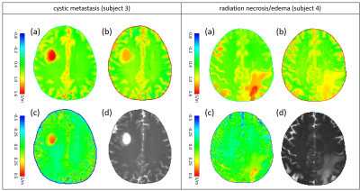 |
9 | Normalization of conductivity maps to support identification of pathologic areas
Ulrich Katscher1 and Khin Khin Tha2
1Philips Research, Hamburg, Germany, 2Hokkaido University, Sapporo, Japan
Electrical tissue conductivity is an emerging quantitative diagnostic parameter, particularly for oncology. Conductivity typically increases with tumor malignancy, however, high conductivity per se is no indication for abnormal tissue, since many tissue types are highly conductive without being abnormal. An empirical rule correlates tissue conductivity with tissue water content. One possibility to analyze abnormality of tissue is thus to compare conductivity as measured with conductivity as expected from water content. The obtained difference yielding “normalized” conductivity might serve as a more direct measure of tissue abnormality. This study presents this concept using pathologic and healthy in vivo example cases.
|
||
3297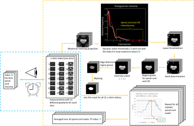 |
10 | Automated Calculation of Optimum z-Shim Gradient Pulses from a Reference Acquisition for Spinal Cord fMRI
Ying Chu1 and Jürgen Finsterbusch1
1Department of Systems Neuroscience, University Medical Center Hamburg-Eppendorf, Hamburg, Germany Slice-specific z-shimming reduces signal losses caused by through-slice dephasing in spinal cord fMRI acquisitions. Typically, the optimum values are determined from a reference acquisition covering a range of z-shim values by a user who identifies the setting with the least signal loss. However, this procedure requires some experience, is time-consuming, and user-dependent. Here, an automated calculation of the optimum values has been developed, implemented on the MR system, and evaluated in phantoms and in vivo. Without compromising the image quality, the approach is faster and user-independent and, thus, may help to facilitate the application of z-shimming in spinal cord fMRI. |
||
3298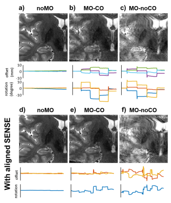 |
11 | Combining navigator-based prospective and data-driven retrospective motion correction for high resolution 7T multi-slice T2W MRI.
Vincent Oltman Boer1, Lucilio Cordero Grande2, Jan Ole Pedersen3, Martin Prener4, Esben Thade Petersen1,5, Olaf B Paulson4,6, Mads Andersen3,7, and Giske Opheim4,8
1Danish Research Centre for Magnetic Resonance, Centre for Functional and Diagnostic Imaging and Research, Copenhagen University Hospital Amager and Hvidovre, Copenhagen, Denmark, 2Biomedical Image Technologies, ETSI Telecomunicación, Universidad Politécnica de Madrid and CIBER-BBN, Madrid, Spain, 3Philips Healthcare, Copenhagen, Denmark, 4Neurobiology Research Unit, Department of Neurology, Copenhagen University Hospital Rigshospitalet, Copenhagen, Denmark, 5Section for Magnetic Resonance, DTU Health Tech, Kgs Lyngby, Denmark, 6Faculty of Health and Medical Sciences, University of Copenhagen, Copenhagen, Denmark, 7Lund University Bioimaging Center, Lund University, Lund, Sweden, 8Department of Radiology, Copenhagen University Hospital Rigshospitalet, Copenhagen, Denmark
High-resolution multi-slice T2W imaging at ultra-high field is highly sensitive to motion due to high tissue-CSF contrast and the k-space filling pattern. In this study we investigated the combination of 3D prospective and in-plane retrospective motion correction to improve image quality. We observed that a combination of navigator based prospective and data-driven retrospective correction is able to correct most severe motion artifacts during scanning, and remove smaller residual artifacts in reconstruction.
|
||
 Back to Meeting Home
 Back to the Program-at-a-Glance
The International Society for Magnetic Resonance in Medicine is accredited by the Accreditation Council for Continuing Medical Education to provide continuing medical education for physicians.
 Back to Meeting Home
 Back to the Program-at-a-Glance
 View the Presentation