Online Gather.town Pitches
Quantitative Imaging
Joint Annual Meeting ISMRM-ESMRMB & ISMRT 31st Annual Meeting • 07-12 May 2022 • London, UK

Online Gather.town Pitches
Quantitative Imaging
| Booth # | ||||
|---|---|---|---|---|
4998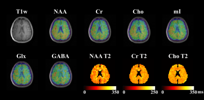 |
1 | High-Resolution Brain Metabolite T2 Mapping Using Optimized Multi-TE MRSI
Zepeng Wang1,2, Yahang Li1,2, and Fan Lam1,2
1Department of Bioengineering, University of Illinois at Urbana-Champaign, Urbana, IL, United States, 2Beckman Institute for Advanced Science and Technology, Urbana, IL, United States
Metabolite T2 is recognized as an important physiological and disease biomarker, whose measurement also benefits metabolite quantification. However, the SNR challenge of MRSI and prolonged scan time for multi-TE acquisition limit the imaging resolution. This work presents an optimized multi-TE MRSI strategy to achieve high-resolution 3D brain metabolite T2 mapping. Specifically, estimation-theoretic TE selection was analyzed for optimized metabolite T2 estimation. An enhanced parameter estimation strategy was proposed. Both simulation and invivo studies were conducted to evaluate our method. The exciting capability of simultaneous high-resolution metabolite, neurotransmitter and T2 mapping is demonstrated for the first time.
|
||
 |
4999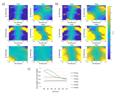 |
2 | MR T1ρ preparations: B1 and B0 inhomogeneity and T2ρ evaluation with Bloch equation-based simulation
Jeehun Kim1,2,3, Qi Peng4, Can Wu5, and Xiaojuan Li1,2,6
1Program of Advanced Musculoskeletal Imaging (PAMI), Cleveland Clinic, Cleveland, OH, United States, 2Department of Biomedical Engineering, Cleveland Clinic, Cleveland, OH, United States, 3Department of Electrical Engineering, Case Western Reserve University, Cleveland, OH, United States, 4Department of Radiology, Albert Einstein College of Medicine and Montefiore Medical Center, Bronx, NY, United States, 5Department of Medical Physics, Memorial Sloan Kettering Cancer Center, New York, NY, United States, 6Department of Diagnostic Radiology, Cleveland Clinic, Cleveland, OH, United States
Quantitative T1ρ mapping is a promising biomarker for detecting tissue compositional changes at early stages of diseases. For reliable and reproducible measurements, T1ρ preparation pulses should be robust to B0 and B1 inhomogeneity. In this work, a Bloch equation based numerical simulation tool was developed and validated. The performance of six different T1ρ preparation schemes were evaluated in terms of their responses to B0 and B1 inhomogeneities using the simulation tool and in phantoms and human subjects. T2ρ measured in phantoms and human subjects were used during simulation.
|
|
5000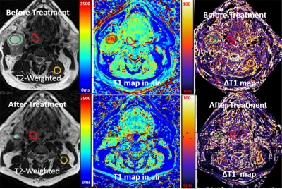 |
3 | 3D Tissue Oxygenation Level Dependence MRI on head and neck cancers by using 3D Stack of Star acquisition and Variable Flip Angle T1 method
Seong-Eun Kim1, John A Roberts1, Eugene Kholmovski1,2, Ying Hitchcock 3, and Yoshimi Anzai 1
1UCAIR, Department of Radiology and Imaging Sciences, University of Utah, Salt Lake City, UT, United States, 2Department of Biomedical Engineering, Johns Hopkins University, Baltimore, MD, United States, 3Department of Radiation Oncology, University of Utah, Salt Lake City, UT, United States
Hypoxia is a common feature of most solid tumors, reflecting an imbalance of oxygen delivery and consumption. Tumor hypoxia is an indicator of a treatment resistance and overall poor prognosis in head and neck cancer. Change in tissue oxygen concentration level (TOLD) induced by breathing pure oxygen produces proportional change in tissue T1 relaxivity. The proposed technique, a motion insensitive 3D TOLD MRI using 3D VFA T1 Stack of Star sampling, has obvious advantages over conventional 3D VFA technique for reliable and accurate T1 measurements and a great potential for the assessment of tumor hypoxia in HNC patients.
|
||
5001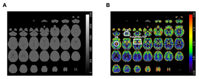 |
4 | High Resolution 3D Ultra-Short Echo Time MRI with Rosette k-Space Pattern for Brain Iron Content Mapping
Xin Shen1, Ali Caglar Özen2, Humberto Monsivais3, Serhat Ilbey2, Antonia Sunjar1, Aparna Karnik4, Mark Chiew5, and Uzay Emir1,3
1Weldon School of Biomedical Engineering, Purdue University, West Lafayette, IN, United States, 2Department of Radiology, Medical Physics, University of Freiburg, Freiburg, Germany, 3Health Science Department, Purdue University, West Lafayette, IN, United States, 4Elmore Family School of Electrical and Computer Engineering, Purdue University, West Lafayette, IN, United States, 5Wellcome Centre for Integrative Neuroimaging, University of Oxford, Oxford, United Kingdom
This study aimed to detect brain iron content with a novel high resolution (0.94 mm isotropic voxel) ultra-short echo time (UTE) MRI based on rosette k-space trajectory. Non-invasive monitoring of brain iron content is beneficial, because the iron concentration increases during normal brain development, but is associated with many neurodegenerative diseases. With the ultra-short echo time (TE=20μs), the fast signal decay caused by high iron concentration can be captured. The relationship between iron concentration and signal intensity was quantified based on phantom study, and was used for modulation of the in vivo data to produce images of iron-rich brain regions.
|
||
5002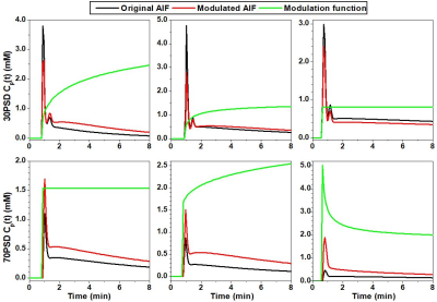 |
5 | Reducing fluctuations in prostate DCE-MRI physiological parameters using muscle as a reference to modulate arterial input function
Xiaobing Fan1, Aritrick Chatterjee1, Grace H. Lee1, Ambereen Yousuf1, Tatjana Antic2, Aytekin Oto1, and Gregory S. Karczmar1
1Radiology, University of Chicago, Chicago, IL, United States, 2Pathology, University of Chicago, Chicago, IL, United States We introduce a method to modulate arterial input functions (AIFs) using gluteal muscle as reference. The method was tested on a split injection protocol for prostate dynamic contrast enhanced (DCE) MRI: first injecting 30% of the standard dose (30PSD), after two minutes, followed by 70% of the standard dose (70PSD) of gadoterate meglumine. The AIFs were measured from the iliac artery for both doses and used to analyze 70PSD data. By assuming gluteal muscle Ktrans=0.1min-1 and ve=0.1 to modulate AIFs for both doses, the fluctuations of calculated physiological parameters were significantly reduced when compared to using the original AIFs. |
||
5003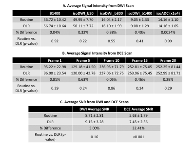 |
6 | Deep Learning Denoising Reconstruction (DLR) Preserves Quantitative DWI and DCE Signal Intensity Measures in Prostate MRI
Hung Phi Do1, Brian Tymkiw1, Dawn Berkeley1, and Mo Kadbi1
1Canon Medical Systems USA, Inc., Tustin, CA, United States
Deep Learning Reconstruction (DLR) has recently been translated into clinical practices as a few DLR methods have received FDA 510-k clearance. Given its novelties, rigorous evaluations are essential to ensure the safety and efficacy of DLR before routine clinical use. This work aims to investigate whether DLR preserves quantitative measures from DWI and DCE prostate MRI. The results show that the average signal intensities measured from routine images are similar (less than 1% difference) to those measured from DLR images.
|
||
5004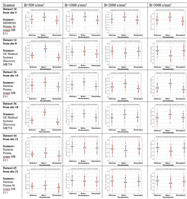 |
7 | Multi-site harmonization of diffusion MRI data from the Adolescent Brain Cognitive Development (ABCD) Study
Suheyla Cetin-Karayumak1, Fan Zhang1, Steve Pieper2, Lauren J. O'Donnell1, and Yogesh Rathi1
1Harvard Medical School and Brigham and Women's Hospital, Boston, MA, United States, 2Isomics, Cambridge, MA, United States
This study presents our harmonization efforts on the multi-site diffusion MRI data from ~11,000 adolescents in the Adolescent Brain Cognitive Development (ABCD) study, collected at 21 sites using Siemens, GE, and Philips scanners. We validated the harmonization of the multi-site dMRI data using several standard and advanced diffusion MRI measures including Fractional Anisotropy (FA), Return-To-Origin Probability (RTOP), Return To the Axis Probability (RTAP), Return To the Plane Probability (RTPP), Mean Squared Displacement (MSD).
|
||
5005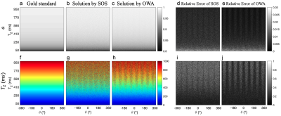 |
8 | Analytical T2 mapping refinement using the bSSFP elliptical signal model
Yiyun Dong1 and Michael Hoff2
1Physics, University of Washington, Seattle, WA, United States, 2Radiology, University of Washington, Seattle, WA, United States
The elliptical signal model (ESM) for bSSFP imaging exhibits potential for quantitative imaging. Recent work proposed the first analytical T2 solution based on the bSSFP ESM, with instability at large T2 values. Instead of using the sum of squares (SOS) to combine multiple intermediate solutions, here a multi-step regional variance weighting methodology is proposed to generate a refined analytical T2 computation. The new method for T2 computation improves T2 computation performance relative to the SOS and near problematic dark bands, inspiring novel clinical applications of bSSFP in quantitative imaging.
|
||
5006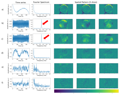 |
9 | Quality Assessment of Mouse fMRI: Evaluating Temporal Stability of Preclinical Scanners
Mila Urosevic1, Jeremie Fouquet2, and M. Mallar Chakravarty1,2,3
1Biological & Biomedical Engineering, McGill University, Montreal, QC, Canada, 2Douglas Mental Health University Institute, Montreal, QC, Canada, 3Department of Psychiatry, McGill University, Montreal, QC, Canada Mouse functional magnetic resonance imaging (fMRI) has interesting potential for basic neuroscience, but there is significant variability in data quality across studies. This variability may be partly explained by differences in scanner stability, however there are no reports of scanner quality assessments in the mouse fMRI field. We evaluated the stability of our preclinical scanner by acquiring an fMRI time-series of an agarose phantom then computing stability metrics. We found that our scanner has acceptable temporal stability but unexpected periodic fluctuations. Our methods are openly available so that other groups can easily implement stability assessments on their respective preclinical scanners. |
||
5007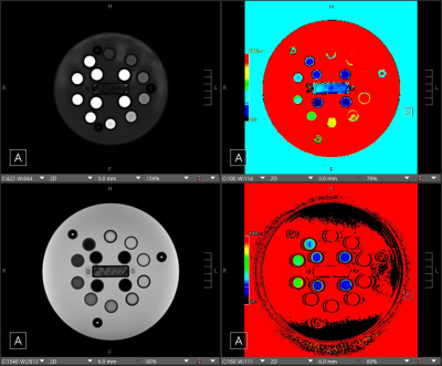 |
10 | Intraplatform Repeatability and Interplatform Reproducibility of T1 and T2 Mapping Using a NIST System Phantom
Justin Yu1, Erika M Rand1, Pamela R Jackson2, Joseph M Hoxworth1, Leland S Hu1, Kristin R Swanson2, and Yuxiang Zhou1
1Radiology, Mayo Clinic Arizona, Phoenix, AZ, United States, 2Neurosurgery, Mayo Clinic Arizona, Phoenix, AZ, United States
Quantitative magnetic resonance imaging, especially T1/T2 relaxation time mapping is increasingly used in both research and clinical practice. The purpose of this study was to assess the precision and cross-platform repeatability and reproducibility of T1 and T2 mapping modules on GE and Siemens MRI platforms, with the goal of standardization across vendors. A standard MRI quantitative phanom was scanned on four scanners, twice per scanner. GE data was processed with a custom script. Our results show high intraplatform repeatability, but showed high inter-platform discrepancies for both T1 and T2 results.
|
||
 Back to Meeting Home
 Back to the Program-at-a-Glance
The International Society for Magnetic Resonance in Medicine is accredited by the Accreditation Council for Continuing Medical Education to provide continuing medical education for physicians.
 Back to Meeting Home
 Back to the Program-at-a-Glance
 View the Presentation