Online Gather.town Pitches
Cardiac II
Joint Annual Meeting ISMRM-ESMRMB & ISMRT 31st Annual Meeting • 07-12 May 2022 • London, UK

| Booth # | ||||
|---|---|---|---|---|
 |
4436 |
1 | Stochastic Fibrosis Signatures from 3D LGE: Novel Threshold-Free Quantification of Left Atrial Fibrosis
Mehri Mehrnia1,2, Eugene Kholmovski3, Rod Passman4, Aggelos Katsaggelos1,5,6, Saman Nazarian7, Daniel Kim1, and Mohammed Elbaz1
1Radiology, Northwestern University, Chicago, IL, United States, 2Biomedical Engineering, Northwestern University, Chicago, IL, United States, 3Johns Hopkins University, Baltimore, MD, United States, 4Cardiology, Northwestern University, Chicago, IL, United States, 5Electrical and Computer Engineering, Northwestern University, Chicago, IL, United States, 6Computer Science, Northwestern Universiy, Chicago, IL, United States, 7Division of Cardiovascular Medicine, Perelman School of Medicine, University of Pennsylvania, Philadelphia, PA, United States Assessment of left atrial (LA) fibrosis in atrial fibrillation (AF) patients from 3D LGE MRI have shown promise in evaluating atrial myopathy for selecting patients for catheter ablation and to predict AF recurrence post intervention. Nevertheless, current methods for fibrosis quantification suffer from lack of standardization and reproducibility as they rely on different thresholds for defining fibrosis. Hence, limiting the clinical translation of 3D LA LGE MRI. Here, we propose the first threshold-free technique to quantify LA fibrosis burden using novel stochastic fibrosis signature technique. We demonstrated feasibility and correlations to four of the previously published methods for fibrosis quantification. |
|
4437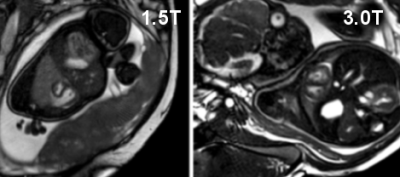 |
2 | CMR of fetal cardiac function and blood flow with doppler ultrasound cardiac gating
Erin K Englund1, Lorna P Browne1, Takashi Fujiwara1, Richard Friesen2, Mehdi H Moghari1, and Alex J Barker1
1Radiology, University of Colorado, Anschutz Medical Campus, Aurora, CO, United States, 2Pediatric Cardiology, University of Colorado, Anschutz Medical Campus, Aurora, CO, United States
Fetal CMR is difficult due to the lack of an ECG signal, fetal motion, small anatomic sizes and high fetal heart rates. Initial results are presented which use a doppler ultrasound gating device to capture fetal cardiac motion in standard cardiac views and blood flow was quantified with 4D flow MRI. bSSFP cine images were scored for image quality by two radiologists and flow in the umbilical vein, ascending aorta and main pulmonary artery were quantified and agreed well with reported values. Unique insight was provided with dynamic imaging regarding the fetal circulation and the impact of congenital disease.
|
||
4438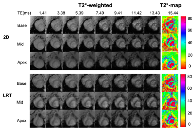 |
3 | Free-breathing Fully Ungated 3D Cardiac T2* MR Mapping using a Low-Rank Tensor Framework
Xingmin Guan1,2, Hsin-Jung Yang1, Zhehao Hu2,3, Nan Wang4, Anthony Christodoulou1, Behzad Sharif5, Debiao Li1, and Rohan Dharmakumar2,5
1Cedars-Sinai Medical Center, Los Angeles, CA, United States, 2University of California, Los Angeles, Los Angeles, CA, United States, 3University of Southern California, Los Angeles, CA, United States, 4Stanford University, Stanford, CA, United States, 5Indiana University, Indianapolis, IN, United States
A 3D fully ungated, free breathing cardiac T2* technique was developed using a low-rank tensor framework. An animal model with iron-oxide contrast-enhanced studies was used to test and validate the proposed technique. T2* images reconstructed from the proposed approach showed better image quality than those from conventional 2D T2* imaging approach. Excellent agreement of septal T2* was found between the proposed approach and conventional 2D approach. Our findings show that the proposed approach can accurately characterize T2* of myocardium under baseline and during iron overload conditions.
|
||
4439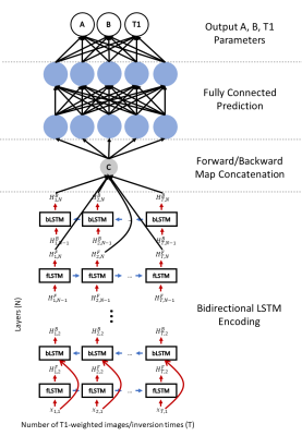 |
4 | Accelerated cardiac T1 mapping with bidirectional LSTMs and cyclic model-based loss
Johnathan V. Le1,2, Jason K. Mendes2, Mark Ibrahim3, Brent D. Wilson3, Edward V.R. DiBella1,2, and Ganesh Adluru1,2
1Department of Biomedical Engineering, University of Utah, Salt Lake City, UT, United States, 2Utah Center for Advanced Imaging Research, University of Utah, Salt Lake City, UT, United States, 3Department of Cardiology, University of Utah, Salt Lake City, UT, United States
Cardiac T1 mapping has been shown to be a promising method for assessing different cardiomyopathies. Most cardiac T1 mapping methods require long breath holds during the acquisition which can be difficult for patients particularly during exercise or pharmacologically induced stress. Here we proposed using a multi-layer bidirectional LSTM with fully connected output and a cyclic model-based loss function to reduce the acquisition time of T1 mapping sequences without significant loss of quality.
|
||
4440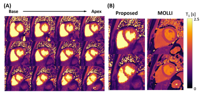 |
5 | Free-running Simultaneous 3D Cardiac T1 Mapping and Cine Imaging Using a Linear Tangent Space Alignment Model
Yanis Djebra1,2, Thibault Marin1, Paul Kyu Han1, Isabelle Bloch3, Georges El Fakhri1, and Chao Ma1
1Gordon Center for Medical Imaging, Department of Radiology, Massachusetts General Hospital, Harvard Medical School, Boston, MA, United States, 2LTCI, Telecom Paris, Institut Polytechnique de Paris, Paris, France, 3LIP6, Sorbonne University, CNRS, Paris, France
Cardiac T1 mapping allows assessment of tissue characteristics and functioning of the heart. However, existing methods are limited in terms of spatial resolution and coverage due to limitations in acquisition speed and the presence of cardiac and respiratory motion. This work proposes a new reconstruction framework for simultaneous, high-resolution 3D cardiac T1 mapping and cine imaging of the heart at 3T from sparsely sampled k-space data.
|
||
4441 |
6 | Restored Torsion and Longitudinal Strain in ACE Inhibitor Treated Hypertension
Alexander Wilson1,2, Gregory B Sands3, Vicky Y Wang2, Beau Pontre4, Daniel B Ennis1,2, Alistair A Young5, Ian J LeGrice3, and Martyn P Nash3,6
1Stanford Cardiovascular Institute, Stanford University, Stanford, CA, United States, 2Department of Radiology, Stanford University, Stanford, CA, United States, 3Auckland Bioengineering Institute, University of Auckland, Auckland, New Zealand, 4Department of Anatomy and Medical Imaging, University of Auckland, Auckland, New Zealand, 5Department of Biomedical Engineering, King's College London, London, United Kingdom, 6Department of Engineering Science, University of Auckland, Auckland, New Zealand Tagged CMR was used to measure torsion and longitudinal strain in a rodent model of hypertensive heart disease. Long-term ACE inhibitor treatment restored ejection fraction, torsion and longitudinal strain by 24 months of age. Longitudinal strain was the first functional measure to be restored, and this may indicate that longitudinal strain is a sensitive imaging biomarker for assessing the efficacy of treatment with regards to reverse remodeling in hypertension. |
||
4442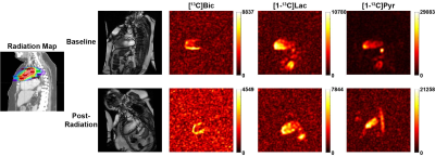 |
7 | Early Detection of Radiation-Induced Cardiotoxicity Using Hyperpolarized Pyruvate
Junjie Ma1, Jun Chen1, Elizabeth Zhang-Velten2, Jayesh Sharma2, Xuliang Wang3, Gabriele Schiattarella3, Thomas Gillette3, Joseph Hill3,4, Craig R. Malloy1,3,5, Vlad G. Zaha1,3, Jae Mo Park1,5,6, and Prasanna Alluri2
1Advanced Imaging Research Center, UT Southwestern Medical Center, Dallas, TX, United States, 2Radiation Oncology, UT Southwestern Medical Center, Dallas, TX, United States, 3Internal Medicine, UT Southwestern Medical Center, Dallas, TX, United States, 4Molecular Biology, UT Southwestern Medical Center, Dallas, TX, United States, 5Radiology, UT Southwestern Medical Center, Dallas, TX, United States, 6Electrical and Computer Engineering, UT Dallas, Richardson, TX, United States
Radiation-induced heart disease is a major source of morbidity and mortality in patients receiving thoracic radiation. In this study, radiation-induced changes in cardiac metabolism is investigated using hyperpolarized [1-13C]pyruvate MRI in animals and patients. Myocardial bicarbonate-to-lactate ratios decreased following radiation treatments while no change was observed in the global strain, suggesting radiation-induced mitochondrial dysfunction in the heart. This translational study demonstrates clinical potential of hyperpolarized 13C pyruvate for early and noninvasive detection of radiation-induced cardiac injury.
|
||
4443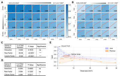 |
8 | Influence of Spatial Resolution in T2* Maps of Intramyocardial Hemorrhage
Xinheng Zhang1,2, Hsin-Jung Yang1, and Rohan Dharmakumar3
1Biomedical Imaging Research Institute, Cedars-Sinai Medical Center, Los Angeles, CA, United States, 2Bioengineering Department, UCLA, Los Angeles, CA, United States, 3Krannert Cardiovascular Research Center, Indiana University School of Medicine, Indianapolis, IN, United States
The influence of spatial resolution on T2*-based characterization of hemorrhage within myocardial infarctions (MI) has not been investigated. We hypothesized that partial volume could play a critical role in the detection and quantification of hemorrhagic remnants. Ex-vivo pig hearts with 8-week old MI were imaged with multi-gradient echo sequences with various in-plane and through-plane spatial resolutions. Our findings show that optimal detection of hemorrhagic remnants with T2* MRI at 3T requires imaging voxels to be between 5 and 7 mm3.
|
||
4444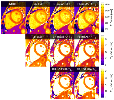 |
9 | Simultaneous T1, T2, and T1ρ Mapping of the Myocardium with Multi-Parametric SASHA
Kelvin Chow1, Hui Xue2, and Peter Kellman2
1Cardiovascular MR R&D, Siemens Healthineers, Chicago, IL, United States, 2National Heart, Lung, and Blood Institute, National Institutes of Health, Bethesda, MD, United States Parametric T1 and T2 mapping can provide valuable insight into myocardial tissue microstructure. T1ρ is a complimentary technique that is sensitive to macromolecular content and may potentially enable non-contrast scar visualization. Characterization with all parameters is desirable as they may have different sensitivities to pathology but is time consuming with separate acquisitions for each. We propose a novel mSASHA sequence with T1, T2, and T1ρ maps in a single 13 HB breath-hold or free-breathing acquisition. mSASHA had superior T1 and T2 accuracy and similar precision to conventional MOLLI and T2p-bSSFP in phantoms and data is presented from 3 volunteers. |
||
4445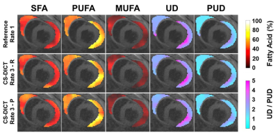 |
10 | Accelerated MRI of epicardial adipose tissue fatty acid composition in mice using a compressed-sensing and dictionary-based reconstruction
Soham Shah1 and Fred H Epstein1
1Biomedical Engineering, University of Virginia, Charlottesville, VA, United States
Epicardial adipose tissue (EAT) and its fatty acid composition (FAC) have been implicated in numerous cardiovascular diseases as saturated fatty acids are known to promote inflammation. FAC MRI techniques, while prominent, have not been applied to EAT due to extended scan times, thus, image acceleration is essential. Here, we demonstrate compressed sensing with a signal model-based dictionary (CS-DICT) to reconstruct psuedo-random undersampled images. Using CS-DICT, we achieve rate-3 acceleration while maintaining accurate EAT FAC maps and estimations in obese mice. These methods facilitate the application of FAC MRI to the EAT.
|
||
4446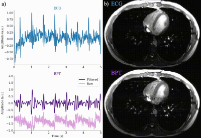 |
11 | Cardiac and Respiratory-Resolved Image Reconstruction with the Beat Pilot Tone
Kathryn Lamar-Bruno1, Suma Anand1, and Michael Lustig1
1Electrical Engineering and Computer Sciences, University of California, Berkeley, Berkeley, CA, United States
We have previously proposed Beat Pilot Tone (BPT), a motion sensing method using ultra-high-frequency RF and preamplifier intermodulation. BPT uses ultra-high-frequencies, which provides greater sensitivity to motion compared to the Pilot Tone (PT). In this work, we use the motion estimates from the BPT to reconstruct respiratory and cardiac-resolved images and compare them to motion-resolved images obtained using PT, respiratory bellows, and electrocardiogram (ECG) data. We show that BPT provides comparable image quality to conventional motion sensing for respiratory and cardiac motion.
|
||
4447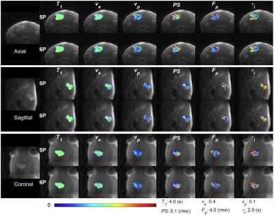 |
12 | Simultaneous estimation of longitudinal relaxation time and intracellular water lifetime using Active Contrast Encoding MRI
Jin Zhang1, Karl Kiser1, Ayesha Bharadwaj Das1, Sawwal Qayyum1, and Gene Kim1
1Weill Cornell Medical College, New York, NY, United States
Dynamic contrast enhanced (DCE)-MRI can be used as a tool to measure intracellular water lifetime (τi) in tumor cells which can be used as a biomarker for tumor aggressiveness and treatment response. Contrast kinetic model analysis in DCE-MRI requires accurate pre-contrast T1 measurement. We used multiple flip angles for active contrast encoding of both T1 and τi during dynamic data acquisition with one injection. The present study demonstrates the feasibility of measuring T1 and τi simultaneously
from the active contrast encoding (ACE) MRI data. ACE-MRI also reduces the scan time by eliminating the need of separate pre-contrast T1 measurement.
|
||
4448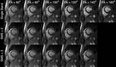 |
13 | High-Performance 0.55T Supports Contrast-Optimal SMS bSSFP Cardiac Imaging
Ye Tian1, Sophia X. Cui2, Yongwan Lim1, Nam G. Lee3, Ziwei Zhao1, and Krishna S. Nayak1
1Ming Hsieh Department of Electrical and Computer Engineering, University of Southern California, Los Angeles, CA, United States, 2Siemens Medical Solutions USA, Inc., Los Angeles, CA, United States, 3Biomedical Engineering, University of Southern California, Los Angeles, CA, United States
Balanced steady-state free precession (bSSFP) cardiac cine MRI at 1.5T and 3T is routinely used for cardiac function assessment. Simultaneous multi-slice (SMS) imaging significantly reduces the number of required breath holds, but is typically performed with suboptimal flip angles (FA) due to SAR constraints and banding artifacts, both of which are significantly relaxed at 0.55T. In this work, we demonstrate blipped-CAIPI bSSFP cine imaging combined with spiral sampling for ventricular function at 0.55T with optimal FA for blood-myocardium contrast (100o-120o) at SMS factors of 2 and 3.
|
||
4449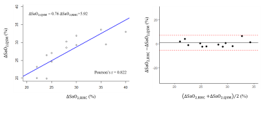 |
14 | Quantitative Susceptibility Mapping for Chamber Blood Oxygenation in Pulmonary Hypertension: Validation using Right Heart Catheterization
Jiahao Li1,2, Katherine Tak3, Rachel Meier3, Pablo Villar-Calle3, Justin Johannesen3, Jiwon Kim3, Yi Wang1,2, Jonathan W. Weinsaft3, and Pascal Spincemaille2
1Biomedical Engineering, Cornell University, New York, NY, United States, 2Radiology, Weill Cornell Medicine, New York, NY, United States, 3Medicine, Weill Cornell Medicine, New York, NY, United States
Pulmonary hypertension (PH) is a progressive and life shortening disorder with increased differential blood oxygen saturation (ΔSaO2) between right and left heart. In this prospective study, we acquired cardiac QSM from patients undergoing clinically indicated right heart catheterization (RHC) for assessment of known or suspected PH. ΔSaO2 estimated from QSM aligned well with RHC oxygenation data, showing QSM as a non-invasive CMR technique can be applied to clinically evaluate heart chamber oxygenation quantification in PH.
|
||
4450 |
15 | Population analysis of B0 magnetic field conditions in the human heart
Yun Shang1, Sebastian Theilenberg1, Boyu Peng2, Sachin R. Jambawalikar1,2, Laura M. Schreiber3,4, and Christoph Juchem1,2
1Department of Biomedical Engineering, Columbia University, New York, NY, United States, 2Department of Radiology, Columbia University Irving Medical Center, New York, NY, United States, 3Section of Medical Physics, Department of Radiology, Mainz University Hospital, Mainz, Germany, 4Chair of Molecular and Cellular Imaging, Comprehensive Heart Failure Center (CHFC), Würzburg, Germany
Cardiac MRI suffers susceptibility-induced artifacts due to B0 inhomogeneity across the heart. The lack of population data in cardiac B0 conditions and the practical inability to obtain such data in large populations impedes the development of optimal cardiac B0 shim strategy. Here, we establish population-based B0 conditions from readily available CT images and simulate cardiac B0 maps of 254 CT subjects with broad demographic parameters. The results are expected to develop optimal subject- and population-specific cardiac B0 shim strategies.
|
||
 Back to Meeting Home
 Back to the Program-at-a-Glance
The International Society for Magnetic Resonance in Medicine is accredited by the Accreditation Council for Continuing Medical Education to provide continuing medical education for physicians.
 Back to Meeting Home
 Back to the Program-at-a-Glance
 View the Presentation