Online Gather.town Pitches
Cardiac & Stroke
Joint Annual Meeting ISMRM-ESMRMB & ISMRT 31st Annual Meeting • 07-12 May 2022 • London, UK

| Booth # | ||||
|---|---|---|---|---|
4783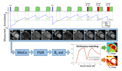 |
1 | Cartesian dictionary-based T1 and T2 mapping of the myocardium
Markus Henningsson1
1Linköping University, Linköping, Sweden
A method for simultaneous T1 and T2 mapping of the myocardium is proposed, termed Multimapping, based on dictionary matching using a simple Cartesian single-shot acquisition across 10 cardiac cycles. The method is evaluated in a phantom, 12 healthy subjects and 43 patients with suspected cardiomyopathy.
|
||
4784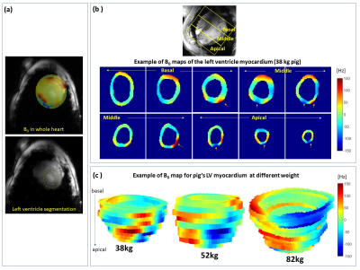 |
2 | B0 shimming for 7T cardiac T2*-weighted MRI in large animals: practical demands and hardware limitations.
Maxim Terekhov1, David Lohr1, Michael Hock1, Maya Bille1, Julia Aures1, Ibrahim A. Elabyad1, Florian Schnitter2, Wolfgang Bauer2, Ulrich Hofmann2, and Laura M. Schreiber1
1Chair of Cellular and Molecular Imaging, Comprehensive Heart Failure Center, University Hospital Würzburg, Comprehensive Heart Failure Center, Wuerzburg, Germany, 2Department of Internal Medicine I, Cardiology, University Hospital Würzburg, Wuerzburg, Germany
Cardiac MRI with T2*-contrast at 7T is of particular interest because it is highly sensitive to cardiac tissue alterations after myocardial injury. A reliable T2*-contrast quantification and usage for tissue characterization require minimization of the inhomogeneity of the B0-field in the heart. In this work, we analyzed the B0-conditions achievable in longitudinal T2* measurements in the hearts of domestic pigs which during the experiment grew from 30 to 90 kg. B0 distribution statistics were summarized for the maps of individual slices. Demands on the 7T-scanners B0-shimming hardware (up
to 3rd-order spherical harmonics) were analyzed.
|
||
4785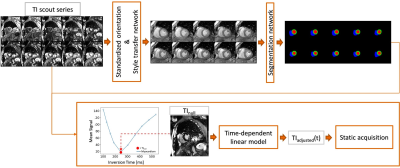 |
3 | Automated Time Adjusted Inversion Time Selection for Late Gadolinium Enhancement Imaging - Validation and Application on Patient Datasets Video Permission Withheld
Seung Su Yoon1,2, Michaela Schmidt2, Manuela Rick2, Teodora Chitiboi3, Puneet Sharma3, Tilman Emrich4,5, Christoph Tillmanns6, Andreas Seitz7, Heiko Mahrholdt7, Solenn Toupin8, Théo Pezel9,10, Jérôme Garot9, Jens Wetzl2, and
Andreas Maier1
1Pattern Recognition Lab, Friedrich-Alexander-Universität Erlangen-Nürnberg, Erlangen, Germany, 2Magnetic Resonance, Siemens Healthcare GmbH, Erlangen, Germany, 3Siemens Medical Solutions USA, Inc, Princeton, NJ, United States, 4Department of Radiology, University Medical Center, Johannes Gutenberg-University Mainz, Mainz, Germany, 5Division of Cardiovascular Imaging, Department of Radiology and Radiological Science, Medical University of South Carolina, Charleston, Germany, 6Diagnostikum Berlin, Berlin, Germany, 7Department of Cardiology, Robert Bosch Medical Center, Stuttgart, Germany, 8Siemens Healthcare France, Saint-Denis, France, 9Institut Cardiovasculaire Paris Sud, Cardiovascular Magnetic Resonance Laboratory, Hôpital Privé Jacques Cartier - Ramsay Santé, Massy, France, 10Division of Cardiology, Johns Hopkins University, Baltimore, MD, United States In cardiac MRI, the Late Gadolinium Enhancement technique is usually performed after inversion recovery scout sequences that are acquired to null the myocardial properly for optimal image contrast. In clinical practice, the selection and adjustment of the inversion time for healthy myocardium nulling are manually performed. To standardize and automate the process, we propose an automated deep-learning-based system combined with a linear regression model, which automates the selection of the inversion time, taking into consideration the time delay between the inversion time scout and LGE sequences. In this work, we validated the system in a large retrospective study (N=765). |
||
4786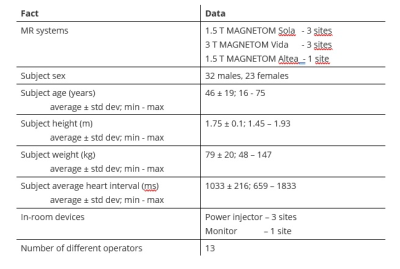 |
4 | Multi-center evaluation of the novel Beat Sensor Cardiac triggering technology
Carmel Hayes1, Yan Tu Huang2, Manuela Rick1, Randall Kroeker1, Mario Bacher1, Yue Pan3, Juliet Varghese3, Orlando Simonetti3, Ning Jin4, Rachel Davids4, Kelvin Chow4, Rudy Vanliedekerke 5, Marieke Vangelder5, Johan Dehem5,
Peter Gatehouse6, Raj Kumar Soundarajan 6, Ricardo Wage6, Alessia Azzu6, Sonia Nielles-Vallespin6, and Peter Speier1
1Siemens Healthcare GmbH, Erlangen, Germany, 2Siemens Shenzhen Magnetic Resonance, Shenzhen, China, 3The Ohio State University, Columbus, OH, United States, 4Siemens Medical Solutions USA, Malvern, PA, United States, 5Jan Yperman Ziekenhuis, Ieper, Belgium, 6Royal Brompton Hospital, London, United Kingdom
A multi-center evaluation of a Pilot Tone-based triggering technique using the BioMatrix Beat Sensor has been performed in healthy volunteers and patients undergoing a cardiac MRI examination. At five different sites, and two different field strengths, a wide range of MR sequences typically used in standard cardiac MRI examinations were successfully triggered using the prototype Beat Sensor Cardiac triggering technology. By including a short signal training and RF calibration step at the start of the examination, the device proved robust across a range of subjects, thereby offering an alternative means to trigger cardiac examinations without the need to attach electrodes.
|
||
4787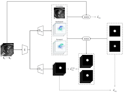 |
5 | Simultaneous learning of group-wise registration and joint segmentation of cardiac cine MRI with sparse annotation
Junwei Yang1,2, Pengfang Qian2, Thomas Kuestner3, Pietro Lio1, Dinggang Shen2,4, and Haikun Qi2
1Department of Computer Science and Technology, University of Cambridge, Cambridge, United Kingdom, 2School of Biomedical Engineering, ShanghaiTech University, Shanghai, China, 3Department of Interventional and Diagnostic Radiology, University Hospital of Tuebingen, Tuebingen, Germany, 4Shanghai United Imaging Intelligence Co., Ltd., Shanghai, China
Numerous deep learning methods have been proposed for cardiac cine MRI segmentation, while most of them require laborious annotation for supervised training. Herein, we propose an approach to perform group-wise registration and joint segmentation of cardiac cine images, by training a registration network in a self-supervised manner to align dynamic images to their mean image space and also a segmentation network in a weakly-supervised manner using sparsely-annotated data and predicted motions from the registration network. By training these two (registration and segmentation) networks simultaneously, our proposed joint learning approach provides better segmentations than the direct segmentation network.
|
||
4788 |
6 | Identification of Hemodynamic Biomarkers for Bicuspid Aortic Valve induced Aortic Dilation using Machine Learning
Pamela Franco1,2,3, Julio Sotelo1,3,4, Andrea Guala5, Lydia Dux-Santoy5, Arturo Evangelista5, José Rodríguez-Palomares5, Domingo Mery6, Rodrigo Salas4, and Sergio Uribe1,3,7
1Biomedical Imaging Center, School of Engineering, Pontificia Universidad Católica de Chile, Santiago, Chile, 2Electrical Engineering Department, School of Engineering, Pontificia Universidad Católica de Chile, Santiago, Chile, 3Millennium Nucleus for Cardiovascular Magnetic Resonance, Santiago, Chile, 4School of Biomedical Engineering, Universidad de Valparaíso, Valparaíso, Chile, 5Department of Cardiology, Hospital Universitari Vall d’Hebron, Vall d’Hebron Institut de Recerca (VHIR), Universitat Autònoma de Barcelona, Barcelona, Spain, 6Deparment of Computer Science, Pontificia Universidad Católica de Chile, Santiago, Chile, 7Radiology Department, School of Medicine, Pontificia Universidad Católica de Chile, Santiago, Chile
Several studies have demonstrated the existence of altered hemodynamics in bicuspid aortic valve (BAV) patients. The objective of this study was to identify which hemodynamic parameters allow an accurate classification between BAV patients with dilated and non-dilated ascending aorta using machine learning (ML) algorithms.
|
||
4789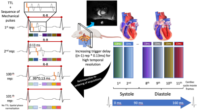 |
7 | Quantification of in-vivo myocardial stiffness in the rat heart using transient mechanical waves Video Permission Withheld
Marco Barbero Mota1, Giacomo Annio1, Guillaume Rucher2, Anna Wittgestein3, David Nordsletten3,4, Jordi Martorell5, and Ralph Sinkus1,3
1LVTS U1148, LVTS U1148 INSERM - Université de Paris, Paris, France, 2FRIM, INSERM - Université de Paris, Paris, France, 3School of Biomedical Engineering and Imaging Sciences, King's College London, London, United Kingdom, 4Biomedical Engineering and Cardiac Surgery, University of Michigan, Ann Arbour, MI, United States, 5Chemical Engineering, IQS, Barcelona, Spain
Heart biomechanics play a crucial role in the diagnosis and prognosis of cardiac diseases. However, current gold standard methods are highly invasive and preclude continuous monitoring of patients’ myocardium stiffness as a biomarker. In this study, we report the first results of a novel magnetic resonance elastography technique that provides with high temporal and spatial resolution enabling accurate transient shear wave speed values quantification, as thereof travel through heart tissue. Although biased due to the thin-plate geometry of the heart, early and mid-late systole, and early diastole preliminary results intuitively match the expected dynamical biomechanics of the human heart.
|
||
4790 |
8 | Pulmonary proton density mapping and cardiac 31P MRS suggest an energetic basis for exercise-induced pulmonary congestion in HFpEF
Jack J. Miller1,2,3, Matthew K Burrage2, Moritz Hundertmark2, Ladislav Valkovič2,4, William D Watson2, Jennifer Rayner2, Nikant Sabharwal5, Vanessa M Ferreira2, Stefan Neubauer2,5, Oliver J Rider2,5, and Andrew J Lewis2,5
1Aarhus University, Aarhus, Denmark, 2OCMR (Oxford Center for Clinical Magnetic Resonance Research), University of Oxford, Oxford, United Kingdom, 3Department of Physics, University of Oxford, Oxford, United Kingdom, 4Department of Imaging Methods, Slovak Academy of Sciences, Bratislava, Slovakia, 5Department of Cardiology, Oxford University Hospitals NHS Trust, Oxford, United Kingdom
We show in 43 patients with 31P MRS, 1H CMR and a novel proton-density mapping sequence that a gradient of myocardial energetic impairment exists across a spectrum of HFpEF phenotypes of increasing clinical severity and worsening diastolic function. A greater degree of myocardial energetic deficit is linked to impaired LV systolic and diastolic functional reserve, to altered RV reserve and RV-PA coupling, and to exercise-induced pulmonary congestion assessed using novel proton density magnetic resonance imaging. A subgroup of HFpEF patients demonstrate transient pulmonary congestion during exercise which can be non-invasively assessed using exercise CMR and proton density mapping.
|
||
4791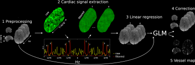 |
9 | Data driven whole-brain cardiac signal regression from highly accelerated fMRI acquisitions
Marco Marino1,2, Nigel Colenbier1,2,3, Nicola Filippini2, Giovanni Pellegrino2, Daniele Marinazzo2,3, and Giulio Ferrazzi2
1Research Center for Motor Control and Neuroplasticity, KU Leuven, Leuven, Belgium, 2IRCCS San Camillo Hospital, Venice, Italy, 3Department of Data Analysis, Faculty of Psychology and Educational Sciences, Ghent University, Ghent, Belgium Cardiac pulsation is a physiological confound of fMRI analysis pipelines and so accurate mapping of cardiac contributions to the BOLD signal is highly warranted. To overcome cardiac aliasing associated with the limited temporal resolution in fMRI, we developed a data-driven methodology to spatially and temporally resolve cardiac contributions from the BOLD signal itself (i.e without the need of processing external physiological recordings such as PPU/ECG signals). This is achieved by combining simultaneous multi-slice imaging and a dedicated hyper-sampling decomposition scheme. The proposed methodology is fully data driven and it does not make specific assumptions on the shape of cardiac pulsation. |
||
4792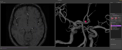 |
10 | AVA Diagnostics - multimodality computer assisted aneurysm management tool
Žiga Bizjak1, Aichi Chien, PhD, FAHA2, Erazem Kos1, and Žiga Špiclin1
1Laboratory of Imaging Technologies, Faculty of Electrical Engineering, University of Ljubljana, Ljubljana, Slovenia, 2Division of Interventional Neuroradiology, Department of Radiological Sciences, Ronald Reagan UCLA Medical Center, David Geffen School of Medicine at UCLA, Los Angeles, CA, United States
Management of (un)ruptured intracranial aneurysms (IAs) through early detection and continuous rupture risk assessment is becoming an integral part of IA treatment. This study showcases the capabilities of AVA Diagnostics tool and its IA workflow including automatic vessel segmentation, detection and isolation, morphologic quantification, future growth prediction and follow-up morphologic analysis. Each of the tools was successfully validated on multimodal MRA, CTA and 3D-DSA scans. The AVA Diagnostics thus aims to simplify the management of IAs through extensive use of computer-assisted image analysis and analytics tools, and enable their application in large-scale studies and in regular clinical workflow.
|
||
4793 |
11 | Brain microstructural changes in stroke patients at subacute/chronic stages by mean of advanced diffusion MRI: a longitudinal study
Shi-Ming Wang1, Guglielmo Genovese2,3,4, Belen Diaz3,5, Fan Huang6, Stéphane Lehericy2,3, Hui Zhang1, Charlotte Rosso3,5, Francesca Branzoli2,3, and Marco Palombo1,7,8
1Centre for Medical Image Computing (CMIC), Department of Computer Science, University College London, London, United Kingdom, 2Paris Brain Institute - ICM, Centre de NeuroImagerie de Recherche - CENIR, Paris, France, 3Sorbonne Université, UMR S 1127, Inserm U 1127, CNRS UMR 7225, ICM, F-75013, Paris, France, 4Center for Magnetic Resonance Research and Department of Radiology, University of Minnesota, Minneapolis, MN, United States, 5Department of Neurology, Pitié-Salpétrière Hospital, Paris, France, 6Department of Medical Physics and Biomedical Engineering, University College London, London, United Kingdom, 7Cardiff University Brain Research Imaging Centre (CUBRIC), School of Psychology, Cardiff University, Cardiff, United Kingdom, 8School of Computer Science and Informatics, Cardiff University, Cardiff, United Kingdom
This study characterises microstructural changes in stroke lesions using NODDI and WMTI, two advanced diffusion MRI techniques, and compares their ability to detect WM alterations on the ipsi-lesional and contralesional sides of 12 patients scanned 2 weeks, 1 month and 3 months after onset. Region-of-interest analysis showed that ODI and FWF from NODDI were significantly altered at the lesion. Significant changes were also found in the WMTI parameters of several ipsi-lesional white matter tracts. Our prospective analysis suggests that the application of enhanced dMRI methods such as NODDI and WMTI can help to further comprehend the biophysical mechanism behind ischemia.
|
||
4794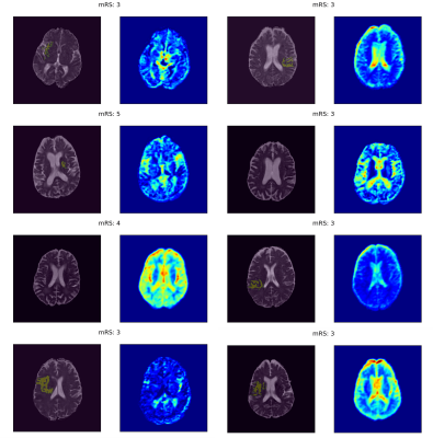 |
12 | A pilot study on the application of explainable deep learning to ADC maps for predicting functional outcome of ischemic stroke patients
Esra Zihni1, Bryony L. McGarry1,2, Jen Guo3, Rani Gupta Sah3, George Tadros3, Philip A. Barber3, and John D. Kelleher1,4
1PRECISE4Q Predictive Modelling in Stroke, Technological University Dublin, Dublin, Ireland, 2School of Psychological Science, University of Bristol, Bristol, United Kingdom, 3Calgary Stroke Program, Department of Clinical Neurosciences, University of Calgary, Calgary, AB, Canada, 4ADAPT Research Centre, ICE Research Institute, Technological University Dublin, Dublin, Ireland
Applying deep learning models to MRI scans of acute stroke patients to extract features indicative of functional outcome could assist a clinician’s treatment decisions. Here, we trained convolutional neural network models on ADC maps from hyper-acute ischemic stroke patients to predict 3-month mRS and used an interpretability technique to highlight regions in the ADC maps that were most important in the prediction of good and poor outcomes. Although the models had poor predictive power, the visual explanations supported our previous findings that predictions might be based not on ischemic regions, but on other relevant information inherent in the image.
|
||
4795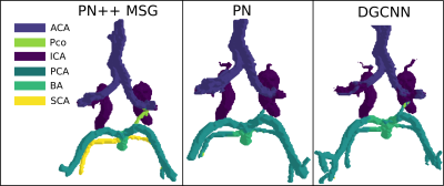 |
13 | Deep learning Circle of Willis arterial labeling strategies for pipeline of cerebrovascular disease analysis
Žiga Bizjak1, Aichi Chien, PhD, FAHA2, Jan Tasič1, and Žiga Špiclin1
1Laboratory of Imaging Technologies, Faculty of Electrical Engineering, University of Ljubljana, Ljubljana, Slovenia, 2Division of Interventional Neuroradiology, Department of Radiological Sciences, Ronald Reagan UCLA Medical Center, David Geffen School of Medicine at UCLA, Los Angeles, CA, United States
The correlation between different variants of the Circle of Willis (CoW) and cerebrovascular disorders such as stroke, aneurysms and mental disorders is not yet well understood. A step towards a better understanding of the role of CoW in the aforementioned diseases is the automatic labeling of main vessels. In this work we tested three different approaches and observed high mIoU value of 0.870. As such the automatic anatomical labelling of the CoW seems feasible for clinical evaluation of the association of different anatomical variants with the risk factors of cerebrovascular pathologies.
|
||
 Back to Meeting Home
 Back to the Program-at-a-Glance
The International Society for Magnetic Resonance in Medicine is accredited by the Accreditation Council for Continuing Medical Education to provide continuing medical education for physicians.
 Back to Meeting Home
 Back to the Program-at-a-Glance
 View the Presentation