Online Gather.town Pitches
MSK III
Joint Annual Meeting ISMRM-ESMRMB & ISMRT 31st Annual Meeting • 07-12 May 2022 • London, UK

| Booth # | ||||
|---|---|---|---|---|
3390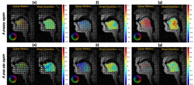 |
1 | Tongue Deformation and Strain Estimation using Tagged RT-MRI and Optical Flow
Ye Tian1, Weiyi Chen1, Dani Byrd2, Shrikanth Narayanan1,2, and Krishna S. Nayak1
1Ming Hsieh Department of Electrical and Computer Engineering, University of Southern California, Los Angeles, CA, United States, 2Department of Linguistics, University of Southern California, Los Angeles, CA, United States
Real-time MRI plays an important role in studying articulator movements during typical and atypical human speech. Recently, real-time tagging methods were developed to reveal inner tongue movements, including deformations in the body of the tongue that were previously unobservable. Here, we demonstrate tracking of tongue deformations and estimation of tongue strain from real-time tagging, using a motion-compensated total generalized variation reconstruction and optical flow. Optical flow tracking has an averaged error of 1.10±0.83mm, compared with manually tracked tagged line intersections, while intra-observer variability is 0.80±0.49mm. The estimated tongue deformation and strain maps provide for linguistically sensible interpretation.
|
||
3391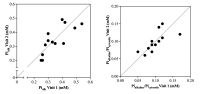 |
2 | Reproducibility of Alkaline Inorganic Phosphate Quantification using 31P-Magnetic Resonance Spectroscopy at 3T
Alexs A Matias1, Corinna F Serviente1,2, Stephen T Decker1, Muhammet E Erol1, Gaia Giuriato 3, Yann Le Fur4, Rajakumar Nagarajan 5, and Gwenael Layec1,6
1Kinesiology, University of Massachusetts at Amherst, Amherst, MA, United States, 2Center for Health and Human Performance, Institute for Applied Life Sciences, University of Massachusetts at Amherst, Amherst, MA, United States, 3Neuroscience, Biomedicine, and Movement Science, University of Verona, Verona, Italy, 4Centre de Resonance Magnetique Biologique et Medicale, Marseille, France, 5Human Magnetic Resonance Center, Institute for Applied Life Sciences, University of Massachusetts at Amherst, Amherst, MA, United States, 6Institute for Applied Life Sciences, University of Massachusetts at Amherst, Amherst, MA, United States Alkaline inorganic phosphate (Pialk) is a promising biomarker of mitochondrial content in vivo that is technically challenging to assess at 3T. The absolute and relative reproducibility of Pialk in the quadriceps muscle across two visits were assessed in healthy sedentary-to-moderately active young adults using 31P-MRS at 3T. The results of this study demonstrate an excellent absolute and relative reproducibility of the quantification of Pialk in the quadriceps muscle at 3T provided that the coil is positioned at the mid-thigh. |
||
3392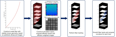 |
3 | Effect of Acquisition and Analysis Conditions on Accuracy of DTI-Based Muscle Architecture Estimates: Predictions Using Numerical Simulations
Xingyu Zhou1,2, Carly Lockard2,3, Melissa Hooijmans4, and Bruce Damon2,3
1Biomedical Engineering, Vanderbilt University, Nashvill, TN, United States, 2Carle Clinical Imaging Research Program, Carle Health, Urbana, IL, United States, 3Stephens Family Clinical Research Institute, Carle Health, Urbana, IL, United States, 4Amsterdam University Medical Center, Amsterdam, Netherlands Diffusion-tensor imaging (DTI) based fiber tractography is a useful tool to study the architecture of human skeletal muscle. However, effects of image acquisition and analysis conditions on the outcome of architectural estimates are challenging to examine in vivo. In this work, we describe a numerical simulation framework where the ground truth of muscle architecture is known and the outcome can be tested under different conditions. Results show that the estimate of fiber curvature is most affected by image noise. While second-order polynomial fitting of fiber tracts is more robust to image noise, third-order fitting performs better on highly curved fibers. |
||
3393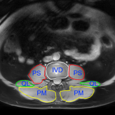 |
4 | R1rho Dispersion in Lumbar Muscles
Alan E. Rivera-Garcia1, Jay D. Turner2, Juan Uribe2, Richard D. Dortch1, John C. Gore3, and Ping Wang1
1Translational Neuroscience, Barrow Neurological Institute, Phoenix, AZ, United States, 2Barrow Neurological Institute, Phoenix, AZ, United States, 3Radiology and Radiological Sciences, Vanderbilt University Medical Center, Nashville, TN, United States
R1rho imaging can provide novel information on dynamic processes within tissues, allowing for a more comprehensive analysis of the parameters of chemical exchange and/or intrinsic microstructure. We measured the dispersion of R1rho with different locking fields in the major lumbar muscles of psoas, paraspinal muscles (multifidus and erector spinae), and quadratus lumborum. The results showed that R1rho dispersion is significant and measurable at 3T which may provide a new way to characterize muscles involved in low back pain.
|
||
3394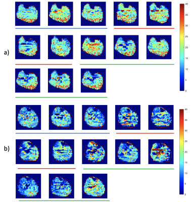 |
5 | Quantitative assessment of peripheral metabolism and perfusion with dynamic 1H MRI in healthy and diabetic individuals
Ryan Wahidi1, Yi Zhang2, Ran Li1, Mohammed A. Zayed3, Mary K. Hastings4, and Jie Zheng1
1Radiology, Washington University School of Medicine, St. Louis, MO, United States, 2Zhejiang University, Hangzhou, China, 3Department of Surgery, Section of Vascular Surgery, Washington University School of Medicine, St. Louis, MO, United States, 4The Program in Physical Therapy, Washington University School of Medicine, St. Louis, MO, United States
The objective of this study is to analyze the efficacy of quantitative PCr MRI in quantifying differing muscle tissue PCr kinetics in healthy individuals and diabetic patients. Healthy subjects and diabetic patients underwent PCr and perfusion imaging in calf muscle at rest, during isometric plantarflexion, and for a recovery period. Only the healthy group displayed moderate correlation between PCr and perfusion. The diabetic patient group displayed attenuated recovery speed of PCr after exercise and no correlation between PCr and perfusion.
|
||
3395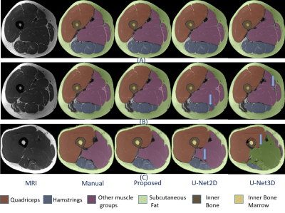 |
6 | Deep Learning Based Automatic Pipeline for Quantitative Assessment of Thigh Muscle Fatty Infiltration in Post-traumatic Osteoarthritis
Sibaji Gaj1, Brendan L. Eck1, Dongxing Xie1, Richard Lartey1, Charlotte Lo1, Mingrui Yang1, Kunio Nakamura1, Carl S. Winalski2, Kurt Spindler3, and Xiaojuan Li1
1Biomedical Engineering, Cleveland Clinic, Cleveland, OH, United States, 2Radiology, Cleveland Clinic, Cleveland, OH, United States, 3Orthopaedics, Cleveland Clinic, Cleveland, OH, United States
Quantitative assessment of thigh muscle morphology and fatty infiltration (FF%) in post-traumatic osteoarthritis is limited. In this work, we developed a deep learning based accurate segmentation method for muscles, bone and adipose tissue from thigh MRI and used these segmentation for automated quantification of FF and cross sectional area(CSA) of these tissues. 16 patients at 10 years after ACL reconstruction were studied. The proposed method showed significant improvement in segmentation metrics (Dice, Average surface distance (ASD)) and CSA compared with popular U-Net based deep learning models. For CSA and FF% quantification, automated methods had similar measurements compared with manual segmentation.
|
||
3396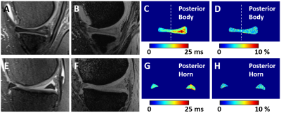 |
7 | Evaluation of Meniscus Degeneration in Patients with Posterior Root Tear of Medial Meniscus Using T2* Mapping at 7T – Comparison with Arthroscopy
Stefan Zbyn1,2, Abdul Wahed Kajabi1,2, Ariel N. Rodriguez3, Gregory J. Metzger1, Robert F. LaPrade3, and Jutta M. Ellermann1,2
1Center for Magnetic Resonance Research, University of Minnesota, Minneapolis, MN, United States, 2Department of Radiology, University of Minnesota, Minneapolis, MN, United States, 3Edina-Crosstown Surgery Center, Twin Cities Orthopedics, Minneapolis, MN, United States
Although T2 mapping allows noninvasive evaluation of meniscus degeneration, its application at 7T requires spin echo sequences with relatively long repetition times to meet the specific absorption rate (SAR) limits which can restrict the resolution of maps. This 7T study evaluates the potential of T2* mapping for the assessment of meniscal degeneration in six patients with arthroscopically verified posterior root tears of the medial meniscus and in healthy volunteers. Increased T2* in degenerated meniscus regions suggest T2* mapping is sensitive to meniscus degeneration. T2* mapping is a promising biomarker of early meniscal degeneration which is less SAR-demanding than T2 mapping.
|
||
3397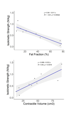 |
8 | MRI quantitative assessment of muscular changes associated to a rehabilitation strategy in FSHD patients
Camille Noël1,2, Constance Patricia Michel1, Amira Trabelsi1, Sébastien Vansteenkiste2, Emmanuelle Salort-Campana3, Maëva Cotinat2,4, Virginie De Bovis Milhe3, Laurent Bensoussan2,4,5, Shahram Attarian3, and David Bendahan1
1Aix Marseille University, Center for magnetic resonance in biology and medecine, Marseille, France, Metropolitan, 2Physical and Rehabilitation Medecine Departement, APHM, Marseille, France, Metropolitan, 3Reference Centre for Neuromuscular Diseases and ALS, APHM, Marseille, France, Metropolitan, 4CNRS, INT UMR 7289, Aix Marseille University, Marseille, France, Metropolitan, 5UGECAM Institut Universitaire de Réadaptation de Valmante Sud, Marseille, France, Metropolitan
Facioscapulohumeral dystrophy is the third genetic myopathy. Given that no therapeutic strategy has proved to be successful, rehabilitation has been considered as an interesting alternative. The aim of this study was to quantitatively evaluate the effects of a personalized rehabilitation strategy using MRI by tracking fat fraction (FF) and contractile volume (CV) over time. The lower limb was scanned before and after a rehabilitation program. FF and CV remained stable over the training period. Clinical parameters evolved positively. FF and CV were correlated to isokinetic strength thereby indicating that both metrics could be considered as biomarkers of the pathology progression.
|
||
3398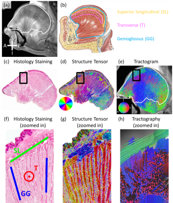 |
9 | Validation of Muscle Fiber Architecture of the Human Tongue Revealed by Diffusion MRI with Histology Verification
Xiao Liang1, Nahla M H Elsaid2, Li Jiang1, Steve Roys1, Maureen Stone3, Jerry L Prince4, Adam C Puche5, Rao P Gullapalli1, and Jiachen Zhuo1
1Department of Diagnostic Radiology and Nuclear Medicine, University of Maryland School of Medicine, Baltimore, MD, United States, 2Department of Radiology and Biomedical Imaging, Yale School of Medicine, New Haven, CT, United States, 3Department of Neural and Pain Sciences and Department of Orthodontics, University of Maryland School of Dentistry, Baltimore, MD, United States, 4Department of Electrical and Computer Engineering, Johns Hopkins University, Baltimore, MD, United States, 5Department of Anatomy and Neurobiology, University of Maryland School of Medicine, Baltimore, MD, United States
Advanced dMRI techniques have been used to resolve the complex tongue muscle architecture. However, dMRI-derived tongue muscle architecture has not been validated with histology. In this study, we validated the dMRI-derived tongue muscle architecture with histology of a tongue specimen. dMRI was acquired for a post-mortem head and a healthy volunteer. dMRI-derived muscle fiber orientations, visualized as the tractogram, were compared against microscopic histology slices of the tongue specimen. Muscle fibers in the tractograms show good correspondence with those appearing in the histology images. The study demonstrates that dMRI can accurately reveal the complex muscle architecture of the human tongue.
|
||
3399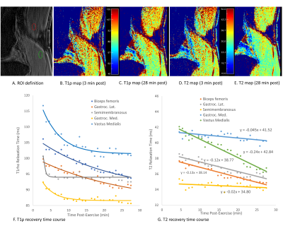 |
10 | Transient Biochemical Recovery of Skeletal Muscles Post-exercise with Simultaneous Dynamic T1rho and T2 Mapping
Esther Rong1, Jason Matakas1, Steven Shamah1, Jenna Le1, Karen Sperling1, Can Wu2, and Qi Peng1
1Department of Radiology, Albert Einstein College of Medicine and Montefiore Medical Center, Bronx, NY, United States, 2Department of Medical Physics, Memorial Sloan Kettering Cancer Center, New York, NY, United States
T1ρ and T2 contrasts provide complementary information on tissue properties. Therefore, their transient recovery after exercise to pre-exercise equilibria is potentially a functional biomarker for muscle disorder diagnosis and treatment evaluation. In this pilot study, we implemented and tested a high-temporal-resolution dynamic MRI sequence for simultaneous T1ρ and T2 mapping immediately after exercise. T1ρ and T2 contrasts presented unique transient recovery time courses in different skeletal muscles. Dynamic information derived from our imaging approach could potentially lead to greater sensitivity and specificity in evaluating muscle conditions compared with traditional methods with either T1ρ or T2 contrast alone.
|
||
3400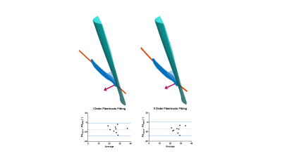 |
11 | A comparison of muscle pennation angles measured with DTI fiber tractography and 3D-ultrasound Video Permission Withheld
Laura Secondulfo1, Moritz Eggelbusch2, Rob C.I. Wust 2,3, Guido Weide3, Richard T. Jaspers3, Aart J. Nederveen4, Melissa Hooijmans1, and Gustav Strijkers1
1Biomedical Engineering and Physics, Amsterdam University Medical Center, Amsterdam, Netherlands, 2Laboratory for Myology, Department of Human Movement Sciences, Faculty of Behavioural and Movement Sciences, Vrije Universiteit Amsterdam, Amsterdam, Netherlands, 3Department of Rehabilitation Medicine, VU University Medical Center Amsterdam, Amsterdam Movement Sciences, Amsterdam, Netherlands, 44 Department of Radiology and Nuclear Medicine, Amsterdam University Medical Centers, University of Amsterdam, Amsterdam, Netherlands
Pennation angle is an important architecture parameter to understand muscle functioning. It is commonly measured using 2D ultrasound. However, it is difficult to infer 3D muscle architecture from 2D imaging. Therefore, we compare the pennation angle measurements obtained with 3D-DTI fiber-tractography and 3D-ultrasound (3D-US). We acquired data of the Vastus Lateralis muscle in 9 healthy subjects. The mean pennation angle with 3D-US was 18.9°± 5.9°, whereas we found 33.3°±6.7° (straight fiber approximation) and 34.5°±4.8°(curved fiber fit) for DTI fiber-tractography. These differences between 3D-US and DTI could be of technical or physiological origin.
|
||
3401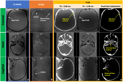 |
12 | Improving cortical bone imaging using a novel Flexible Ultra Short Echo time (FUSE) sequence for optimal synthetic CT generation
Lumeng Cui1, Emily J. McWalter1,2, Gerald R. Moran3, and Niranjan Venugopal4
1Division of Biomedical Engineering, University of Saskatchewan, Saskatoon, SK, Canada, 2Department of Mechanical Engineering, University of Saskatchewan, Saskatoon, SK, Canada, 3Research Collaboration Manager, Siemens Healthcare Limited, Oakville, ON, Canada, 4Department of Radiology, University of Manitoba, Winnipeg, MB, Canada
Ultrashort echo time (UTE) pulse sequences are used in synthetic computed tomography (sCT) generation for MR-based radiation treatment planning (RTP). This study used a novel Flexible UTE sequence (FUSE) for improving cortical bone imaging. Using a human skull, we applied the optimized UTE acquisition techniques and parameters and verified the improvements in overall image quality of cortical bone in comparison to a product UTE sequence. Also, we have shown the feasibility and potential of our FUSE sequence for generating the sCT data that is clinically equivalent to the traditional CT for RTP.
|
||
3402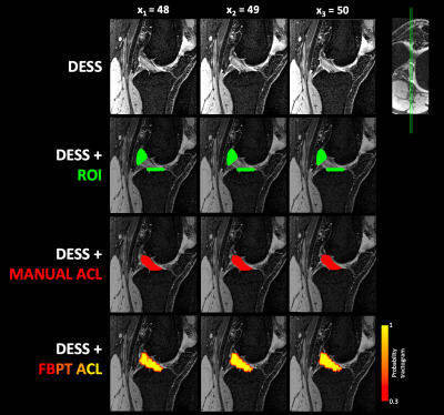 |
13 | Bilateral characterization of fractional anisotropy within the anterior cruciate ligament: A step toward clinical use of DTI in the knee
Allen A Champagne1, Andrew McGuire2, Kaden Shearer1, Don Brien3, Paul A Martineau4, and Davide D Bardana2
1School of Medicine, Queen's University, Kingston, ON, Canada, 2Department of Orthopedic Surgery, Queen's University, Kingston, ON, Canada, 3Center for Neuroscience Studies, Queen's University, Kingston, ON, Canada, 4Department of Orthopedic Surgery, McGill University, Montréal, QC, Canada
Diffusion tensor imaging (DTI) has emerged as biomarker to characterize the ligamentization process of the anterior cruciate ligament (ACL), following surgical reconstruction. One major limitation to this approach is the necessity for pre-injury baseline measurements to be used as a frame of reference, in order to inform clinical decision making with respect to recovery of the graft, and return-to-activity (RTA). Here, we explore the linear relationship for anisotropy between knees bilaterally, in healthy participants, to provide a methodological framework that allows for the clinical integration of DTI to monitor recovery of the tendon autograft, following surgical reconstruction, and inform RTA.
|
||
3403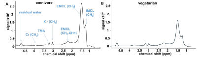 |
14 | In vivo metabolic characterization of vegetarian diet effects on skeletal muscle using magnetic resonance spectroscopy Video Permission Withheld
Joevin Sourdon1,2, Tangi Roussel1,2, Pierre Daude1,2, Monique Bernard1,2, Frank Kober1,2, and Stanislas Rapacchi1,2
1Aix-Marseille Université, CNRS, CRMBM, Marseille, France, 2AP-HM, Hôpital universitaire Timone, CEMEREM, Marseille, France Vegetarian nutrition may contribute for metabolic differences that we proposed to explore non-invasively. 6 omnivores and 6 lacto-ovo-vegetarians underwent a 7T MRI exam including proton and phosphorus magnetic resonance spectroscopy to measure creatine (tCr), phosphocreatine (PCr) and triglyceride components in the soleus muscle. tCr and unsaturated fatty acids concentration were reduced in vegetarians but similar PCr/γ-ATP ratios were found. We show that vegetarian diet induces lower skeletal muscle creatine and different lipid profiles, which was not associated with altered bioenergetic reserve. This protocol opens new avenues to explore the impact of diet in skeletal muscle. |
||
3404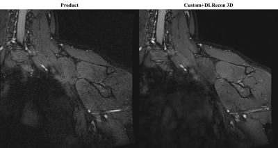 |
15 | Using DL Reconstruction and 3D Directionally-Selective Crusher Gradients to Improve 3D Brachial Plexus Magnetic Resonance Neurography
Yan Wen1, Ek Tsoon Tan2, Marc Lebel3, Suryanarayanan Kaushik3, Maggie Fung1, and Darryl B. Sneag2
1GE Healthcare, New York, NY, United States, 2Department of Radiology and Imaging, Hospital for Special Surgery, New York, NY, United States, 3GE Healthcare, Waukesha, WI, United States To improve vascular suppression in 3D brachial plexus MR neurography (MRN) with FSE readouts, 3D directionally-selective crusher gradient technique was implemented. 3D Deep learning reconstruction (DLRecon 3D) with denoising and sharpening properties was applied to improve nerve conspicuity. Preliminary data showed that using both DLRecon 3D reconstruction and 3D crusher gradients resulted in improved image quality, image sharpness, and vascular suppression. |
||
 Back to Meeting Home
 Back to the Program-at-a-Glance
The International Society for Magnetic Resonance in Medicine is accredited by the Accreditation Council for Continuing Medical Education to provide continuing medical education for physicians.
 Back to Meeting Home
 Back to the Program-at-a-Glance
 View the Presentation