Online Gather.town Pitches
Processing & Analysis I
Joint Annual Meeting ISMRM-ESMRMB & ISMRT 31st Annual Meeting • 07-12 May 2022 • London, UK

Online Gather.town Pitches
Processing & Analysis I
| Booth # | ||||
|---|---|---|---|---|
3196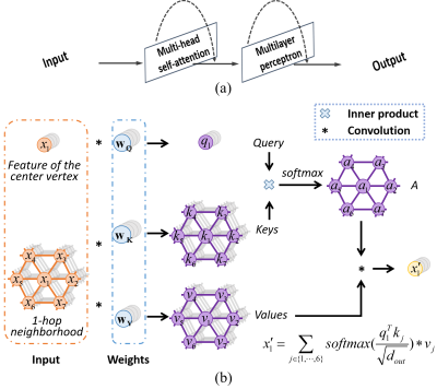 |
1 | Quality Assessment of Pediatric Cortical Surfaces with Spherical Transformer
Jiale Cheng1,2, Xin Zhang1, Fenqiang Zhao2, Zhengwang Wu2, Ya Wang2, Ying Huang2, Weili Lin2, Li Wang2, and Gang Li2
1School of Electronic and Information Engineering, South China University of Technology, Guangzhou, China, 2Department of Radiology and BRIC, University of North Carolina at Chapel Hill, Chapel Hill, NC, United States
Quality assessment of cortical surfaces, which is a crucial step and prerequisite in surface-based large-scale neuroimaging studies, aims to identify the low-quality surfaces and exclude them in the subsequent analysis. Convolutional neural network-based methods have achieved great success in image quality assessment, but they are inherently inapplicable for objects presented in non-Euclidean spaces, such as the brain cortical surfaces. To this end, we propose a transformer-based network, which describes the local information based on feature correspondences among vertices, thus enabling itself to be applied directly onto a spherical manifold. Extensive experiments on 1,860 infant cortical surfaces validated its superior performance.
|
||
3197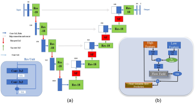 |
2 | Accurate Prostate Segmentation in MR Images Guided by Semantic Flow
Yousuf Babiker M. Osman1, Cheng Li1, Zhenzhen Xue1, Hairong Zheng1, and Shanshan Wang1
1Paul C. Lauterbur Research Center for Biomedical Imaging, Shenzhen Institute of Advanced Technology, Chinese Academy of Sciences, Shenzhen, China
To enlarge the receptive field, downsampling is frequently utilized in deep learning (DL) models. Consequently, there exists one common issue for DL-based image segmentation – the misalignment between high-resolution features and high-semantic features. To this end, decoding or upsampling has been proposed and promising performances have been achieved. However, upsamling without explicit pixel-wise localization guidance may introduce errors. To address this issue, we propose a semantic flow-guided prostate segmentation method. By guiding the upsampling process with semantic flow calculated from both high-resolution and high-semantic features, more accurate segmentation results are generated.
|
||
3198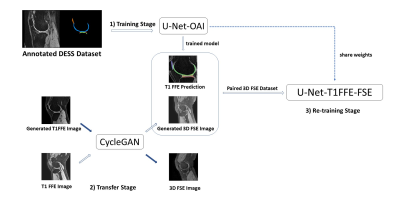 |
3 | Unsupervised Domain Adaptation via CycleGAN for knee joint Segmentation in MR Images
Siyue LI1, Sheheryar Khan2, Fan XIAO1, Shutian ZHAO1, Junru ZHONG1, Dόnal G. Cahill1, James F. Griffith1, and Weitian CHEN1
1The Chinese University of Hong Kong, Hong Kong, Hong Kong, 2The City University of Hong Kong, Hong Kong, Hong Kong
Knee joint tissues segmentation is necessary for quantitative analysis of musculoskeletal diseases like knee osteoarthritis. Three-dimensional Fast Spin Echo (3D FSE) imaging is a potential MRI technique for routine clinical knee imaging. Thus, segmentation based on 3D FSE has valuable clinical application. However, the conventional deep learning-based segmentation requires manually annotating 3D knee images which is time-consuming. In this work, we proposed a domain adaption-based unsupervised approach for cartilage and meniscus segmentation on 3D FSE images without the need for annotating images. We demonstrated that the proposed method improved the quality of segmentation.
|
||
3199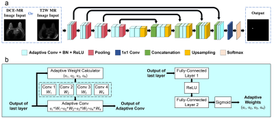 |
4 | Adaptive convolution kernels for breast tumor segmentation in multi-parametric MR images
Cheng Li1, Hui Sun1, Zhenzhen Xue1, Xin Liu1, Hairong Zheng1, and Shanshan Wang1
1Paul C. Lauterbur Research Center for Biomedical Imaging, Shenzhen Institute of Advanced Technology, Chinese Academy of Sciences, Shenzhen, China
Multi-parametric magnetic resonance imaging (mpMRI) provides high sensitivity and specificity for breast cancer diagnosis. Accurate breast tumor segmentation in mpMRI can help physicians achieve better clinical managements. Existing deep learning models have presented promising performances. However, the effective exploitation and fusion of information provided in mpMRI still need further investigation. In this study, we propose a convolutional neural network (CNN) with adaptive convolution kernels (AdaCNN) to automatically extract and absorb the useful information from multiple MRI sequences. Extensive experiments are conducted, and the proposed method can generate better breast tumor segmentation results than those obtained by CNNs with normal convolutions.
|
||
3200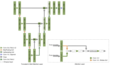 |
5 | WRIST CARTILAGE SEGMENTATION USING U-NET CONVOLUTIONAL NEURAL NETWORKS ENRICHED WITH ATTENTION LAYERS
Nikita A. Vladimirov1, Ekaterina A. Brui1, Anatoliy G. Levchuk1, Aleksandr Y. Efimtsev1,2, and David Bendahan1,3
1Faculty of Physics, ITMO University, Saint-Petersburg, Russian Federation, 2Federal Almazov North-West Medical Research Center, Saint-Petersburg, Russian Federation, 3Aix-Marseille Universite, CNRS, Centre de Résonance Magnétique Biologique et Médicale, UMR, Marseille, France
Detection of cartilage loss is crucial for the diagnosis of osteo- and rheumatoid arthritis. An automatic tool for wrist cartilage segmentation may be of high interest as the corresponding manual procedure is tedious. U-Net is a convolution neural network, which has been largely used for biomedical images, but its performance in segmenting wrist cartilage images is modest. Here, we assessed whether adding attention layers to U-Net architecture would improve the segmentation performance. A truncated version of U-Net with attention layers showed the best performance(3D DSC - 0.811), as well as in the accuracy of cartilage cross-section measurements (bias - 6.4 mm2).
|
||
3201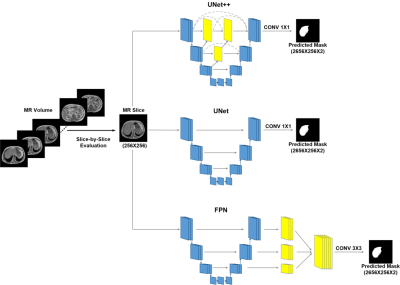 |
6 | Deep learning based liver segmentation using T1 weighted abdominal MRI
Md Sakib Abrar Hossain1, Muhammad E. H. Chowdhury1, Enamul H. Bhuyian2, Tawsifur Rahman3, Zaid B. Mahbub4, Amith Khandakar3, Anas Tahir3, Md Shafayet Hossain5, and M. Salman Khan6
1Electrical Engineering, Qatar University, Doha, Qatar, 2Biomedical Engineering and Imaging Institute, Icahn School of Medicine at Mount Sinai, New York, NY, United States, 3Electrical Engineering,, Qatar University, Doha, Qatar, 4Dept. of Physics and Mathematics, North South University, Dhaka, Bangladesh, 5Dept. of Electrical, Electronics and Systems Engineering, Universiti Kebangsaan Malaysia, Bangi, Malaysia, 6Department of Electrical Engineering, University of Engineering and Technology, Peshawar, Pakistan
MR scans are preferred by clinicians for liver pathology diagnosis over volumetric abdominal CT scans, due to their superior resolution for soft tissues. Nevertheless, deep learning based automated liver segmentation from abdominal MRI is challenging as the liver exhibits variable characteristics. This study investigates multiple state-of-the-art segmentation architectures (UNet, UNet++, and FPN) with varying encoder and decoder backbones. Here, T1 weighted MR images are investigated as it demonstrates brighter fat content. Among the investigated networks UNet++ with DenseNet backbone demonstrates top performance for the liver segmentation with a DSC and IoU of 94.3% and 91.0%, respectively.
|
||
3202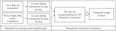 |
7 | Combination of Echo Planar Imaging Correction and Compressed SENSE Framework for enhanced Diffusion weighted imaging
Zhigang Wu1, Peng Sun1, Yajing Zhang2, Xiuquan Hu1, Jing Zhang1, Guangyu Jiang3, Yan Zhao3, and Jiazheng Wang1
1Philips Healthcare, Beijing, China, 2MR Clinical Science, Philips Healthcare (Suzhou), Suzhou, China, 3MR R&D, Philips Healthcare (Suzhou), Suzhou, China
Diffusion-weighted imaging (DWI) using single-shot EPI (ssEPI) has suffered from distortion, blurring, and signal loss caused by B0 inhomogeneity. SENSE can be used to reduce distortion. However, it also suffers from noise breakthrough issues when the accelerator factor is high. It’s still a challenge to get DWI images with reduced distortion and high SNR without significantly increasing the scan time. We propose a framework that combines FSL top-up technique, Compressed Sensing and SENSE framework simultaneously to overcome these challenges. This framework allows a new solution for ssEPI based diffusion imaging with high resolution, low distortion, and without noise breakthrough issue.
|
||
3203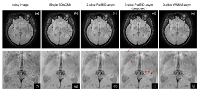 |
8 | Improved Parallelized Blind MR Image Denoising using Asymmetric Weighting coefficients
Satoshi ITO1 and Kazuki YAMATO1
1Utsunomiya university, Utsunomiya, Japan
Parallelized blind image denoising (ParBID) is an improved CNN based denoising method in which weighted average of slice images are denoised using blind denoising CNN followed by image separation. To further improve the denoising performances, weighting coefficients (wc) of slice image averaging was examined. Negative value wc resulted in a significant change in the noise distribution, resulting in a change in the noise distribution and consequently changes the degree of noise reduction. Experimental studies showed that the PSNR was improved compared to the previous method using positive value wc at all noise levels.
|
||
3204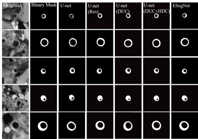 |
9 | A Semantic Segmentation Method with Emphasizing Edge information for Automatic Vessel Wall Analysis
Wenjing Xu1,2, Qing Zhu1, Guanxun Cheng3, Liwen Wan2, Lei Zhang2, Qiang He4, Yongming Dai4, Dong Liang2, Ye Li2, Hairong Zheng2, Xin Liu2, and Na Zhang2
1Faculty of Information Technology, Beijing University of Technology, Beijing, China, 2Paul C. Lauterbur Research Center for Biomedical Imaging, Shenzhen Institute of Advanced Technology, Chinese Academy of Sciences, Shenzhen, China, 3Department of Radiology, Peking university shenzhen hospital, Shenzhen, China, 4United Imaging Healthcare, Shanghai, China
Edge information is essential for medical image analysis, especially for image segmentation. This paper aims to develop a precise semantic segmentation method with emphasizing the edges for automated segmentation of arterial vessel wall and plaque based on the convolutional neural network (CNN) for facilitating the quantitative assessment of plaque in patients with ischemic stroke. An end-to-end architecture network that can emphasize the edge information is proposed. The results suggest that the proposed segmentation method improves segmentation accuracy effectively and will facilitate the quantitative assessment on atherosclerosis.
|
||
 Back to Meeting Home
 Back to the Program-at-a-Glance
The International Society for Magnetic Resonance in Medicine is accredited by the Accreditation Council for Continuing Medical Education to provide continuing medical education for physicians.
 Back to Meeting Home
 Back to the Program-at-a-Glance
 View the Presentation