Online Gather.town Pitches
Processing & Analysis IV
Joint Annual Meeting ISMRM-ESMRMB & ISMRT 31st Annual Meeting • 07-12 May 2022 • London, UK

Online Gather.town Pitches
Processing & Analysis IV
| Booth # | ||||
|---|---|---|---|---|
4069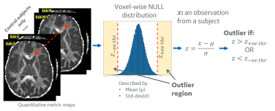 |
1 | Subject-specific Analysis for Diffusion and Kurtosis Changes in mild Traumatic Brain Injury (mTBI) Patients Video Permission Withheld
Chitresh Bhushan1, Nastaren Abad1, Radhika Madhavan2, Luca Marinelli1, H. Doug Morris3, Maureen Hood3,4, J Kevin DeMarco3, Robert Y Shih3,4, Ante Zhu1, Gail Kohls3, Eric Fiveland1, Kimbra Kenney3,4, Vincent B Ho3,4, and
Thomas K Foo1,4
1GE Research, Niskayuna, NY, United States, 2GE Healthcare, Niskayuna, NY, United States, 3Walter Reed National Military Medical Center, Bethesda, MD, United States, 4Uniformed Services University of the Health Sciences, Bethesda, MD, United States
Advanced neuroimaging capabilities enabled by the MAGNUS (ultra-high performance head gradient) system was leveraged in this study to assess subject-specific microstructural changes with acute and chronic mild traumatic brain injury. MAGNUS allows for a four-fold increase in maximum gradient-amplitude compared to current clinical whole-body scanners and enables identification of new imaging biomarkers for improved detection of microstructural changes in the brain. We present an outlier analysis approach that allows study of microstructure differences in individual subjects rather than solely on a group basis. Preliminary results demonstrate identification of several white-matter regions in patient groups that differ from healthy controls.
|
||
4070 |
2 | Combined Effect of Iron Overload and Steatosis on Liver R2* Using Morphological Modeling and MRI Signal Synthesis via Monte Carlo Simulations
Utsav Shrestha1, Juan Pablo Esparza1, Sanjaya Satapathy2, Jason Vanatta3, and Aaryani Tipirneni-Sajja1,4
1Biomedical Engineering, The University of Memphis, Memphis, TN, United States, 2Department of Medicine, North Shore University Hospital/Northwell Health, Manhasset, NY, United States, 3College of Medicine, University of Tennessee Health Science Center, Memphis, TN, United States, 4St. Jude Children’s Research Hospital, Memphis, TN, United States Chemical-shift based multi-spectral fat-water models accounting for single or dual R2* correction are used to assess hepatic iron concentration (HIC) and fat fraction (FF). In this study, we developed a Monte-Carlo based approach for simulating steatosis and iron overload models mimicking the in-vivo characteristics and synthesizing MRI signal to compare the accuracy of the R2* models to estimate FF in the presence of iron. Our results show that R2* estimated by single R2* model is highly governed by iron whereas dual R2* model predicts R2*-FF relationship close to the in-vivo calibration. Nevertheless, FF predicted by both the models were close to the true FF. |
||
4071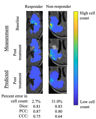 |
3 | Forecasting glioblastoma response to anti-angiogenic therapy via image-driven mathematical models
Tarini Thiagarajan1, Thomas E Yankeelov2,3,4,5,6,7, and David A Hormuth, II2,3
1Aerospace Engineering, The University of Texas at Austin, Austin, TX, United States, 2Oden Institute for Computational Engineering and Sciences, The University of Texas at Austin, Austin, TX, United States, 3Livestrong Cancer Institutes, The University of Texas at Austin, Austin, TX, United States, 4Biomedical Engineering, The University of Texas at Austin, Austin, TX, United States, 5Diagnostic Medicine, The University of Texas at Austin, Austin, TX, United States, 6Oncology, The University of Texas at Austin, Austin, TX, United States, 7Imaging Physics, MD Anderson Cancer Center, Houston, TX, United States
A fundamental challenge in the care of patients with recurrent high-grade gliomas is the selection of appropriate salvage therapies to control further disease progression. To address this challenge, we have developed a biology-based mathematical model of tumor growth and response initialized and calibrated from patient-specific MRI data to predict which patients will respond to anti-angiogenic therapy. We evaluated the predictive accuracy of this image-driven modeling framework in an initial cohort of four patients. The model accurately predicted future total tumor cell number with an average error of 15%.
|
||
4072 |
4 | Simulation of a Virtual Liver Iron-overload Model and Estimation of R2* Using Complex Fat-Water Models
Prasiddhi Neupane1, Utsav Shrestha1, and Aaryani Tipirneni-Sajja1
1Biomedical Engineering, The University of Memphis, Memphis, TN, United States Multi-spectral fat-water-R2* modeling techniques fit either a single R2* or estimate independent R2* for water and fat molecules. In this study, a virtual liver model with hepatic iron overload was created based on true histological data and MRI signals were synthesized using Monte Carlo simulations. Our results demonstrate that the dual R2* values predicted by the ARMA model exhibit relaxivity behavior similar to in vivo and thus can be used for R2* estimation in vivo in varying concentrations of iron overload. |
||
4073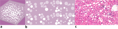 |
5 | Comparison of Single- and Dual-R2* Relaxivity and Estimation of Fat Fraction Using Steatosis Modeling and Monte Carlo Simulations
Utsav Shrestha1, Juan Pablo Esparza1, Sanjaya Satapathy2, Jason Vanatta3, and Aaryani Tipirneni-Sajja1,4
1Biomedical Engineering, The University of Memphis, Memphis, TN, United States, 2Department of Medicine, North Shore University Hospital/Northwell Health, Manhasset, NY, United States, 3College of Medicine, University of Tennessee Health Science Center, Memphis, TN, United States, 4St. Jude Children’s Research Hospital, Memphis, TN, United States MRI multi-spectral fat-water models assuming single or independent R2* for fat (R2*F) and water (R2*F) are non-invasive fat fraction (FF) quantification techniques, but there is no consensus on which is more accurate. Monte Carlo simulations allowed correlation of single R2*, R2*F, and R2*W with FF and assessment of the R2* models. MRI signal was synthesized by creating a virtual hepatic steatosis model from extracted characteristics of fat droplets (FD) obtained using histology. R2*W and single R2* were within the confidence bound of the in-vivo calibration and both R2* models predicted FF with high accuracy which confirms the suitability of Monte-Carlo model to mimic steatosis condition. |
||
4074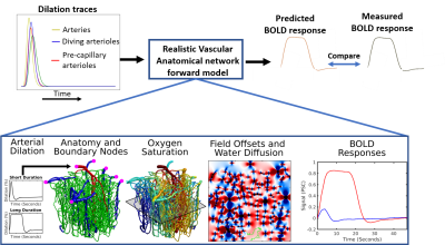 |
6 | Biophysical modeling of abnormal BOLD responses in Cerebral Amyloid Angiopathy due to reduced arteriolar reactivity
Divya Varadarajan1, Joerg P. Pfannmoeller1, Grant A. Hartung1, and Jonathan R. Polimeni1
1Massachusetts General Hospital, Boston, MA, United States
A consistently slower and weaker blood-oxygenation-level-dependent (BOLD) fMRI response is consistently seen in patients with cerebral amyloid angiopathy (CAA).However, our mechanistic understanding of why this occurs in CAA is lacking. To improve our mechanistic understanding, we propose a hypothesis testing framework for simulating the relationship between of impaired microvascular function and the associated abnormal fMRI response. We use BOLD biophysical modeling to link microvascular dynamics with fMRI signals. Here we test whether reduced arteriolar reactivity predicts abnormal BOLD response in early- and late-stage CAA.
|
||
4075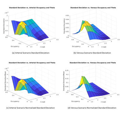 |
7 | The dependence of the resting-state macrovascular fMRI signal power on vascular volume and orientation: A simulation study
Xiaole Zhong1,2 and J. Jean Chen1,2
1Rotman Research Institute, Baycrest Health Sciences, Toronto, ON, Canada, 2Department of Medical Biophysics, University of Toronto, Toronto, ON, Canada
The resting-state fMRI (rs-fMRI) signal fluctuation amplitude is widely used as the resting-state fMRI marker, but it could be biased by the contribution of macrovessels. We demonstrate the dependence of the macrovascular contribution on blood oxygenation, vascular occupancy and orientation. This work paves the way for more appropriate interpretation of rs-fMRI signal amplitude given different vascular morphology across brain regions and populations.
|
||
4076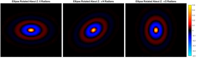 |
8 | Initial Evaluation of a Transverse Isotropic Finite Difference Model for Training Learned Inversion
Jonathan Trevathan1, Jonathan Scott1, Joshua Trzasko1, Armando Manduca1, John Huston1, Richard Ehman1, and Matthew Murphy1
1Mayo Clinic, Rochester, MN, United States While most existing inversion algorithms used in MR elastography assume that the mechanical properties of tissue are isotropic, many tissues exhibit spatial anisotropy in structure that is not accommodated by these algorithms.1,2 In this work we present a framework for developing a learned inversion to address transverse isotropy, the simplest anisotropic case. A transversely isotropic stiffness matrix was used in a feed forward finite difference model to generate simulated displacements. The squared wave speeds anisotropic inclusions were calculated using direct inversion to validate the model against the theoretical wave speeds.3 |
||
4077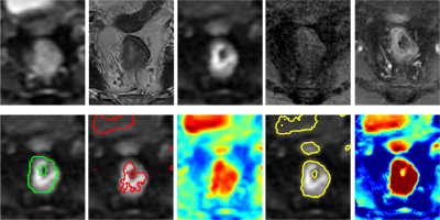 |
9 | Deep-learning based rectal tumor localization and segmentation on multi-parametric MRI
Yang Zhang1,2, Liming Shi3, Weiwen Zhou3, Xiaonan Sun3, Ning Yue1, Min-Ying Su2, and Ke Nie1
1Department of Radiation Oncology, Rutgers-Cancer Institute of New Jersey, Rutgers-Robert Wood Johnson Medical School, New Brunswick, NJ, United States, 2Department of Radiological Sciences, University of California, Irvine, CA, United States, 3Department of Radiation Oncology, Sir Run Run Shaw Hospital, Zhejiang University School of Medicine, Hangzhou, China
Two deep learning methods using the convolutional neural network (CNN) was implemented to segment the rectal cancer in 197 LARC patients, with tumor ROI outlined by a radiologist. For each patient, six frames, including T2, 2 DWI sequences and 3 LAVA sequences, were used for training and validation. The Dice similarity coefficient (DSC) value were used to compare the results of the proposed algorithm and the ROI outlined by reader. The mean DSC was 0.67 and 0.78 from each these 2 method respectively. The proposed algorithms especially the combined serials of U-Net showed improved performance compared to prior published work with individual sequence only. Our work showed the deep-learning with combined image sequence can provide as a promising tool for fully automatic tumor localization and segmentation for rectal cancer.
|
||
4078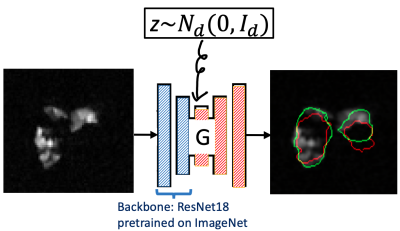 |
10 | Automatic lung segmentation for hyperpolarized gas MRI using transferred generative adversarial network and three-view aggregation
Shih-Kang Chao1, Ummul Afia Shammi2, Lucia Flors-Blasco3, Talissa Altes4, John Mugler5,6, Craig Meyer5,6, Jaime Mata6, Wilson Miller6, and Robert Thomen2,4
1Department of Statistics, University of Missouri, Columbia, MO, United States, 2Department of Biomedical, Biological & Chemical Engineering, University of Missouri, Columbia, MO, United States, 3Keck School of Medicine, University of Southern California, Los Angeles, CA, United States, 4Department of Radiology, University of Missouri, Columbia, MO, United States, 5Department of Biomedical Engineering, University of Virginia, Charlottesville, VA, United States, 6Department of Radiology and Medical Imaging, University of Virginia, Charlottesville, VA, United States
We evaluate an automatic lung segmentation approach that aggregates the predicted mask of coronal, axial, and sagittal views generated by a deep conditional generative adversarial network (GAN) whose only input is the hyperpolarized gas (HPG) MRI. On five test subjects with ventilation defect percentages [VDP] of 25-38%, our method achieved an average Dice score of 87.72, and above 90 on a healthy control subject. The slice-wise Dice score had an average correlation of 0.72 with the human expert and a median correlation of -0.79 with VDP, and both are significant for 4 out of 5 test patients at level 1%.
|
||
4079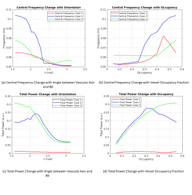 |
11 | The frequency dependence of the resting-state fMRI signal on macrovascular volume and orientation
Xiaole Zhong1,2 and J. Jean Chen1,2
1Rotman Research Institute, Baycrest Health Sciences, Toronto, ON, Canada, 2Department of Medical Biophysics, University of Toronto, Toronto, ON, Canada
Frequency analysis of the BOLD fMRI signal is increasingly finding application, including in evaluating denoising procedures and biological brain changes. In resting-state fMRI (rs-fMRI), the signal frequency can be biased by contributions from large vessels, but this phenomenon is uninvestigated. In this work, we show that the rs-fMRI frequency content varies with both vascular volume fraction and orientation. Moreover, arteries and veins contribute differently to these variations.
|
||
4080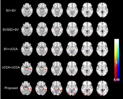 |
12 | Group-level adaptive-analysis of task fMRI data
Xiaowei Zhuang1,2, Zhengshi Yang1, Tim Curran3, Rajesh Nandy4, Mark Lowe5, and Dietmar Cordes1,3
1Lou Ruvo Center for Brain Health, Cleveland Clinic, Las Vegas, NV, United States, 2Interdisciplinary neuroscience PhD program, University of Nevada, Las Vegas, Las Vegas, NV, United States, 3University of Colorado Boulder, Boulder, CO, United States, 4University of North Texas Health Science Center at Fort Worth, Fort Worth, TX, United States, 5Cleveland Clinic, Cleveland, OH, United States
A task-fMRI group-level analysis method is proposed to incorporate spatial covariance structures in fMRI data using the subject-level steerable filter smoothing with various full-wide-half-maximums followed by a group-level one-step optimization. Subject-level smoothed time series are further orthogonalized to guarantee non-overlapped contributions to group-level activations. Using the proposed method, we are able to detect more accurate activations in both simulated data and during a real-fMRI episodic memory task.
|
||
4081 |
13 | Addressing Acquisition Variability in DWI on Prostate Cancer with Unsupervised Methods
Batuhan Gundogdu1, Jay M Pittman2, Aritrick Chatterjee2, Milica Medved2, Roger Engelmann2, Aytekin Oto2, and Gregory S Karczmar2
1Radiology, University of Chicago, Chicago, IL, United States, 2University of Chicago, Chicago, IL, United States
Diffusion-weighted MR images are typically obtained as multiple acquisitions with multiple diffusion-sensitizing gradient directions. Due to molecular motion, some acquisitions suffer from signal loss at random locations. This affects cancer conspicuity and degrades the diagnostic efficacy of DWI. We propose an agglomerative clustering-based unsupervised method to address this. The model automatically rejects acquisitions of voxels that are likely to be corrupted by bulk motion and lack coherence with the rest of the acquisitions. We observed that this method both reduces the DWI signal variability and enhances the cancer detection accuracy.
|
||
4082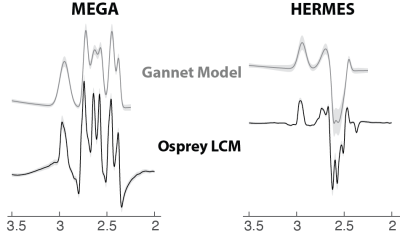 |
14 | Importance of linear-combination modeling for quantification of GABA and glutathione levels using Hadamard-edited MRS
Yulu song1,2, Helge J. Zöllner 1,2, Steve C.N. Hui1,2, Georg Oeltzschner1,2, James J. Prisciandaro3, and Richard A.E. Edden1,2
1Russell H Morgan Department of Radiology and Radiological Science, The Johns Hopkins University School of Medicine, Baltimore, MD, United States, 2F.M. Kirby Center for Functional Brain Imaging, Kennedy Krieger Institute, Baltimore, MD, United States, 3Department of Psychiatry and Behavioral Sciences, Addiction Sciences Division, Center for Biomedical Imaging, Medical University of SC, Charleston, SC, United States
There are two main approaches for modeling edited spectra: simple Gaussian modeling, and linear-combination modeling based on simulated metabolite basis functions. The simple Gaussian modeling of GSH in Gannet was reported to be less reproducible for HERMES than for MEGA-PRESS. Recent consensus recommended linear-combination modeling for quantification of edited MRS. Here we compared the performance of simple Gaussian and linear-combination modeling on a test-retest dataset to display the improvement of the reproducibility of metabolite quantification with HERMES.
|
||
4083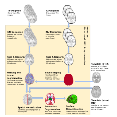 |
15 | NiBabies: A robust preprocessing workflow tailored for neonate and infant MRI
Mathias Goncalves1, Christopher J. Markiewicz1, Martin Styner2, Lucille A. Moore3, Kathy Snider4, Eric A. Earl3, Christopher D. Smyser5, Lilla Zöllei6, Russel A. Poldrack1, Oscar Esteban7, Eric Feczko4, and Damien A. Fair4
1Stanford University, Stanford, CA, United States, 2University of North Carolina School of Medicine, Chapel Hill, NC, United States, 3Oregon Health & Science University, Portland, OR, United States, 4University of Minnesota, Minneapolis, MN, United States, 5Washington University in St. Louis, St. Louis, MO, United States, 6Harvard Medical School, Boston, MA, United States, 7University of Lausanne, Lausanne, Switzerland
Advances in both data acquisition and processing methods have given magnetic resonance imaging researchers (MRI) a plethora of options on how best to clean and standardize data before statistical analysis. Recently, there has been a surge in standardized data processing workflows, but special populations, such infants, require modified techniques not normally found in general pipelines. Here we introduce NiBabies, a robust and open-source structural and functional MRI preprocessing pipeline designed for infant populations.
|
||
 Back to Meeting Home
 Back to the Program-at-a-Glance
The International Society for Magnetic Resonance in Medicine is accredited by the Accreditation Council for Continuing Medical Education to provide continuing medical education for physicians.
 Back to Meeting Home
 Back to the Program-at-a-Glance
 View the Presentation