Online Gather.town Pitches
New Systems & Devices V
Joint Annual Meeting ISMRM-ESMRMB & ISMRT 31st Annual Meeting • 07-12 May 2022 • London, UK

Online Gather.town Pitches
New Systems & Devices V
| Booth # | ||||
|---|---|---|---|---|
 |
5038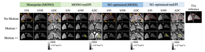 |
1 | Validation of motion-robust, blood-suppressed, reduced-distortion liver diffusion techniques in a pulsatile motion phantom
Ruiqi Geng1,2, James Rice1,3, Yuxin Zhang1,2, David R. Rutkowski1,3, Alejandro Roldán-Alzate1,3, Arnaud Guidon4, and Diego Hernando1,2,5,6
1Radiology, University of Wisconsin-Madison, Madison, WI, United States, 2Medical Physics, University of Wisconsin-Madison, Madison, WI, United States, 3Mechanical Engineering, University of Wisconsin-Madison, Madison, WI, United States, 4GE Healthcare, Boston, MA, United States, 5Electrical and Computer Engineering, University of Wisconsin-Madison, Madison, WI, United States, 6Biomedical Engineering, University of Wisconsin-Madison, Madison, WI, United States
Conventional liver diffusion MRI acquisitions suffer from several challenges including pulsatile motion-induced signal voids and B0-induced distortions. In this study, we validated motion-robust, blood-suppressed, reduced-distortion liver diffusion techniques in an anatomically accurate liver phantom including controlled pulsatile motion. By combining optimized motion-compensated, blood-suppressed diffusion waveforms (i.e. MODI) with msEPI acquisitions, superior image quality and quantitative performance can be achieved in the presence of various degrees of compressive tissue motion.
|
|
5039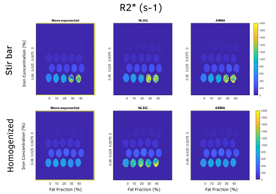 |
2 | Impact of Fat Droplet Sizes on R2* in Fat-Iron phantoms for Accurate Assessment of Hepatic Steatosis and Iron Overload Using MRI
Sarah Caroline Brasher1, Cara Morin2, and Aaryani Tipirneni-Sajja1,2
1Biomedical Engineering, University of Memphis, Memphis, TN, United States, 2St. Jude Children's Research Hospital, Memphis, TN, United States
R2* correction is necessary for improving the accuracy of fat fraction quantification in assessing steatosis. However, the dephasing effects of concurrent iron overload may be dependent on the size of iron and fat particles. In this study, we controlled the size of fat droplets in fat-iron emulsion phantoms by traditional stir bar methods and homogenization. R2* and far fraction (FF) values were estimated with multi-spectral models that assume a common or independent R2* for water and fat. Our results show that R2* was slightly reduced in homogenized phantoms at higher fat fractions compared to stir bar phantoms.
|
||
| 5040 | 3 | Acceptance Procedure for the MRI Component of the 1.5T MRI-Linac
Jie Deng1, Chenyang Shen1, Justin Visak1, and Andrew Godley1
1Radiation Oncology, UT Southwestern Medical Center, Dallas, TX, United States
On two Unity MRI-Linac systems simultaneously installed in our institution in May 2021, we developed and implemented the acceptance and commissioning testing for the MRI component independently. We also worked together with the vendors during troubleshooting and re-testing processes. The comprehensive acceptance testing consisted of 4 phases: basic testing using vendor-provided tools, advanced testing, Linac system influence on MRI, and end-to-end testing for treatment planning and dose delivery. Both Unity machines successfully passed the MRI component acceptance testing in consideration of influence of the Linac component. MR-to-MV alignment defect on one system was fixed after major work of troubleshooting.
|
||
5041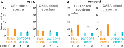 |
4 | Impact of gradient scheme and shimming on out-of-voxel echo artefacts in edited MRS
Yulu Song1,2, Helge J. Zöllner1,2, Steve C.N. Hui1,2, Georg Oeltzschner1,2, and Richard A.E. Edden1,2
1Department of Radiology and Radiological Science, Johns Hopkins University School of Medicine, Baltimore, MD, United States, 2F.M. Kirby Research Center for Functional Brain Imaging, Kennedy Krieger Institute, Baltimore, MD, United States
Out-of-voxel (OOV) signal is a common spurious echo artifact, which interferes with spectral quantification. These signals arise from either incomplete suppression or accidental refocusing. Dephasing Optimization Through Coherence Order Pathway Selection (DOTCOPS) is an optimized gradient scheme that can be used to suppress unwanted signals. Here we explored the impact of gradient scheme and shimming-order on OOV artefacts in Hadamard-encoded edited MRS.
|
||
5042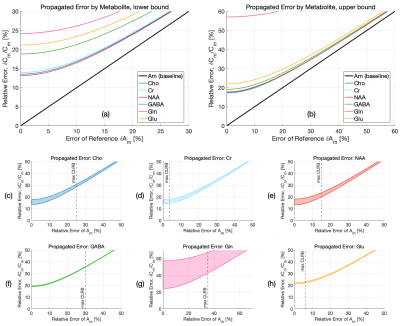 |
5 | Error Propagation in Absolute Metabolite Quantification for MR Spectroscopy of the Human Brain Video Permission Withheld
Ronald Instrella1 and Christoph Juchem1,2
1Biomedical Engineering, Columbia University, New York, NY, United States, 2Radiology, Columbia University, New York, NY, United States
Spectral quantification methods provide absolute concentration estimates of metabolites from data acquired using in vivo MR spectroscopy. Cramér-Rao Lower Bounds (CRLB) are commonly employed as a minimum estimate of concentration error; however, absolute quantification relies on more than simply the uncertainty of Linear Combination Modeling (LCM). The uncertainties of both metabolite (e.g. T1, T2) and sequence-specific parameters (e.g. TR, TE), are generally ignored, leading to a systemic overestimation of accuracy. In this study, we present an analysis using the propagation of uncertainty to derive a more comprehensive estimate of the overall error of metabolite quantification.
|
||
5043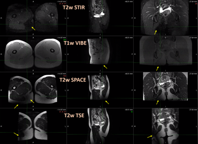 |
6 | Evaluation of a prototype MR conditional hoverboard and stirrups system for MR-guided brachytherapy at 3T
Evangelia Kaza1, Bret Nicholson2, Kevin Anderson2, Jesse Drake2, Alex Marques1, Steven Hatch1, Jeremy Bredfeldt1, Martin King1, and Atchar Sudhyadhom1
1Radiation Oncology, Brigham and Women's Hospital, Dana-Farber Cancer Institute, Harvard Medical School, Boston, MA, United States, 2Diacor Inc, West Valley City, UT, United States
MR conditional stirrups would be beneficial in MR-guided gynecological and prostate brachytherapy but have not been clinically available. Evaluation of a prototype Diacor MR conditional board and stirrups system revealed no significant projectile or heating risks at 3T. The maximum temperature measured on its metallic surfaces after scanning was 22 ⁰C, relating to a 3 ⁰C increase. The stainless steel blades of the stirrup joints caused signal cancellation artifacts which extended radially up to 23cm on phantom images. On volunteer scans, artifacts were observed in posterior hip areas only. Pelvic organ visualization was considered suitable for brachytherapy treatment planning.
|
||
5044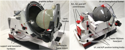 |
7 | Mock TcMRgFUS Transducer for Off-site Technological Development
Rock Hadley1, Robb Merrill1, Henrik Odeen1, and dennis parker1
1University of Utah, Salt Lake City, UT, United States
A portable device that mimics the shape, size, and configuration of the Insightec transcranial MR guided focused ultrasound (tcMRgFUS) assembly is presented. This Mock system provides a water bath and conductive ground plane that accurately emulates that of the Insightec system. The system is shown to generate the same well-known banding artifact as the Insightec system. The construction details of the Mock system and image comparison to the Insightec system are presented.
|
||
5045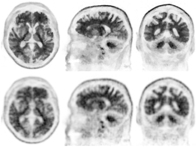 |
8 | PET Recon with MR Priors: Application in Ambiguously Rated Amyloid PET Scans
Mehdi Khalighi1, Greg Zaharchuk1, Michael Zeineh1, Guido Davidzon1, Gabriel Kennedy2, Christina Young1, Michael Greicius2, Kathleen Poston2, and Elizabeth Mormino2
1Radiology, Stanford University, Stanford, CA, United States, 2Neurology, Stanford University, Stanford, CA, United States
A recently developed PET recon with MR priors and motion correction method is applied to a set of ambiguously rated amyloid PET scans and is compared with regular TOF-OSEM PET recon using SUVR. The same comparison is done in a set of scans which were matched by clinical diagnosis. By using PET recon with MR priors, lower SUVRs for negative scans compared to increased SUVRs for positive scans were measured, which shows improvement in the ability to distinguish intermediate amyloid levels.
|
||
5046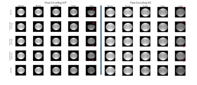 |
9 | Concomitant Field Effects on Asymmetric Gradient Systems: Impacts on Slow Flow Imaging
Nastaren Abad1, Seung-Kyun Lee1, Yihe Hua1, J. Kevin DeMarco2,3, Robert Shih2,3, Maureen Hood2,3, H Doug Morris2, Vincent B Ho2,3, Myung-Ho In4, Matt A Bernstein4, and Thomas KF Foo1
1GE Global Research Center, Niskayuna, NY, United States, 2Walter Reed National Military Medical Center, Bethesda, MD, United States, 3Uniformed Services University of the Health Sciences, Bethesda, MD, United States, 4Department of Radiology, Mayo Clinic, Rochester, MN, United States This study investigates concomitant gradient field effects on gradient echo phase contrast imaging when visualizing very slow (<50 mm/s) flow. The impact of real-time concomitant field compensation and the consequence of uncompensated higher-ordered terms is shown. |
||
5047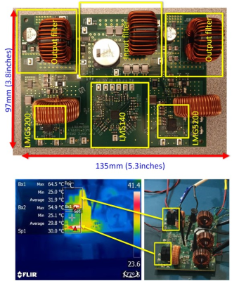 |
10 | In Scan Room No-Interference High Efficiency Switching Power Supply with GaN
Juan A Sabate1, Logan A Snow1, Huan A Hu1, and Randy Buchwald2
1GE Research, Niskayuna, NY, United States, 2MR-Eng-Systems, GE Healthcare, Waukesha, WI, United States
A switching regulator implemented with GaN power semiconductors and air core inductors was shown not to create any noise that affected the scanner imaging. This was due to the selected switching frequency of 2.489MHz, which avoided harmonics at imaging frequencies of interest. The switching regulator size and thermal management at 90% does not require any additional support systems in the scan room. This demonstrates that GaN-based switching regulators can be a good solution to regulate voltages in the scan room and also significantly reduce the cabling and simplify installation.
|
||
5048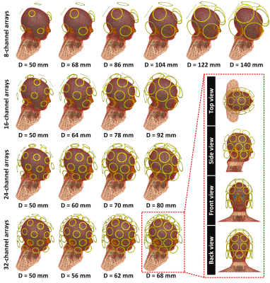 |
11 | Comparison of simulated parallel transmit head arrays at 7T using excitation uniformity, local SAR, and global SAR
Ehsan Kazemivalipour1,2, Lawrence L. Wald1,2,3, and Bastien Guerin1,2
1A.A. Martinos Center for Biomedical Imaging, Department of Radiology, Massachusetts General Hospital, Charlestown, MA, United States, 2Harvard Medical School, Boston, MA, United States, 3Harvard-MIT Division of Health Sciences Technology, Cambridge, MA, United States
We investigate the performance of parallel transmission arrays with 8, 16, 24, and 32 channels and varying loop sizes (18 coils total) for brain imaging at 7T. We compare RF-shimming performance of the arrays using the L-curve method showing optimal tradeoffs between excitation uniformity (slice), local-SAR, global-SAR and peak-power. For the numbers of channels simulated here, the coils with larger loops achieved a better local-SAR vs. excitation uniformity than those with smaller loops (this was true for axial/sagittal slice orientations). At constant local-SAR constraint, the 24-channel arrays showed the best performance in improving the excitation uniformity in the coronal/axial slices.
|
||
5049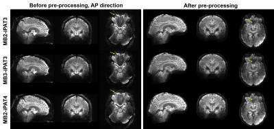 |
12 | High-resolution whole-brain diffusion MRI at 7 Tesla using 63-channel signal reception
Xiaodong Ma1, Andrea Grant1, Matt Waks1, Edward Auerbach1, Gregor Adriany1, Kamil Ugurbil1, and Xiaoping Wu1
1Center for Magnetic Resonance Research, Radiology, Medical School, University of Minnesota, Minneapolis, MN, United States
There has been an increasing interest to acquire high-resolution diffusion MRI at ultrahigh field (≥7 Tesla) due to the increased SNR and improved parallel imaging performance. To fully capitalize on the benefit of ultrahigh field, it is desirable to image with many receiver coils. In this study, we acquired slice-accelerated whole-brain 1.05-mm isotropic diffusion images on an FDA-approved 7 Tesla scanner (Siemens Terra) using a homemade 63-channel head RF array with various slice and in-plane accelerations. We found that the use of 63 channels can achieve higher acceleration factors (up to 9-fold acceleration in total) while maintaining the image quality.
|
||
5050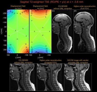 |
13 | Higher-order image reconstruction with integrated gradient nonlinearity correction using a low-rank encoding operator
Nam G. Lee1, Kübra Keskin2, Ziwei Zhao2, and Krishna S. Nayak2
1Biomedical Engieering, University of Southern California, Los Angeles, CA, United States, 2Electrical Engieering, University of Southern California, Los Angeles, CA, United States
Conventional MR image reconstruction relies on the assumption of perfectly linear gradient fields. However, the gradient fields contain spatially varying nonlinear components. We present a higher-order image reconstruction method that incorporates a theoretical model of gradient nonlinearity without any external field monitoring device. This approach utilizes the separability of Fourier encoding in Cartesian imaging and employs a low-rank approximation only to the higher-order readout encoding matrix, allowing a memory-efficient implementation suitable for large FOVs. Image distortions due to gradient nonlinearity were successfully mitigated by the proposed method using axial/sagittal/coronal 2D Cartesian datasets acquired on a prototype 0.55T MRI system.
|
||
 Back to Meeting Home
 Back to the Program-at-a-Glance
The International Society for Magnetic Resonance in Medicine is accredited by the Accreditation Council for Continuing Medical Education to provide continuing medical education for physicians.
 Back to Meeting Home
 Back to the Program-at-a-Glance
 View the Presentation