Online Gather.town Pitches
Fast, Novel & Robust Acquisitions II
Joint Annual Meeting ISMRM-ESMRMB & ISMRT 31st Annual Meeting • 07-12 May 2022 • London, UK

Online Gather.town Pitches
Fast, Novel & Robust Acquisitions II
| Booth # | ||||
|---|---|---|---|---|
4796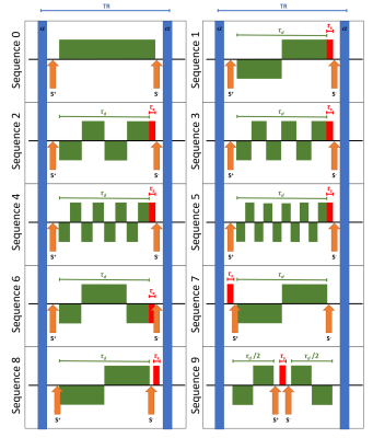 |
1 | On the evaluation of different variants of diffusion-weighted double-echo steady-state (dwDESS) sequences
Ulrich Katscher1, Bjoern Steinhorst1, Jakob Meineke1, and Jochen Keupp1
1Philips Research, Hamburg, Germany Double-echo steady-state (DESS) sequences are a promising candidate for diffusion weighted imaging (DWI) free of geometric distortions. The design of diffusion-weighted DESS (dwDESS) sequences allows waveform variations for the diffusion weighting gradient GD. While dwDESS sequences were originally introduced with unipolar GD, also bipolar or higher order GD are possible. A balanced GD (i.e., with nulling zeroth momentum) typically necessitates a spoiler gradient to suppress banding artefacts, which additionally increases the degrees of freedom in the design of dwDESS sequences. This study explores different dwDESS sequence variants according to predefined optimization criteria. |
||
4797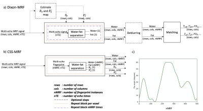 |
2 | A versatile framework for chemical species separation in Dixon MR Fingerprinting
Elizabeth Huaroc Moquillaza1, Manuel Baumann2, Kilian Weiss3, Thomas Amthor2, Peter Koken2, Benedikt Schwaiger4, Markus R. Makowski1, Mariya Doneva2, and Dimitrios C. Karampinos1
1Department of Diagnostic and Interventional Radiology, School of Medicine, Technical University of Munich, Munich, Germany, 2Philips Research Lab, Hamburg, Germany, 3Philips Healthcare, Hamburg, Germany, 4Department of Diagnostic and Interventional Neuroradiology, School of Medicine, Technical University of Munich, Munich, Germany
Dixon Magnetic Resonance Fingerprinting (Dixon-MRF) is being increasingly used to measure multiple quantitative parameters with effective fat suppression across body tissues. Dixon-MRF processing is typically performed in two steps: water-fat separation and dictionary matching. The present work introduces a versatile formulation for chemical species separation in Dixon-MRF (CSS-MRF) which allows to process a multi-echo fingerprint in a single step obtaining a water fingerprint, a fat fingerprint, a B0 value and a R2* value as outputs. CSS-MRF allows an easy and flexible definition of the parameters to be estimated and enables the option of a varying fat spectrum across the MRF-dimension.
|
||
4798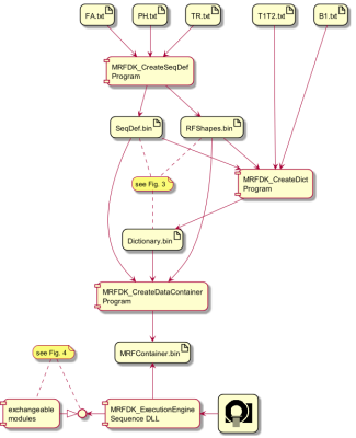 |
3 | A Comprehensive MR Fingerprinting Development Kit (MRFDK)
Thomas Kluge1, Mathias Nittka1, Stephan Kannengiesser1, Gregor Koerzdoerfer1, Christina Grund1, Guido Buonincontri2, Jianing Pang3, Rasim Boyacioglu4, Yong Chen4, and Mark Griswold4
1Siemens Healthcare GmbH, Erlangen, Germany, 2Siemens Healthcare s.r.l, Milan, Italy, 3Siemens Medical Solutions USA Inc., Chicago, IL, United States, 4Department of Radiology, Case Western Reserve University, Cleveland, OH, United States
Magnetic Resonance Fingerprinting (MRF), as an approach for multi-parametric, quantitative imaging, imposes new paradigms for sequence design in terms of optimized signal encoding and dictionary matching. In this work, we present a novel comprehensive framework and tool chain for rapid and efficient MRF prototyping. Key concepts are modularization and abstraction of the MRF experiment related to spin-physics, spatial encoding, and scanner hardware.
|
||
4799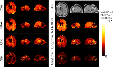 |
4 | Whole-brain high-resolution MRSI at 7T with non-Cartesian FID-ECCENTRIC in glioma patients
Antoine Klauser1,2, Bernhard Strasser3, Wolfgang Bogner3, Lukas Hingerl3, Claudiu Schirda4, Bijaya Thapa5, Daniel Cahill6, Tracy Batchelor7, Francois Lazeyras1, and Ovidiu Andronesi5
1University of Geneva, Geneva, Switzerland, 2CIBM Center for Biomedical Imaging, Geneva, Switzerland, 3Medical University of Vienna, Vienna, Austria, 4University of Pittsburgh Medical Center, Pittsburgh, PA, United States, 5Athinoula A. Martinos Center for Biomedical Imaging, Boston, MA, United States, 6Massachusetts General Hospital, Boston, MA, United States, 7Brigham and Women Hospital, Boston, Switzerland
MR spectroscopic imaging (MRSI) can map specific metabolic alterations of glioma brain tumors. Increasing structural details for spatial distribution of metabolites is needed to probe tumor margins and heterogeneity with higher sensitivity and specificity. Here we evaluate the performance of ECCENTRIC, a newly developed non-Cartesian compressed sense MRSI method, to realize fast and high-resolution metabolic imaging at 7T ultra-high field in glioma patients. This is expected to improve diagnosis, prognostication, planning, guidance, and response assessment of treatment in glioma patients and other brain diseases.
|
||
4800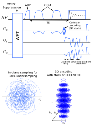 |
5 | Fast adiabatic spin-echo with whole-brain ECCENTRIC for glioma metabolic imaging at 7T
Antoine Klauser1,2, Bernhard Strasser3, Wolfgang Bogner3, Bijaya Thapa4, Jorg Dietrich5, Erik Uhlmann5, Tracy Batchelor6, Daniel Cahill5, Francois Lazeyras1,2, and Ovidiu Andronesi4
1University of Geneva, Geneva, Switzerland, 2CIBM Center for Biomedical Imaging, Geneva, Switzerland, 3Medical University of Vienna, Vienna, Austria, 4Athinoula A. Martinos Center for Biomedical Imaging, Boston, MA, United States, 5Massachusetts General Hospital, Boston, MA, United States, 6Brigham and Women Hospital, Boston, MA, United States
Metabolic alterations specific to glioma can be imaged by MR spectroscopic imaging (MRSI). Adiabatic spin-echo (ASE) MRSI enables spectral editing for specific metabolites with uniform excitation over whole-brain but needs long TR at 7T due to specific absorption rate which results in long acquisition times for high spatial resolution. Acceleration of ASE can be obtained with non-cartesian compressed sense MRSI (ECCENTRIC) acquisition. Here we evaluate the performance of ASE-ECCENTRIC metabolic imaging in glioma patients. We expect that enhanced spectral sensitivity, specificity and high spatial resolution of ASE-ECCENTRIC will improve diagnosis, prognostication, and treatment response monitoring in glioma patients.
|
||
4801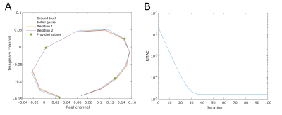 |
6 | Accelerating phase-cycled bSSFP using sparsity across the phase-cycling dimension
Michael van Rijssel1, Cornelis van den Berg1, Stan Noordman1, and Astrid van Lier1
1Radiotherapy, UMC Utrecht, Utrecht, Netherlands
Banding artifacts in balanced steady state free precession images can be resolved by applying radiofrequency phase cycling. Unfortunately, the scan time increases linearly with the amount of phase cycles acquired, hindering clinical adoption of this sequence. We aimed to reduce the amount of phase cycles that need to be acquired by employing a signal model in an iterative reconstruction. Preliminary validation of this algorithm was performed in-silico and in a phantom. Results show excellent agreement in-silico (relative mean absolute error, RMAE, 0.0045%) and in phantom tubes with T1 and T2 values in the physiological range (RMAE 3-4%).
|
||
4802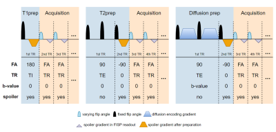 |
7 | Optimal experimental design of MR Fingerprinting for simultaneous quantification of T1, T2, and ADC
Siyuan Hu1, Debra McGivney1, Mark Griswold2, and Dan Ma1
1Biomedical Engineering, Case Western Reserve University, Cleveland, OH, United States, 2Radiology, Case Western Reserve University, Cleveland, OH, United States
The MRF framework has been recently investigated to estimate T1, T2 and apparent diffusion coefficient (ADC) from a single scan with b-tensor encoding schemes. However, the current human-designed experiment protocol for multi-dimensional MRF is still subject to limited measurement accuracy. Here we propose to adapt the Cramer-Rao Bounds to optimize multi-dimensional MRF scan for simultaneous quantification of relaxation and diffusion. The optimization framework explores all possible combinations of MRF sequence parameters, including flip angles, TRs, b-values, and preparation modules to seek the optimal tissue parameter encoding scheme. The optimized sequence simultaneously improves the measurement precision of T1, T2 and ADC.
|
||
 Back to Meeting Home
 Back to the Program-at-a-Glance
The International Society for Magnetic Resonance in Medicine is accredited by the Accreditation Council for Continuing Medical Education to provide continuing medical education for physicians.
 Back to Meeting Home
 Back to the Program-at-a-Glance
 View the Presentation