Online Gather.town Pitches
Diffusion II
Joint Annual Meeting ISMRM-ESMRMB & ISMRT 31st Annual Meeting • 07-12 May 2022 • London, UK

| Booth # | ||||
|---|---|---|---|---|
4743 |
1 | Improving estimation of cell size distribution using Fourier expansion-based deconvolution for microstructural parameters mapping (FED-MPM)
Diwei Shi1, Sisi Li2, Yishi Wang3, Li Chen1, Xiaoyu Jiang4,5, Junzhong Xu4,5,6,7, Quanshui Zheng1, and Hua Guo2
1Center for Nano and Micro Mechanics, Department of Engineering Mechanics, Tsinghua University, Beijing, China, 2Center for Biomedical Imaging Research, Tsinghua University, Beijing, China, 3Philips Healthcare, Beijing, China, 4Institute of Imaging Science, Vanderbilt University Medical Center, Nashville, TN, United States, 5Department of Radiology and Radiological Sciences, Vanderbilt University Medical Center, Nashville, TN, United States, 6Department of Physics and Astronomy, Vanderbilt University, Nashville, TN, United States, 7Department of Biomedical Engineering, Vanderbilt University, Nashville, TN, United States Cellular microstructural parameters mapping can provide quantitative information for tumor diagnosis and treatment monitoring. Particularly, voxel-wise cell size distribution may provide critical clinical biomarkers to characterize heterogeneity of tumors. Currently, MRI-cytometry may measure such a distribution, but it “blurs” the distribution peaks and prevents differentiating different cell populations with different cell sizes. In this work, we develop a new approach to improve this and hence make it more practical to distinguish different cells. Detection of T cell infiltration for assessment of early response to immunotherapy may be a potential application of this method.
|
||
4744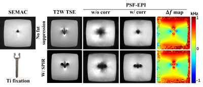 |
2 | Metallic Artifact Suppression in DWI Using Efficient 2D Imaging
Sisi Li1, Yishi Wang2, Yajing Zhang3, and Hua Guo1
1Center for Biomedical Imaging Research, Tsinghua University, Beijing, China, 2Philips Healthcare, Beijing, China, 3MR Clinical Science, Philips Healthcare, Suzhou, China
Diffusion-weighted imaging (DWI) near metal remains a technical challenge although several 3D multispectral imaging methods have achieved effective metallic artifacts correction in anatomical MRI. Additionally, considering scan time and scan flexibility, 2D multi-slice acquisition approaches are typically used in DWI. In this study, we adopted 2D Point-Spread-Function Encoded EPI to generate a series of echo-shifted in-plane distortion-corrected images and introduced an off-resonance frequency-informed reconstruction framework to further reduce metallic artifacts in DWI by through-slice signal registration. The efficacy of this method was validated in phantom experiments, in-vivo brain DTI and spinal cord DWI.
|
||
4745 |
3 | Distortion-Free DWI using PSF-EPI at High-Performance 0.5T MRI
Simin Liu1, Yunhao Xie2, Hai Luo2, Yishi Wang1, Sisi Li1, Jieying Zhang1, Ziyi Pan1, Ziyue Wu2, and Hua Guo1
1Center for Biomedical Imaging Research, Department of Biomedical Engineering, School of Medicine, Tsinghua University, Beijing, China, 2Marvel Stone Healthcare Co., Ltd., Wuxi, China
Low-field MR systems with state-of-the-art hardware may improve the accessibility of MRI. DWI is valuable for diagnosing diseases, while it requires multiple averages at low-field MRI considering SNR. Given similar acquisition time, multi-shot EPI DWI can be considered alternatively. Particularly, the tilted-CAIPI accelerated multi-shot PSF-EPI has been proposed in recent years to achieve fast distortion-free DWI. This study tested the feasibility of PSF-EPI on a high-performance 0.5T MR scanner, and proposed a method for further noise reduction using B0 field information. The results show that PSF-EPI eliminates distortions and improves structure fidelity at tissue boundaries compared with interleaved EPI.
|
||
4746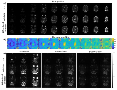 |
4 | Exploiting slice-by-slice B1 adjusted adiabatic refocusing RF pulses to improve diffusion weighted imaging at 7T
Yasmin Blunck1,2, Daniel Staeb3, Rebecca Glarin1, Leigh Johnston1,2, Jin Jin4, and Kieran O'Brien4
1Melbourne Brain Centre Imaging Unit, The University of Melbourne, Melbourne, Australia, 2Department of Biomedical Engineering, The University of Melbourne, Melbourne, Australia, 3Siemens Healthcare Pty Ltd, Melbourne, Australia, 4Siemens Healthcare Pty Ltd, Brisbane, Australia
The application of diffusion weighted imaging (DWI) at ultra-high field is hampered by severe B1 inhomogeneities leading to signal dropouts in certain parts of the brain. Utilising TR-FOCI pulses in a twice-refocussed spin echo DWI sequence can effectively recover signal in these areas, albeit at the cost of increased SAR. Maintaining this enhanced B1 homogeneity, this work demonstrates a ~30% reduction in SAR based on a slice-by-slice RF amplitude optimization.
|
||
4747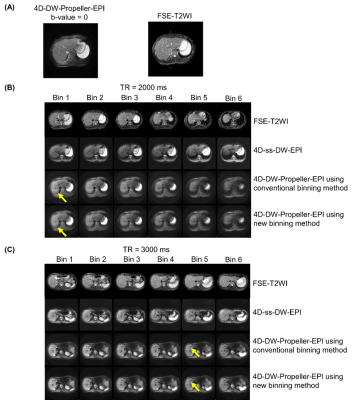 |
5 | Evaluation of k-space uniformity and blade number-optimized (K-B-optimized) binning method for abdominal 4D-DW-Propeller-EPI
Lu Wang1, Tian Li2, Jing Cai2, Liyuan Liang1, and Hing-Chiu Chang3
1Department of Diagnostic Radiology, University of Hong Kong, Hong Kong, China, 2Department of Health Technology and Informatics, The Hong Kong Polytechnic University, Hong Kong, China, 3Department of Biomedical Engineering, Chinese University of Hong Kong, Hong Kong, China Four-dimensional diffusion-weighted imaging (4D-DWI) attracts lots of attention in MRI-guided radiotherapy since it can provide functional information and better delineate the tumor. However, the previously reported 4D-DW-Propeller-EPI technique used amplitude data binning with continuous interval to sort the blade data that might lead to non-uniform distribution of k-space sampling, therefore being suboptimal for subsequent Propeller-EPI reconstruction. Thus, we propose a new (K-B-optimized) binning method to consider the requirement of propeller reconstruction during the sorting of blade data, thereby improving the k-space distribution. Simulation and in-vivo studies demonstrated that the new binning method can improve the data quality of 4D-DW-Propeller-EPI. |
||
4748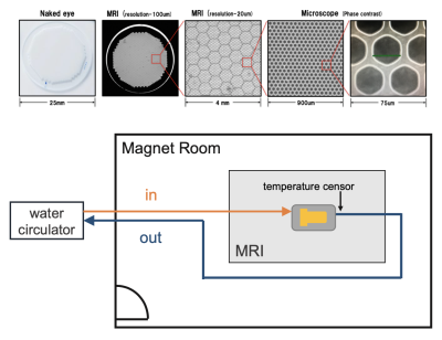 |
6 | Characteristics of three factors that influence the diffusion signal with PGSTE and OGSE Video Permission Withheld
Hinako Oshiro1,2, Junichi Hata1,2,3,4, Daisuke Nakashima3, Yawara Haga1,2, Naoya Hayashi1,2, Daisuke Yoshimaru2,3,4, Kei Hagiya2, and Hideyuki Okano2,3
1Tokyo Metropolitan University, Tokyo, Japan, 2RIKEN Center for Brain Science, Saitama, Japan, 3Keio University School of Medicine, Tokyo, Japan, 4The Jikei University School of Medicine, Tokyo, Japan We investigated the characteristics of diffusion quantification values in a restricted structure with a capillary phantom in PGSTE and OGSE. Smaller structure sizes require shorter diffusion times for structure estimation. In OGSE, we observed a transition of diffusion coefficients under 10 ms for capillary sizes from 6-12 μm, but no differences for those from 25-100 μm. Moreover, it was necessary to consider the temperature dependence of the diffusion measurement at low temperatures. We developed a system to measure the effects on diffusion quantification values at three phases of different temperatures, structure sizes, and diffusion times. |
||
4749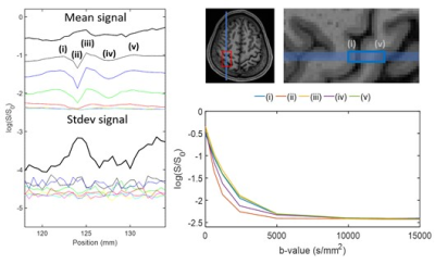 |
7 | Investigating the dot-compartment using diffusion MRI line scanning
Viktor Vegh1,2, Thomas Barrick3, Qianqian Yang4, Qiang Yu1, and Martijn Cloos1,2
1Centre for Advanced Imaging, University of Queensland, Brisbane, Australia, 2ARC Training Centre for Innovation in Biomedical Imaging Technology, Brisbane, Australia, 3Neuroscience Research Centre, St George's, University of London, London, United Kingdom, 4School of Mathematical Sciences, Queensland University of Technology, Brisbane, Australia Diffusion MRI provides opportunities in probing tissue microstructure based on mm-scale measurements. Generally, an MRI diffusion model is used to infer biologically meaningful information from diffusion MRI measurements. Recently, there has been increasing interest in mapping the so-called dot-compartment in the human brain. The dot-compartment has been ascribed to a tissue compartment which has highly restricted diffusion. Using a custom diffusion MRI line scan we investigated the ability to obtain high b-value signals using 3T MRI equipped with 80mT/m gradients. It appears that the dot-compartment is elusive when probed using clinical standard scanners even when b-values of 15000s/mm2 are achieved. |
||
4750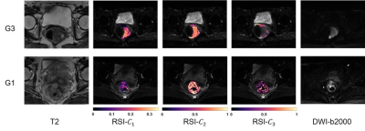 |
8 | Discriminating Rectal Cancer Grades using Restriction Spectrum Imaging
Zhongyan Xiong1, Zhijun Geng2, Shanshan Lian2, Shaohan Yin2, Guixiao Xu2, Yunfei Zhang3, Yongming Dai3, Jing Zhao2, Lidi Ma2, Xin Liu1, Hairong Zheng1, Chuanmiao Xie2, and Chao Zou1
1Paul C. Lauterbur Centre for Biomedical Imaging, Shenzhen Institutes of Advanced Technology, Chinese Academy of Sciences, Shenzhen, China, 2Department of Radiology, Sun Yat-sen University Cancer Center, State Key Laboratory of Oncology in Southern China, Guangzhou, China, 3Central Research Institute, United Imaging Healthcare, Shanghai, China
Restriction spectrum imaging (RSI) is a novel diffusion model that captures the distinct diffusion behavior of tumors. It separates water diffusion into several microscopic compartments. The restricted compartment correlating to the tumor cellularity is expected to be a potential indicator of rectal cancer aggressiveness. To assess the ability of RSI model for rectal tumor grading, we applied a three-compartment RSI model to DWI images of patients with different histopathological grades of rectal cancer. The RSI model demonstrated its ability to discriminate the rectal cancer of low and high grades, and the results outperforms the traditional ADC model and DKI model.
|
||
4751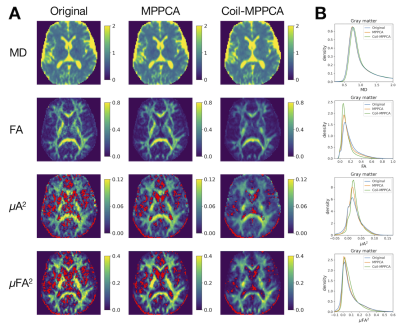 |
9 | MPPCA denoising before GRAPPA reconstruction improves the precision of microscopic anisotropy in the gray matter
Kouhei Kamiya1,2,3, Issei Fukunaga1, Syo Murata1,4, Tomoko Maekawa1, Shimpei Kato1,3, Katsutoshi Murata5, Thorsten Feiweier6, Koji Kamagata1, Masaaki Hori1,2, and Shigeki Aoki1
1Department of Radiology, Juntendo University, Tokyo, Japan, 2Department of Radiology, Toho University, Tokyo, Japan, 3Department of Radiology, The University of Tokyo, Tokyo, Japan, 4Department of Radiological Sciences, Komazawa University, Tokyo, Japan, 5Siemens Healthcare K.K., Tokyo, Japan, 6Siemens Healthcare GmbH, Erlangen, Germany
The ability of double diffusion encoding to estimate tissue microscopic anisotropy has gained increasing attention in clinical studies. However, the estimation of smaller values of microscopic anisotropy is known to be less precise, posing a challenge for clinical translation because many tissues of interest including the gray matter exhibit smaller values. In this study, we adopted the recently proposed denoising strategy where Marcenko‐Pastur principal component analysis (MPPCA) is applied to coil data before GRAPPA reconstruction. The results suggested this strategy is effective for improving the scan-rescan repeatability of microscopic anisotropy in the gray matter.
|
||
4752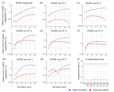 |
10 | An efficient approach for fast signal computation of restricted diffusion with arbitrary gradient waveforms
Diwei Shi1, Sisi Li2, Li Chen1, Quanshui Zheng1, Hua Guo2, and Junzhong Xu3,4,5,6
1Center for Nano and Micro Mechanics, Department of Engineering Mechanics, Tsinghua University, Beijing, China, 2Center for Biomedical Imaging Research, Tsinghua University, Beijing, China, 3Institute of Imaging Science, Vanderbilt University Medical Center, Nashville, TN, United States, 4Vanderbilt University Medical Center, Nashville, TN, United States, 5Department of Physics and Astronomy, Vanderbilt University, Nashville, TN, United States, 6Department of Biomedical Engineering, Vanderbilt University, Nashville, TN, United States Quantitative microstructural imaging based on diffusion MRI usually replies on some simple gradient waveforms, with which analytical expressions can be derived e.g., for fitting cell size. However, it is challenging for this approach for modified irregular gradient waveforms that are increasingly used. Inspired by Callaghan’s matrix formalism, we propose an efficient approach for signal computation with arbitrary gradient waveforms. It can accelerate computation by three orders of magnitude with maintained accuracy, making it feasible in practical data fittings. This work paves the way for quantitative microstructural imaging with arbitrary diffusion gradient waveforms in practice.
|
||
4753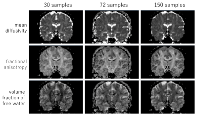 |
11 | Single-Shell Free Water Imaging by Synthetic Q-Space Learning
Yoshitaka Masutani1 and Koh Sasaki1,2
1Graduate School of Information Sciences, Hiroshima City University, Hiroshima, Japan, 2Hiroshima Heiwa Clinic, Hiroshima, Japan
Free water imaging (FWI) belongs to the dMRI family, and is an extention of the DTI model by adding the isotropic diffusion compartment. Conventionally, FWI parameters have been obtained by numerical fitting to measured signal values of DWI of single-shell or multi-shell. It has been reported that it is harder to obtain robust results in single shell data. Recently, machine learning techniques have shown promising results in dMRI parameter inference. In this study, we aimed at FWI parameter inference from single-shall dMRI data by using synthetic Q-space learning. Several validation experiments by quantitative and visual assessments were performed.
|
||
4754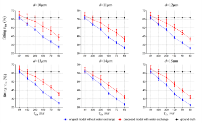 |
12 | A general framework to incorporate transcytolemmal water exchange in diffusion MRI-based microstructural imaging
Sisi Li1, Diwei Shi2, Li Chen2, Quanshui Zheng2, Hua Guo1, and Junzhong Xu3,4,5,6
1Center for Biomedical Imaging Research, Tsinghua University, Beijing, China, 2Center for Nano and Micro Mechanics, Department of Mechanics Engineering, Tsinghua University, Beijing, China, 3Institute of Imaging Science, Vanderbilt University Medical Center, Nashville, TN, United States, 4Department of Radiology and Radiological Sciences, Vanderbilt University Medical Center, Nashville, TN, United States, 5Department of Biomedical Engineering, Vanderbilt University, Nashville, TN, United States, 6Department of Physics and Astronomy, Vanderbilt University, Nashville, TN, United States Quantitative microstructural imaging based on diffusion MRI provides a non-invasive means to measure microstructural parameters, e.g., cell size. However, due to the remarkable complexity of incorporating transcytolemmal water exchange into diffusion biophysical models, water exchange is usually ignored. This leads to biased estimation of microstructural parameters, particularly intracellular volume fraction. Here, we propose a new approach to incorporate water exchange naturally in diffusion biophysical models with arbitrary gradient waveforms, making it possible to fit cell size, density, and intracellular water lifetime simultaneously. This establishes a general framework to incorporate transcytolemmal water exchange in any diffusion MRI-based microstructural imaging models.
|
||
4755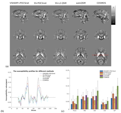 |
13 | Deep-Learning-regularized Single-step Quantitative Susceptibility Mapping (QSM) Quantification
Zuojun Wang1, Henry Ka-Fung Mak1, and Peng Cao1
1Department of Diagnostic Radiology, The University of HongKong, Hong Kong, China
We develop a deep-learning-regularized single-step QSM quantification to generate QSM directly from the total phase map. A deep-learning-regularized dipole inversion network, named POCSnet, was deployed to a single-step QSM (SS-POCSnet) network, which combined a variable-SHARP (VSHARP) and the POCSnet. Meanwhile, SS-POCSnet showed improved accuracy compared with conventional single-step QSM methods. We also demonstrated the generalizability of SS-POCSnet on different datasets in vivo.
|
||
4756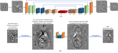 |
14 | A deep learning dipole inversion method for QSM of arbitrary head orientation and image resolution
Zhuang Xiong1, Yang Gao1, Steffen Bollmann1, and Hongfu Sun1
1School of ITEE, the University of Queensland, Brisbane, Australia
Due to the intrinsic data-driven property, many existing deep learning QSM methods can only be applied to local field maps with FOV orientation and image resolution consistent with the training data. This work proposes a novel and robust deep learning approach to reconstruct QSM of arbitrary head orientation and image resolution. Experiments are conducted on both simulated and in vivo human brain data to verify the proposed approach.
|
||
4757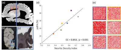 |
15 | Microstructural Basis of Fractional Anisotropy Difference Associated with b-value in Diffusion MRI
Junye Yao1, Zihan Zhou1, Benjamin C. Tendler2, Karla L. Miller2, Hao Lei3, Lei Zhang4,5, Aimin Bao4,5, Jianhui Zhong1,6, and Hongjian He1
1Center for Brain Imaging Science and Technology, College of Biomedical Engineering and Instrumental, Zhejiang University, Hangzhou, China, 2Wellcome Centre for Integrative Neuroimaging, FMRIB, Nuffield Department of Clinical Neurosciences, University of Oxford, London, United Kingdom, 3State Key Laboratory of Magnetic Resonance and Atomic and Molecular Physics, Innovation Academy for Precision Measurement Science and Technology, Chinese Academy of Sciences, Wuhan, China, 4Department of Neurology in Second Affiliated Hospital, Key Laboratory of Medical Neurobiology of Zhejiang Province, and Department of Neurobiology, Zhejiang University, Hangzhou, China, 5National Human Brain Bank for Health and Disease, School of Brain Science and Brain Medicine, Zhejiang University, Hangzhou, China, 6Department of Imaging Sciences, University of Rochester, Rochester, NY, United States
The microstructural basis of b-value dependence on FA was investigated in the human brain, using in vivo MRI and a formalin-fixed human brain hemisphere. Consistent decreases of FA with increasing b-values were observed both in vivo and ex vivo. The percentage difference of FA (pdFA) between high and low b-value datasets was found to increase with neurite density estimated from NODDI, which was further validated using histology and a multi-compartment model simulation. Results indicate that the b-value dependence of FA can be explained by compartmental differences between intra- and extra-neurites.
|
||
 Back to Meeting Home
 Back to the Program-at-a-Glance
The International Society for Magnetic Resonance in Medicine is accredited by the Accreditation Council for Continuing Medical Education to provide continuing medical education for physicians.
 Back to Meeting Home
 Back to the Program-at-a-Glance
 View the Presentation