Online Gather.town Pitches
White Matter & Nervous System III
Joint Annual Meeting ISMRM-ESMRMB & ISMRT 31st Annual Meeting • 07-12 May 2022 • London, UK

Online Gather.town Pitches
White Matter & Nervous System III
| Booth # | ||||
|---|---|---|---|---|
3354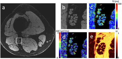 |
1 | Comparison of high-resolution quantitative peripheral nerve imaging in CMT1A and healthy controls using Double-Echo in Steady-State at 7 Tesla
Bragi Sveinsson1,2, Robert Barry1,2,3, Olivia Rowe1,2, Jason Stockmann1,2, Daniel Park1, Peter Lally4, Matthew S Rosen1,2,5, and Reza Sadjadi6
1Radiology, A. A. Martinos Center, Massachusetts General Hospital, Boston, MA, United States, 2Radiology, Harvard Medical School, Boston, MA, United States, 3Harvard-MIT Health Sciences and Technology, Cambridge, MA, United States, 4Brain Sciences, Imperial College London, London, United Kingdom, 5Physics, Harvard University, Cambridge, MA, United States, 6Neurology, Massachusetts General Hospital, Boston, MA, United States
The ability to estimate neurological disease progression based on objective quantitative measurements could be of great value for assessing treatment efficacy. Here, we demonstrate the feasibility of acquiring high-resolution anatomic and quantitative images at 7T in the sciatic, tibial, and fibular nerves, allowing quantitative assessment of individual fascicles. We also measure the quantities of fascicle cross-sectional area, T2, ADC, and fat fraction for CMT1A patients and healthy controls and investigate any between-group differences.
|
||
3355 |
2 | Pain and the NeuroMatrix in the Brain: Feasibility and Safety Study of 3T DWI in a Chronic Low Back Pain Patient Treated with Spinal Cord Stimulation
Isaiah Ailes1, KiChang Kang1, Anish Sathe1, Jingya Miao1, Leonard A. Frizon2, Laura Krisa1, Kristen Fleming1, Peter Natale1, Kiran Talekar1, Devon Middleton1, Feroze Mohamed1, Ashwini Sharan1, and Mahdi Alizadeh1
1Thomas Jefferson University, Philadelphia, PA, United States, 2Faculdades Pequeno Príncipe, Curitiba, Brazil
Spinal cord stimulation (SCS) in patients with failed back surgery syndrome aims to reduce neuropathic pain. However, minimal studies show a safe procedure using 3T diffusion weighted imaging (DWI) MRI for patients with SCS. Additionally, more studies looking at the neurological SCS treatment responses can lead to better optimization of SCS. It was our objective to safely develop a systematic procedure for 3T DWI MRI while also generating a map of the neural network connectivity in structures associated with pain. Long term application of this protocol aims to provide an objective assessment to SCS treatment.
|
||
3356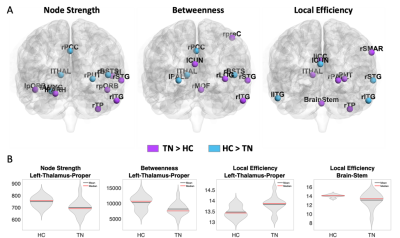 |
3 | Aberrant Structural Brain Network Hierarchy in Trigeminal neuralgia
Yael Jacob1, Gaurav Verma1, Judy Alper1, Bradley Delman1, Alan Seifert1, Raj Shrivastava1, and Priti Balchandani1
1Icahn School of Medicine at Mount Sinai, New York, NY, United States
Trigeminal neuralgia (TN) is a physically and mentally incapacitating disorder characterized by extreme, sporadic, sudden facial pain. TN is associated with the trigeminal sensory pathway, however, the etiology of TN remains unclear. Implementing a graph-theory analysis using diffusion MRI we tested whether whole brain network structural connectivity hierarchies differentiate between TN patients and healthy controls. We found aberrant centrality measures of various brain regions in TN. Specifically, this whole-brain data-driven network analysis was able to depict two major regions of the trigeminal sensory circuit, the brainstem and thalamus, and highlight their dysfunction role in the whole brain network.
|
||
3357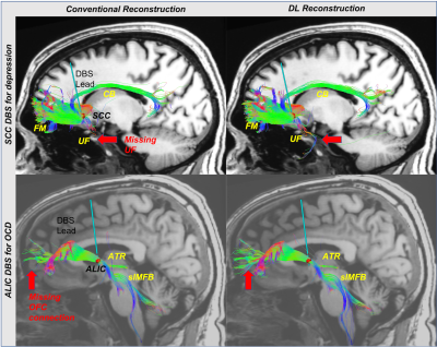 |
4 | Evaluation of the efficacy of a Deep Learning-based Reconstruction in the Connectomic Deep Brain Stimulation
Ki Sueng Choi1, Martijn Figee2, Robert Marc Lebel3, Maggie Fung4, Suchandrima Banerjee5, Helen S Maybeg6, and Jaemin Shin4
1Radiology / Neurosurgery, Icahn School of Medicine at Mount Sinai, New York, NY, United States, 2Psychiatry, Icahn School of Medicine at Mount Sinai, New York, NY, United States, 3GE Healthcare, Alberta, AB, Canada, 4GE Healthcare, New York, NY, United States, 5GE Healthcare, Menlo Park, CA, United States, 6Neurology, Icahn School of Medicine at Mount Sinai, New York, NY, United States
The connectomic DBS approach, stimulation tractographically defined white mater pathways, has been successfully employed in functional neurosurgery, and it demonstrated the feasibility of clinical utility. However, this approach is limited in the clinical environment due to low SNR and various artifacts of DWI. The recent development of deep learning-based MR reconstruction allows us to improve SNR and reduce artifacts. This study evaluated the DL reconstruction method in the field of connectomic DBS using deterministic and probabilistic tractography. Tractography results from DL reconstruction show higher sensitivity for delineating WM pathways in specific DBS targets.
|
||
3358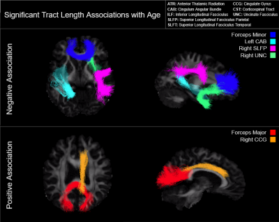 |
5 | Age-related differences in white matter tract length assessed via TRACULA
Tyler Robinson1, Paul Chang1, and Jean Chen1
1Rotman Research Institute, Baycrest, Toronto, ON, Canada
We examined differences in median tract length with age across eighteen major white matter tracts in 541 subjects of the Human Connectome Project in Aging. We observed a significant negative age effect on tract length in the forceps minor, left cingulum angular bundle, right superior longitudinal fasciculus parietal, and right uncinate fasciculus. Paradoxically, we also report a significant positive age effect on tract length in the forceps major and right cingulate gyrus. Restricted analyses suggest that these effects are driven by female subjects.
|
||
3359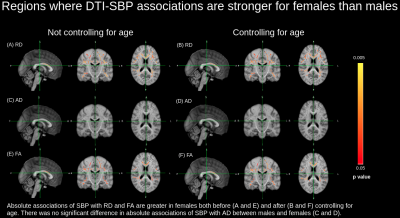 |
6 | Are vascular risk factors associated with white matter health equally between men and women?
Arjun Patel1, Jordan A. Chad1,2, and J. Jean Chen1,2
1Rotman Research Institute, Baycrest Health Sciences, Toronto, ON, Canada, 2Department of Medical Biophysics, University of Toronto, Toronto, ON, Canada
White matter microstructural degeneration is an early marker of declining brain health, and vascular risk factors such as high blood pressure and adipose fat deposition have been found to be significantly associated with white matter microstructural degeneration in healthy adults. In this study, using diffusion tensor imaging, we find that the vascular-risk influences on white matter are more pronounced in women than in men. This study therefore calls attention to the consideration of sex differences in studying brain vulnerability to vascular risk factors.
|
||
3360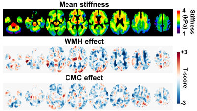 |
7 | Associations between vascular health and brain stiffness
Matthew C Murphy1, Prashanthi Vemuri1, Matthew L Senjem1, Clifford R Jack, Jr.1, Richard L Ehman1, and John Huston, III1
1Mayo Clinic, Rochester, MN, United States
Vascular health is a predictor of cognitive outcomes, but the relationship between the two remains incompletely understood. Here we tested the hypothesis that brain stiffness is significantly associated with vascular health as assessed by white matter hyperintensity (WMH) load and the presence of systemic risk factors. Through voxel-wise mapping, WMH was associated with decreased stiffness in periventricular regions, while systemic risk factors were associated with widespread, lower-amplitude stiffness decreases. Examining the partial correlations between stiffness, WMH and vascular risk factors, stiffness was significantly associated with both measures, while WMH and vascular risk were not significantly correlated after controlling for stiffness.
|
||
3361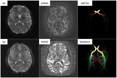 |
8 | Initial Clinical Experience with MAGNUS Ultra-High-Performance Gradient Coil for Diffusion Microstructure Imaging of Intracranial Pathology
Robert Shih1,2, Ante Zhu3, J Kevin DeMarco1,2, H Douglas Morris1,2, Maureen Hood1,2, Nastaren Abad3, Radhika Madhavan3, Luca Marinelli3, Thomas Foo3, and Vincent Ho1,2
1Uniformed Services University, Bethesda, MD, United States, 2Walter Reed National Military Medical Center, Bethesda, MD, United States, 3GE Research, Niskayuna, NY, United States The MAGNUS ultra-high-performance gradient coil delivers simultaneous 200 mT/m and 500 T/m/s performance on each axis, with higher PNS thresholds than whole-body gradient coils, which is particularly useful for diffusion microstructure imaging. Our initial clinical experience with the MAGNUS research scanner has successfully identified white matter abnormalities (? intra-axonal edema) using multi-shell DTI (bmax = 4000 s/mm2) and OGSE (fmax = 100 Hz) in an acute symptomatic mTBI subject. It has also successfully identified differences in intracellular volume fraction using multi-shell multi-frequency OGSE (bmax = 2000 s/mm2, fmax = 100 Hz) between a low-grade diffuse astrocytoma and a high-grade glioblastoma. |
||
3362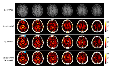 |
9 | Myelin Water Imaging using Dimensionality Reduction Video Not Available
Jae Eun Song1, Shreyas Vasanawala2, and Dong-Hyun Kim3
1Radiology, Stanford University, Stanford, CA, United States, 2Department of Radiology, Stanford University, Stanford, CA, United States, 3Department of Electrical and Electronic Engineering, Yonsei University, Seoul, Korea, Republic of
Multi-echo gradient-echo (mGRE)-based myelin water fraction (MWF) mapping is a promising myelin water imaging (MWI) modality but is vulnerable to noise and artifact corruption. The linear dimensionality reduction (LDR) method has recently shown improvements with regard to these challenges. However, the magnitude value based low rank operators have been shown to misestimate the MWF for regions with T2* anisotropy. This paper presents a nonlinear dimensionality reduction (NLDR) method to estimate the MWF map better by encouraging nonlinear low dimensionality of mGRE signal sources.
|
||
3363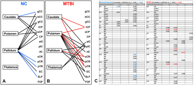 |
10 | Unique Alterations in the Pattern of Iron Deposition in Deep Gray Matter Relative to White Matter Microstructure in Mild Traumatic Brain Injury
Sohae Chung1, Els Fieremans1, Dmitry S. Novikov1, Prin X. Amorapanth2, Joseph F. Rath2, Steven R. Flanagan2, and Yvonne W. Lui1
1Department of Radiology, NYU Grossman School of Medicine, New York, NY, United States, 2Department of Rehabilitation Medicine, NYU Grossman School of Medicine, New York, NY, United States
Primary and secondary injury are both believed to play important roles in the pathogenesis of disease after mild traumatic brain injury (MTBI). Here we investigate the relationships between white matter microstructure and deep gray matter iron deposition after MTBI, which may shed light on primary WM injuries and potential secondary changes in brain iron. Our results show different patterns of correlation between deep gray matter iron content as measured by QSM and WM microstructure as measured by diffusion MRI in MTBI compared with normal controls.
|
||
 Back to Meeting Home
 Back to the Program-at-a-Glance
The International Society for Magnetic Resonance in Medicine is accredited by the Accreditation Council for Continuing Medical Education to provide continuing medical education for physicians.
 Back to Meeting Home
 Back to the Program-at-a-Glance
 View the Presentation