Online Gather.town Pitches
Pediatrics, Normal Development & Aging I
Joint Annual Meeting ISMRM-ESMRMB & ISMRT 31st Annual Meeting • 07-12 May 2022 • London, UK

Online Gather.town Pitches
Pediatrics, Normal Development & Aging I
| Booth # | ||||
|---|---|---|---|---|
 |
3775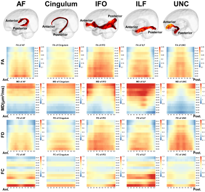 |
1 | Developmental pattern of association fibers and their interaction with associated cortical microstructures in 0-5 month-old infants
Tingting Liu1, Yuqing You2, Zhiyong Zhao1, Zuozhen Cao1, Ruike Chen1, Mingyang Li1, Ying Lv3, Mingyan Li3, Fusheng Gao2, Hongxi Zhang2, Chai Ji3, and Dan Wu1
1Key Laboratory for Biomedical Engineering of Ministry of Education, Department of Biomedical Engineering, College of Biomedical Engineering & Instrument Science, Zhejiang University, Hangzhou, China, 2Department of Radiology, Children's Hospital, Zhejiang University School of Medicine, Hangzhou, China, 3Department of Child Health, Children's Hospital, Zhejiang University School of Medicine, Hangzhou, China
The present study aimed to investigate the spatiotemporal developmental pattern of association fibers in infants aged 0-5 months and the interaction between these fibers and the associated cortex utilizing FBA based on HARDI data. We found that the C-shaped fibers demonstrate an approximately symmetrical along-track pattern with more advanced development in the middle segments than the extremities; the horizontally oriented fibers manifest that the anterior segments started later but developed faster than the posterior segments. Mediation analysis revealed the mediation effect of cortical GM on the development of WM was more prominent than that of WM on GM.
|
|
3776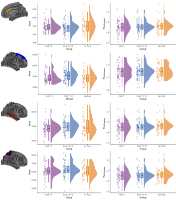 |
2 | Effects of low-moderate prenatal alcohol exposure on brain development in childhood
Claire Kelly1,2,3, Thijs Dhollander2, Evelyne Muggli1,4, Jane Halliday4,5, Elizabeth Elliott6,7, Anthony Penington4,8, Alicia Spittle1,9, Della A Forster10,11, Sharon Lewis4,5, Stephen Hearps12, Deanne K Thompson1,2,4, and Peter J Anderson1,3
1Victorian Infant Brain Studies (VIBeS), Murdoch Children's Research Institute, Melbourne, Australia, 2Developmental Imaging, Murdoch Children's Research Institute, Melbourne, Australia, 3Turner Institute for Brain and Mental Health, School of Psychological Sciences, Monash University, Melbourne, Australia, 4Department of Paediatrics, The University of Melbourne, Melbourne, Australia, 5Reproductive Epidemiology, Murdoch Children's Research Institute, Melbourne, Australia, 6Child and Adolescent Health, Faculty of Medicine and Health, The University of Sydney, Sydney, Australia, 7Sydney Children's Hospitals Network, Westmead, Sydney, Australia, 8Royal Children’s Hospital, Melbourne, Australia, 9Department of Physiotherapy, The University of Melbourne, Melbourne, Australia, 10The Royal Women's Hospital, Melbourne, Australia, 11Judith Lumley Centre, La Trobe University, Melbourne, Australia, 12Brain and Mind, Murdoch Children's Research Institute, Melbourne, Australia
Effects of low-moderate prenatal alcohol exposure (PAE) on brain development have been infrequently studied. In a general antenatal population cohort, we compared brain structure between 6-8-year-old children with no PAE (n=41), PAE during trimester 1 (n=44), and PAE throughout gestation (n=58). Most brain regions did not differ between groups, however the caudal anterior cingulate cortex area, and striato-cortical tract cross-sectional area, were significantly smaller in the group exposed to alcohol throughout gestation relative to the other groups. Overall, low-moderate PAE is not strongly associated with brain structure, except low-moderate PAE throughout gestation is associated with specific brain regional alterations.
|
||
3777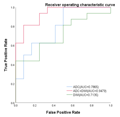 |
3 | Diffusion weighted imaging-Based Radiomic Features of Distincting Low and High Grade of Pediatric Brain Tumors Video Permission Withheld
TING YI1, Qiuhong MA1, QIFANG CAI1, Huiting Zhang2, Weian Wei1, Bing Gao1, and KE JIN1
1Hunan Children's Hospital, Changsha, China, 2Siemens Healthineers, Wuhan, China
This study investigated the feasibility of diffusion weighted imaging (DWI)-Based radiomic features in differential diagnosis of low- and high-grade pediatric brain tumors. The results showed that the DWI and apparent diffusion coefficient (ADC) combined model can effectively distinct low- and high-Grade pediatric brain tumors. This suggests that DWI sequence may facilitate brain tumor grading.
|
||
3778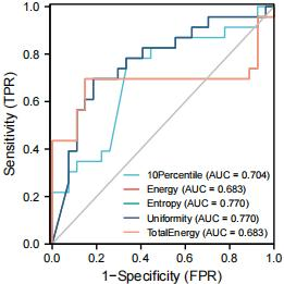 |
4 | The application value of T2 weight imaging histogram in assessing the Placenta Accreta
songhong Yue1, jie Li1, fengxian Fan1, haoyuan Li1, xiao Ling1, yupin Bai1, Kai A2, and jing Zhang1
1Lanzhou University second hospital, lanzhou, China, 2Philips Healthcare, Xian, China
Accurate evaluation of placenta accreta before parturition can effectively avoid adverse maternal and infantile outcomes. Based on the T2 weighted imaging (T2WI) histogram, this study analyze the placenta quantitatively. We compared the difference in T2WI histogram parameters between the placenta accreta group (23 patients) and the non-placenta accreta group (27 patients) to evaluate the diagnostic efficacy. Among these parameters, the difference of 10 Percentile, Energy, Entropy, Total Energy, Uniformity was statistically significant (P<0.05). Uniformity and Entropy have the largest area under the curve (AUC). We speculate T2WI based histogram is useful for noninvasive assessment of placental implantation.
|
||
3779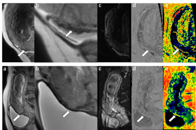 |
5 | Intravoxel Incoherent Motion MR Imaging-based Virtual Elastography for the Assessment of Placenta Accreta Video Not Available
jiaojiao lu1, ting liu1, junjun li1, xianjun li1, and jian yang1
1Department of Radiology, the first Affiliated Hospital of Xi'an Jiaotong University, xi'an, China
Placenta accreta may lead to life-threatening complications and is difficult to distinguish by traditional MRI sequences. In this study, IVIM-based virtual elastography was used for the first time to detect the stiffness of the placenta, and it was found that the virtual (IVIM) stiffness values for the AP-ROIs were mostly higher than those for the NP-ROIs; the IR-ROIs were also significantly higher than the NIR-ROIs. Thus, the implanted regions of the placenta could be significantly stiffer than those of the non-implanted regions. That is helpful for adding a new method for clinical diagnosis of placenta accreta.
|
||
3780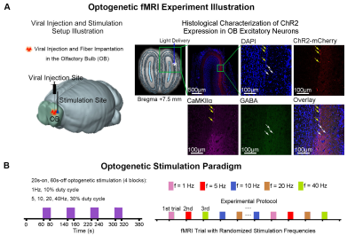 |
6 | Dissecting Brain-wide Olfactory Dysfunctions in Accelerated Aging Rodent Model
Teng Ma1,2,3, Xunda Wang1,2, Linshan Xie1,2, Pit Shan Chong4, Venice Sin1,2, Peng Cao3, Pek-Lan Khong3, Lee Wei Lim4, Alex T. L. Leong1,2, and Ed. X Wu1,2,4
1Laboratory of Biomedical Imaging and Signal Processing, The University of Hong Kong, Hong Kong SAR, China, 2Department of Electrical and Electronic Engineering, The University of Hong Kong, Hong Kong SAR, China, 3Department of Diagnostic Radiology, Li Ka Shing Faculty of Medicine, The University of Hong Kong, Hong Kong SAR, China, 4School of Biomedical Sciences, Li Ka Shing Faculty of Medicine, The University of Hong Kong, Hong Kong SAR, China
The olfactory system plays a pivotal role in driving behavioral responses that are critical to survival. In particular, the decline in ability to detect and discriminate odors in aged humans lead to an overall decrease in quality of life. However, our present understanding of olfactory dysfunction in aging brains beyond the cellular and micro-circuit level is scarce and incomplete. In this study, we deployed optogenetic fMRI to reveal the changes of brain-wide odor-associated regions brought about by aging in an accelerated aging rat model. We found diminished activations brain-wide indicating dysfunction at the systems level across multiple long-range olfactory pathways.
|
||
3781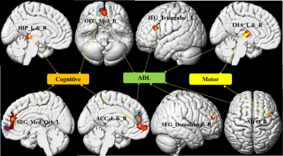 |
7 | Gray Matter Atrophy and Neurological Deficits in Patients with Carbon Monoxide Poisoning
Yanli Zhang1, Tianhong Wang2, Shuaiwen Wang2, Yuling Gao2, Shaoyu Wang3, Shunlin Guo2, and Junqiang Lei2
1The first hospital of lanzhou university, Lanzhou, China, 2The first hospital of Lanzhou university, Lanzhou, China, 3MR scientific marketing, Siemens Healthineers, Xi an, China
White matter damage in patients with carbon monoxide (CO) poisoning have been extensively reported in previous studies, but neurological deficits-associated gray matter volume (GMV) changes are not well explored. Forty-one CO poisoning patients and 36 healthy controls were enrolled in this study. All subjects underwent 3D T1WI and cognition and motor function assessment. GMV change patterns of patients were explored using Voxel-based morphometry processing. This study would provide a simple neuroimaging biomarker to identify the brain anatomical signatures underlying neurological deficits in CO poisoning, and thus facilitate the ongoing effort of knowing of the neuropathologic mechanism happened in these patients.
|
||
3782 |
8 | Virtual Stiffness of Gray Matter in Normal Adult Brain Assessed by Virtual Magnetic Resonance Elastography Video Not Available
Chenyue Liu1, Chunxu Da1, Jian Yang1, and Xianjun Li1
1The First Affiliated Hospital of Xi’an Jiaotong University, Shaanxi, Xi'an, China The mechanical properties of biological tissue provide information related to the strength and integrity of the cellular microstructure. Recently, mechanical properties of the brain have been visualized and measured non-invasively with magnetic resonance elastography (MRE). Virtual MRE (VMRE) is based on non-Gaussian distribution model, which can be regarded as an indicator to reflect the complexity and change of different brain microstructure. The purpose of this study is to explore the capability of diffusion–based VMRE in the characterization of the gray matter shear stiffness, discuss the cause of the differences of gray matter encephalic in normal human brains.
|
||
3783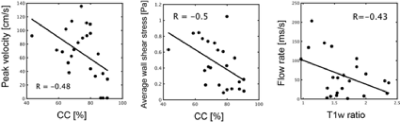 |
9 | Evaluation of Blood Flow and Plaque Vulnerability in Carotid Artery Stenosis Focusing on Morphological and Component Characteristics
Yuki Matsumoto1, Yuki Kanazawa1, Yuki Kinjo2, Masafumi Harada1, Tosiaki Miyati3, Hiroaki Hayashi3, Mitsuharu Miyoshi4, Naoki Maeda2, Yasuhisa Kanematsu5, Yasushi Takagi5, and Akihiro Haga1
1Institute of Biomedical Sciences, Tokushima University Graduate School, Tokushima, Japan, 2Graduate school of Health Science, Tokushima University, Tokushima, Japan, 3Faculty of Health Sciences, Institute of Medical, Pharmaceutical and Health Sciences, Kanazawa University, Kanazawa, Japan, 4Global MR Applications and Workflow, GE Healthcare Japan, Hino-city, Japan, 5Department of Neurosurgery, Tokushima University Graduate School of Biomedical Sciences, Tokushima, Japan
Our purpose was to assess plaque vulnerability under the influence of blood flow in terms of morphological and component characteristics using MRI.Twenty-three patients with symptomatic and asymptomatic carotid stenosis, all of whom provided informed consent, underwent 3T MRI scan (GE Healthcare). We evaluated the consistency between the flow patterns and plaque features in the carotid artery stenosis, using 4D-flow MRI and 3D-FSE-MSDE sequences. During the vulnerability analysis, it was observed that there was a negative correlation between 3D-FSE-MSDE and 4D-flow parameter (R = -0.43, P < 0.001).
|
||
3784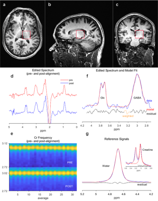 |
10 | Sex-related Changes in GABA+ Signaling in the Right Thalamus in Patients with Knee Osteoarthritis through Acupuncture Stimulation
Nan Gao1, Sheng Hu1,2, Xiaoxiao Wang1, and Bensheng Qiu1
1Hefei National Laboratory for Physical Sciences at the Microscale and Center for Biomedical Engineering, University of Science and Technology of China, Hefei, China, 2School of Medical Information Engineering, Anhui University of Chinese Medicine, Hefei, China This study aimed to investigate GABA variation between KOA patients and healthy people without therapy and how GABA changes through acupuncture stimulation. MEGA-PRESS sequence was used to detect GABA signaling in the right thalamus before and after acupuncture stimulation on EX-LE5 acupoint of the left leg. Our study demonstrated a sex-related GABA+/Cr difference in patients with KOA chronic pain, proving the quantitative sex differences in human pain processing. Moreover, the therapeutic effect of acupuncture stimulation was positively proved and quantified by measuring GABA+/Cr. |
||
3785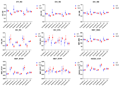 |
11 | Striatum-thalamus microstructural alterations in levodopa-induced dyskinesia using four advanced diffusion models Video Not Available
Tingting Yuan1, Ying Zhang2, Hongchao Wang1, Yueluan Jiang3, Chenglong Wang4, Chunjie Guo1, Guang Yang4, and Huiting Zhang3
1Department of Radiology, The First Hospital of Jilin University, Changchun, China, 2Department of Neurology and Neuroscience Center, The First Hospital of Jilin University, Changchun, China, 3MR Scientific Marketing, Siemens Healthineers, Beijing, China, 4Shanghai Key Laboratory of Magnetic Resonance, East China Normal University, Shanghai, China
The major complication of dopaminergic therapy in Parkinson’s disease (PD) is dyskinesia which affects patients' quality of life. The pathogenesis of levodopa-induced dyskinesia (LID) remains unclear. In this study, we aimed to investigate striatum-thalamus alterations in PD patients with and without LID using advanced diffusion magnetic resonance imaging (dMRI) techniques (DTI, DKI, NODDI and MAP).
|
||
3786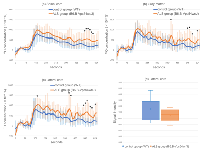 |
12 | Increased Vascular Permeability of 17O-labeled Water in the Amyotrophic Lateral Sclerosis Model Mice using Indirect Proton MRI
Hiroyuki Kameda1,2, Yuji Komaki3, and Kohsuke Kudo4,5
1Department of Diagnostic and Interventional Radiology, Hokkaido University Hospital, Sapporo, Japan, 2Faculty of Dental Medicine, Department of Radiology, Hokkaido University, Sapporo, Japan, 3Live Imaging Center, Central Institute for Experimental Animals, Kawasaki, Japan, 4Department of Diagnostic Imaging, Hokkaido University Graduate School of Medicine, Sapporo, Japan, 5Global Center for Biomedical Science and Engineering, Hokkaido University Faculty of Medicine, Hokkaido University, Sapporo, Japan
We evaluated the increased vascular permeability of 17O-labeled Water in the amyotrophic lateral sclerosis (ALS) mice using indirect proton MRI. The spinal cord of nine ALS mice and ten wild-type (WT) mice were scanned with 7T MRI with the T2-weighted RARE sequence. In the femoral vein, 17O-labeled water was intravenously injected. In ROIs of the spinal cord, the 17O concentration increased more in ALS mice than in the control mice. This latest method can detect the abnormalities in water kinetics probably caused by the impairments in blood–spinal cord barrier of ALS mice.
|
||
 |
3787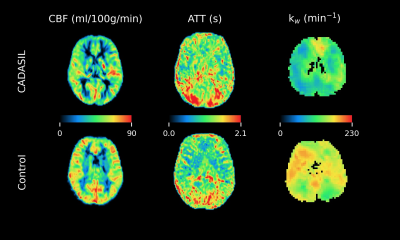 |
13 | Pseudo-continuous arterial spin labeling reveal altered cerebral hemodynamics and blood-brain barrier dysfunction in CADASIL patients
Jinyuan Zhang1,2,3, Zihao Zhang1,2,3, Chen Ling4,5, Xingfeng Shao6, Zhixin Li1,2,3, Huilou Liang1,2,3, Jing An7, Qi Yang8, Rong Xue1,2,3, Yan Zhuo1,2,3, Zhaoxia Wang4,5, Yun Yuan4,5, and Danny JJ Wang6
1State Key Laboratory of Brain and Cognitive Science, Institute of Biophysics, Chinese Academy of Sciences, Beijing, China, 2University of Chinese Academy of Sciences, Beijing, China, 3The Innovation Center of Excellence on Brain Science, Chinese Academy of Sciences, Beijing, China, 4Department of Neurology, Peking University First Hospital, Beijing, China, 5Beijing Key Laboratory of Neurovascular Disease Discovery, Peking University First Hospital, Beijing, China, 6Mark & Mary Stevens Neuroimaging and Informatics Institute, Keck School of Medicine, University of Southern California, Los Angeles, CA, United States, 7Siemens Shenzhen Magnetic Resonance Ltd., Shenzhen, China, 8Department of Radiology, Chaoyang Hospital, Capital Medical University, Beijing, China Cerebral autosomal dominant arteriopathy with subcortical infarcts and leukoencephalopathy (CADASIL) is an inherited form of cerebral small vessel disease (cSVD). In this study, we investigated the changes of cerebral blood flow (CBF), arterial transit time (ATT) and water exchange rate (kw) measured by multi-delay pseudo-continuous arterial spin labeling (pCASL) and diffusion prepared pCASL. Increased ATT and decreased kw were found in CADASIL patients compared with healthy controls. The results indicated modified cerebral hemodynamics and dysfunction of blood-brain barrier in CADASIL patients. MD-pCASL and DP-pCASL are promising for evaluating hemodynamics and BBB function of cSVD. |
|
3788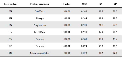 |
14 | 3D texture analyses of quantitative susceptibility maps to differentiate patients with Wilson’s disease from healthy controls
Gaiying Li1, Rong Wu2, Yasong Du3, Yang Song4, Yi Wang5, Xiaoping Wang2, and Jianqi Li1
1Shanghai Key Laboratory of Magnetic Resonance, School of Physics and Electronic Science, East China Normal University, Shanghai, China, 2Department of Neurology, Shanghai Tong-Ren Hospital, Shanghai Jiao Tong University School of Medicine, Shanghai, China, 3Shanghai Mental Health Center, Shanghai Jiao Tong University School of Medicine, Shanghai, China, 4MR Scientific Marketing, Siemens Healthineers, Shanghai, China, 5Department of Radiology, Weill Medical College of Cornell University, New York, NY, United States
Abnormal metal accumulation in deep gray matter (DGM) nuclei of patients with Wilson’s disease (WD) could be detected using quantitative susceptibility mapping (QSM), yet no study has quantitatively evaluated how the textures of susceptibility maps might evolve with WD. The aim of this study was to evaluate texture features extracted from susceptibility maps of DGM nuclei for differentiating WD from healthy controls (HC). The results showed that part of the texture parameters was significantly different between WD and HC, meanwhile the receiver operating characteristic curve revealed that some second-order texture parameters were more suitable and sensitive for diagnosis of WD.
|
||
3789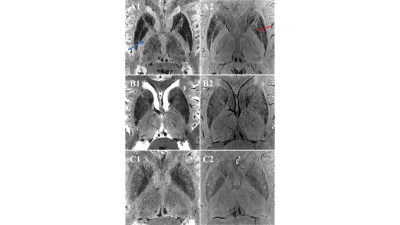 |
15 | Hyperintense globus pallidus rim sign could be a novel hallmark of Wilson disease at 7T MRI: a pilot case-control study
Su Dongning1,2, Zhang Zhe2,3, Zhang Zhijin1,2, Zhu Wanlin2,3, Sui Binbin2,3, Zhang Yingkui2,3, Bi Jingfeng2,3, Kong Qingle 3,4, Gan Yawen1,2, Yan Rui1,2, Wang Xuemei1,2, Wang Zhan1,2, Wang Yongjun 1,2,3, Wu Tao1,2,
Jing Jing1,2,3, and Feng Tao1,2
1Department of Neurology, Beijing Tiantan Hospital,Captital Medical University, Beijing, China, 2China National Clinical Research Center for Neurological Diseases, Beijing, China, 3Tiantan Neuroimaging Center of Excellence, Beijing Tiantan Hospital, Capital Medical University, Beijing, China, 4MR Collaboration, Siemens Healthineers Ltd., Beijing, China
To explore specific neuroimaging characteristics of iron deposition in WD, we conducted a small sample, prospective case-control study to compare the 7T MRI manifestation of iron deposition in the basal ganglia among PD, WD patients and healthy controls. The hyperintense globus pallidus rim sign could be a specific neuroimaging characteristic of WD in 7T T2*-weighted and SWI images, which may be related to the unique iron deposition pattern in the striatum in WD patients. Internal capsule appears to be hyperintensity with a clear border only observed in WD patients. These findings have the potential to provide diagnostic biomarkers for WD.
|
||
3790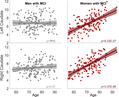 |
16 | Sex-dependent Pathological Aging Effect on Caudate Functional Connectivity in Mild Cognitive Impairment Video Permission Withheld
Zhengshi Yang1,2, Jessica Z.K. Caldwell1, Jeffrey L Cummings2, Aaron Ritter1, Jefferson W Kinney2, and Dietmar Cordes1,2,3
1Cleveland Clinic Lou Ruvo Center for Brain Health, Las Vegas, NV, United States, 2University of Nevada Las Vegas, Las Vegas, NV, United States, 3University of Colorado, Boulder, CO, United States
Resting-state fMRI was used to investigate the aging effect on caudate function in mild cognitively impaired participants, with the focus of sex-dependent effect. Graph theory analysis was conducted to derive caudate nodal strength, which was then input to linear mixed effect model to evaluate its association with age. A striking aging effect was observed only in women with MCI but not in men with MCI, which was closely related to cognitive decline in woman participants. This finding suggests that caudate may be critical for alleviating cognitive decline in women with MCI.
|
||
 Back to Meeting Home
 Back to the Program-at-a-Glance
The International Society for Magnetic Resonance in Medicine is accredited by the Accreditation Council for Continuing Medical Education to provide continuing medical education for physicians.
 Back to Meeting Home
 Back to the Program-at-a-Glance
 View the Presentation