Online Gather.town Pitches
Multiple Sclerosis, Alzheimer's & Dementia II
Joint Annual Meeting ISMRM-ESMRMB & ISMRT 31st Annual Meeting • 07-12 May 2022 • London, UK

Online Gather.town Pitches
Multiple Sclerosis, Alzheimer's & Dementia II
| Booth # | ||||
|---|---|---|---|---|
3977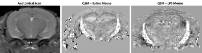 |
1 | Decrease in magnetic susceptibility correlates with reduction in cortical blood flow in a model of systemic inflammation: A 9.4T in-vivo study
Qandeel Shafqat1, Rania Muhammed1, Hongfu Sun2, Ying Wu1, A. Max Hamilton1, Mada Hashem1, and Jeff F. Dunn1
1Department of Radiology, University of Calgary, Calgary, AB, Canada, 2School of Information Technology and Electrical Engineering, University of Queensland, Brisbane, Australia
Brain hypoxia is present in multiple sclerosis, a condition associated with inflammation. Hypoxia may be related to inflammation. Quantitative susceptibility mapping (QSM) is sensitive to deoxyhemoglobin and so hypoxia. Here, we combined perfusion MRI with QSM to assess hypoxia and cerebral blood flow (CBF) in the lipopolysaccharide (LPS) inflammatory model. We report a reduction in susceptibility, as well as a significant reduction in CBF, in the cortex of the LPS model. As reduced tissue susceptibility was observed in more hypoxic subjects, this could be consistent with hypoxia in the inflammatory model, and QSM might be a possible biomarker.
|
||
3978 |
2 | Feasibility of $$$R_2^*$$$ /QSM susceptibility source separation for application in multiple sclerosis
Alexey Dimov1, Thanh Nguyen1, Kelly Gillen1, Melanie Marcille2, Pascal Spincemaille1, Pitt David3, Susan Gauthier2, and Yi Wang1
1Radiology, Weill Cornell Medicine, New York, NY, United States, 2Neurology, Weill Cornell Medicine, New York, NY, United States, 3Neurology, Yale School of Medicine, New Haven, CT, United States
For determination of dia- and paramagnetic sources, quantitative susceptibility mapping (QSM) of phase is combined with a R2* model of magnitude of multiecho gradient echo data, which is advantageous over R2’ model that requires additional T2 mapping, Magnetic source separation was performed in in vivo (17 multiple sclerosis (MS) patients) and ex vivo (a whole brain and 3 MS brain tissue blocks). Both R2* and R2’ methods were found to be well correlated in vivo data. R2* based positive and negative susceptibility sources estimation in ex vivo MS lesions correlated with optical density of lesion histology.
|
||
3979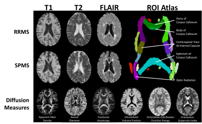 |
3 | Assessing Tissue Structure Differences Between RRMS and SPMS Patients Using Advanced Diffusion and MRI Texture Analysis
Olayinka Oladosu1, Wei-Qiao Liu2, Lenora Brown2,3, Bruce Pike2,3,4, Marcus Bruce Koch2,3, Luanne Metz2,3, and Yunyan Zhang2,3,4
1Neuroscience, University of Calgary, Calgary, AB, Canada, 2Clinical Neurosciences, University of Calgary, Calgary, AB, Canada, 3Hotchkiss Brain Institute, University of Calgary, Calgary, AB, Canada, 4Radiology, University of Calgary, Calgary, AB, Canada
Tissue structure changes underlying disease development and progression in MS are not fully understood. We investigated tissue structure in 29 RRMS and SPMS patients using advanced diffusion imaging and MRI texture measures using analyses over 3 spatial scales: white matter histogram analysis, tract ROI analysis, and along-tract statistics. We found that phase congruency texture analysis best differentiated RRMS and SPMS patients in histogram analysis. Diffusion measures differentiated the subtypes across all analyses in multiple tracts. The findings by advanced measures of tissue structure may further research into disease progression in MS.
|
||
3980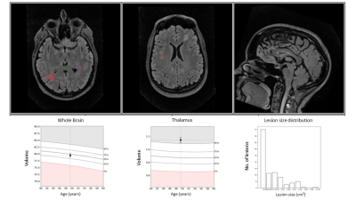 |
4 | Association Between Common MRI Metrics of Disease Activity in MS and Spinal Cord Atrophy
Burcu Zeydan1,2,3, Nur Neyal1, Jiye Son2, Holly A. Morrison3, Elizabeth J. Atkinson4, John D. Port2,3, Kejal Kantarci2, and Orhun H. Kantarci1,3
1Neurology, Mayo Clinic, Rochester, MN, United States, 2Radiology, Mayo Clinic, Rochester, MN, United States, 3Center for Multiple Sclerosis and Autoimmune Neurology, Mayo Clinic, Rochester, MN, United States, 4Quantitative Health Sciences, Mayo Clinic, Rochester, MN, United States
Cervical spinal cord atrophy is an important MRI measure that correlates with progressive MS. We have previously shown that the cranio-caudal loss of cervical cord area correlates with the evolution in MS disease continuum. In this prospective study, in addition to age, we showed that thalamic volumes may correlate with cervical cord area measurements, possibly through a Wallerian degeneration mechanism, while total brain lesion volumes do not. Our findings suggest that spinal cord area measurements in MS is potentially an independent metric from lesion load measurements but can be complemented by thalamic volume measurements as imaging outcomes in clinical trials.
|
||
3981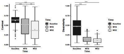 |
5 | Texture analysis of clinical MRI detects characteristics of tissue repair in lesions of multiple sclerosis after treatment with domperidone
Zahra Hosseinpour1, Olayinka Oladosu2, Mahshid Soleymani1, Wei-qiao Liu3, G Bruce Pike3,4,5, Marcus Werner Koch3,5, V. Wee Yong3,5, Luanne Metz3,5, and Yunyan Zhang3,4,5
1Biomedical Engineering Graduate Program, Schulich School of Engineering, University of Calgary, Calgary, AB, Canada, 2Department of Neurosciences, University of Calgary, Calgary, AB, Canada, 3Department of Clinical Neurosciences, University of Calgary, Calgary, AB, Canada, 4Department of Radiology, University of Calgary, Calgary, AB, Canada, 5Hotchkiss Brain Institute, University of Calgary, Calgary, AB, Canada MS pathology is dynamic, involving damage and repair. We used statistical texture analysis to assess changes in MS lesions seen in sequential brain MRI of 16 RRMS patients after treatment with domperidone. We focused on 2 histology-verified texture measures: contrast and dissimilarity, which characterize tissue coarseness and heterogeneity, respectively. Lower contrast and dissimilarity were detected in both acute and total lesion groups at weeks 16 and 32 compared to baseline, and at week 32 compared to week 16. The observed changes in texture suggest remyelination in MS lesions following treatment. |
||
3982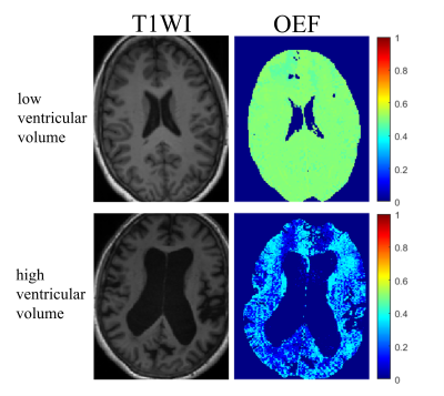 |
6 | Brain Oxygen Extraction Fraction in Patients with Normal Pressure Hydrocephalus Video Permission Withheld
Hangwei Zhuang1,2, Junghun Cho2, Gloria Chia-Yi Chiang2, Ilhami Kovanlikaya2, Linda A Heier2, and Yi Wang1,2
1Biomedical Engineering, Cornell University, New York, NY, United States, 2Department of Radiology, Weill Medical College of Cornell University, New York, NY, United States
Oxygen extraction fraction (OEF) maps were obtained from multi-echo gradient echo (mGRE) using a novel MRI biophysics model called QSM+qBOLD (QQ) in Normal Pressure Hydrocephalus (NPH) patients. OEF of the whole brain, cortical gray matter, caudate, and pallidum were significantly negatively correlated with ventricular volume. The finding demonstrates the feasibility of OEF mapping using MRI in NPH patients and suggests global and local neurodegeneration.
|
||
3983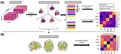 |
7 | Higher functional activity in the somatomotor network is associated with functional reorganization in the early stage of multiple sclerosis
Ceren Tozlu1, Sophie Card2, Keith Jamison1, Susan Gauthier3, and Amy Kuceyeski1
1Department of Radiology, Weill Cornell Medicine, New York, NY, United States, 2Horace Greeley High School, Chappaqua, NY, United States, 3Department of Neurology, Weill Cornell Medicine, New York, NY, United States
Multiple Sclerosis (MS) is a disease that causes neuroinflammation and neurodegeneration in central nervous system. Neuroimaging techniques may enable us to better understand the neuropathological mechanisms of MS and how the brain may compensate the pathological changes. We identified the recurring dynamic brain states using fMRI data and investigated the energy required for the transition between those states using the network control theory approach. Our main findings are the dynamic states were visual, somatomotor, and frontoparietal states and the transition energy averaged over all transitions was significantly greater in the MS patients with disability compared to those without disability.
|
||
3984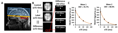 |
8 | Longitudinal changes in brain oxygen extraction fraction (OEF) in older adults: relationship to vascular and Alzheimer’s pathology
Zixuan Lin1, Chantelle Lim2, Dengrong Jiang1, Anja Soldan3, Corinne Pettigrew3, Kumiko Oishi1, Peiying Liu1,4, Marilyn Albert3, and Hanzhang Lu1
1Department of Radiology, Johns Hopkins University School of Medicine, Baltimore, MD, United States, 2Department of Biomedical Engineering, Johns Hopkins University School of Medicine, Baltimore, MD, United States, 3Department of Neurology, Johns Hopkins University School of Medicine, Baltimore, MD, United States, 4Department of Diagnostic Radiology and Nuclear Medicine, University of Maryland School of Medicine, Baltimore, MD, United States
Oxygen extraction fraction (OEF) has been suggested to be differentially affected by Alzheimer’s and vascular pathology in older adults. We aimed to investigate age-related OEF change with a longitudinal study design. 138 elderly participants were recruited with a 2-year follow-up. Individuals with higher vascular risks showed significant elevation in OEF but not those with lower vascular risks. Higher OEF was also associated with a faster growth in white matter hyperintensities, but not with any Alzheimer’s pathology or APOE gene. The results suggested a prominent effect of vascular pathology on OEF in aging.
|
||
3985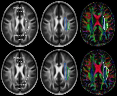 |
9 | Glymphatic System Evaluation in Obstructive Sleep Apnea Adults using Diffusion Tensor Imaging
Alba Nunez1, Bhaswati Roy1, Ravi S. Aysola2, Daniel W Kang2, and Rajesh Kumar1,3,4,5
1Anesthesiology, University of California Los Angeles, Los Angeles, CA, United States, 2Medicine, University of California Los Angeles, Los Angeles, CA, United States, 3Radiological Science, University of California Los Angeles, Los Angeles, CA, United States, 4Bioengineering, University of California Los Angeles, Los Angeles, CA, United States, 5Brain Research Institute, University of California Los Angeles, Los Angeles, CA, United States
OSA is characterized by recurrent episodes of complete or partial collapse of the upper airway during sleep, leading to sleep fragmentation. The altered sleep architecture can potentially hinder the extracellular waste removal system known, the glymphatic system, which is known for clearing interstitial solutes, including beta amyloids. In this study, we evaluated the glymphatic system using non-invasive MRI based diffusion tensor imaging data analyses along the perivascular space (DTI-ALPS) in OSA patients and healthy controls, and show impaired glymphatic system in the condition.
|
||
3986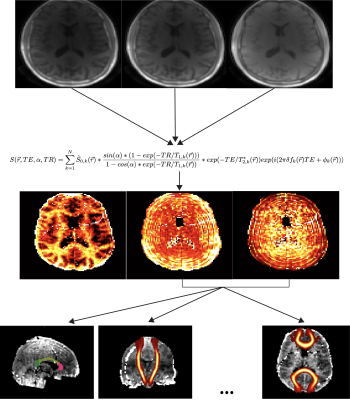 |
10 | Brain Ultrashort-T2* component measurements in patients with multiple sclerosis
Nikhil Deveshwar1,2, Eduardo Caverzasi1, Jingwen Yao1, Ari Green3, Roland Henry2,3, and Peder E. Z. Larson1,2
1Department of Radiology and Biomedical Imaging, University of California, San Francisco, San Francisco, CA, United States, 2UC Berkeley - UCSF Graduate Program in Bioengineering, Berkeley and San Francisco, CA, United States, 3Department of Neurology, University of California, San Francisco, San Francisco, CA, United States
This work presents a study measuring and characterizing the brain ultrashort-T2* component in patients suffering from MS. Our results show that the measured T2* relaxation time of the ultrashort-T2* component is significantly lower in almost all white matter ROIs measured. Fitted signal curves for the fractional component suggest the fractional component fits well in certain patients but not as well in others suggesting a more robust fitting model may be necessary for using this UTE relaxometry technique in patients with MS.
|
||
3987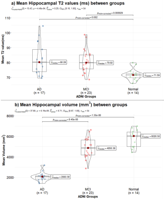 |
11 | T2 mapping in Alzheimer’s Disease from Standard Sequences
Gitanjali Chhetri1, Kelly C. McPhee2, and Alan H. Wilman1
1Biomedical Engineering, University of Alberta, Edmonton, AB, Canada, 2Medical Physics, CancerCare Manitoba, Winnipeg, MB, Canada
T2 mapping was applied retrospectively to healthy subjects, mild cognitive impairment (MCI) and Alzheimer’s disease (AD) by deriving the T2 from modelling standard dual-echo proton density and T2-weighted fast spin echo sequence, as used in the Alzheimer’s disease and Neuroimaging Initiative (ADNI-1). T2 differences were compared to volume changes in hippocampus and thalamus. In both regions, T2 mapping showed significant differences between MCI and healthy subjects while volume measures did not. Volume was more effective for distinguishing MCI from AD, due to profound hippocampal atrophy. By modelling actual refocusing angles, T2 mapping revealed differences between healthy, MCI and Alzheimer’s subjects.
|
||
3988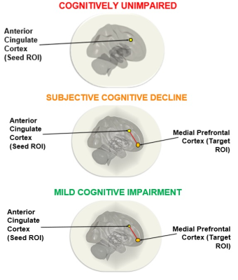 |
12 | Differences in functional connectivity in older adults with mild cognitive impairment and subjective cognitive decline
Arunan Srikanthanathan1, Susan Vandermorris1, Nicolaas Paul Verhoeff1, Jean Chen1, Nathan Herrmann2, and Linda Mah1
1Rotman Research Institute , Baycrest Health Sciences, North York, ON, Canada, 2Sunnybrook Research Institute, Toronto, ON, Canada Subjective cognitive decline (SCD) is a preclinical stage of Alzheimer’s disease (AD) that may precede mild cognitive impairment (MCI), a prodrome of AD. We used resting-state functional magnetic resonance imaging to examine the pattern of functional connectivity in anterior brain regions that support memory processes in SCD and MCI. We found reduction of posterior cingulate cortex functional connectivity with medial temporal lobe structures in MCI and SCD, and associated increases in functional connectivity in the anterior brain regions, although direct group comparisons of the latter did not yield statistically significant differences. |
||
3989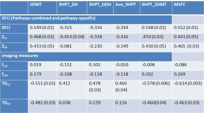 |
13 | Association between SFCI, a robust pathway-combined connectivity metric and neurological scores in multiple sclerosis
Pallab Bhattacharyya1, Robert Fox1, Jian Lin1, Ken Sakaie1, and Mark Lowe1
1Cleveland Clinic Foundation, CLEVELAND, OH, United States
Structural and functional connectivity index (SFCI) is a composite metric that combines structural and functional connectivity along different functionally specific pathways in multiple sclerosis. Performing functional connectivity and diffusion tensor imaging scans, we show that motor and cognitive pathway-combined SFCI is closely associated with multiple sclerosis functional composite, a composite neurological metric. Pathway-combined SFCI shows stronger association with MSFC than motor or cognitive pathway-specific composite metrics. Results from this study support the use of SFCI and composite pathway-based imaging measures as surrogate biomarkers for MS disease status and progression.
|
||
3990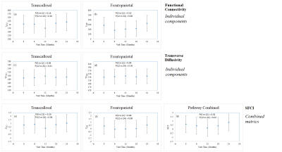 |
14 | Tracking motor and cognitive network connectivity in MS over 2 years using SFCI, a pathway-combined connectivity metric
Pallab Bhattacharyya1, Robert Fox1, Jian Lin1, Ken Sakaie1, and Mark Lowe1
1Cleveland Clinic Foundation, CLEVELAND, OH, United States
Motor and cognitive network integrity in multiple sclerosis over 2 years in patients receiving the same therapy was tracked using structural and functional connectivity index (SFCI), a metric that combines functional and structural connectivity along function-specific pathways and then forms a pathway-combined composite metric. Changes of the individual measures, functional connectivity and transverse diffusivity along transcallosal motor and frontoparietal cognitive pathways, were also tracked over 2 years. Pathway-combined SFCI was more efficient in tracking network integrity than the individual imaging measures or pathway-specific composite metric.
|
||
3991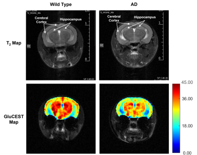 |
15 | Mapping of Early-Stage Changes in Cerebral Glutamate in APPNL-F/NL-F Mouse Model of Alzheimer’s Disease Using GluCEST MRI
Narayan Datt Datt Soni1, Ravi Prakash Reddy Nanga1, Juul Halvor1, and Ravinder Reddy1
1Department of Radiology, Center for Advanced Metabolic Imaging in Precision Medicine, Perelman School of Medicine, University of Pennsylvania, Philadelphia, PA, United States
Alzheimer’s disease (AD) is characterized by progressive loss of cognitive abilities. Glutamate being the major excitatory neurotransmitter in mammalian brain, regulates various cognitive functions. Reports suggesting compromised cerebral glutamate homeostasis in AD indicates the potential of glutamate mapping in early diagnosis of AD. In the current study, we have used glutamate weighted Chemical Exchange Saturation Transfer (GluCEST) MRI to investigate changes in cerebral glutamate in 6-month-old APPNL-F/NL-F mouse model of AD. Our findings suggest reductions in the levels of cortical and hippocampal glutamate in APPNL-F/NL-F mice. A longitudinal study is ongoing to understand the pattern of perturbation with disease progression.
|
||
3992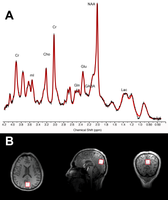 |
16 | Administration of ketone beta-hydroxybutyrate modulates 1H-MRS cortical GABA and Glu in healthy adults Video Permission Withheld
Antoine Hone-Blanchet1, Botond Antal2, Liam McMahon1, Andrew Lithen1, Helena van Nieuwenhuizen2, Nathan Smith3, Alexander Lin1, Yi-Fen Yen1, Bruce Jenkins1, Liliane Mujica-Parodi2, and Eva Maria Ratai1
1Harvard Medical School/Massachusetts General Hospital, Boston, MA, United States, 2Biomedical Engineering, Stony Brook University, Stony Brook, NY, United States, 3Children's National Hospital, Washington D.C., DC, United States
Brain aging is highly correlated with deficiencies in glucose metabolism. Ketone beta-hydroxybutyrate (d-βHB) may represent an alternative fuel for energy metabolism in brain aging. This study assesses the neurochemical effect of an acute administration of d-βHB in the healthy human brain using MR spectroscopy. Results show that levels of GABA and Glu were significantly reduced in the posterior cingulate region after administration of d-βHB, but not glucose. Additionally, older age significantly predicted higher decreases in levels of both GABA and Glu. This suggests that administration of d-βHB alters normal cerebral metabolic profile in physiological brain aging.
|
||
3993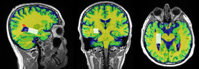 |
17 | Hippocampal Glx in RRMS: A potential therapeutic indicator in fingolimod and injectables
Oun Al-iedani1,2, Rodney Lea2, Jameen Arm1, Saadallah Ramadan1,2, and Jeannette Lechner-Scott2,3,4
1School of Health Sciences, University of Newcastle, Newcastle, Australia, 2Hunter Medical Research Institute, Newcastle, Australia, 3Department of Neurology, John Hunter Hospital, Newcastle, Australia, 4School of Medicine and Public Health, University of Newcastle, Newcastle, Australia
This novel longitudinal study evaluates the hippocampal metabolic and morphologic effects of different DMTs on the RRMS brain. RRMS patients were on fingolimod(N=36), injectables(N=29) and HCs cohort(N=44). MRS was acquired from hippocampus. Findings revealed that fingolimod is associated with a larger statistically significant reduction in hippocampal Glx (p=0.003) compared to injectable (p=0.01) and trending lower compared to HCs (p=0.09). Hippocampal NAA levels showed statistically significant increase in the fingolimod cohort (p≤0.0001) compared to HCs over the 2-years follow-up. These results demonstrate that fingolimod has a more potent effect on hippocampal Glx and NAA profiles than injectable DMTs.
|
||
 Back to Meeting Home
 Back to the Program-at-a-Glance
The International Society for Magnetic Resonance in Medicine is accredited by the Accreditation Council for Continuing Medical Education to provide continuing medical education for physicians.
 Back to Meeting Home
 Back to the Program-at-a-Glance
 View the Presentation