Online Gather.town Pitches
Molecular Imaging II
Joint Annual Meeting ISMRM-ESMRMB & ISMRT 31st Annual Meeting • 07-12 May 2022 • London, UK

Online Gather.town Pitches
Molecular Imaging II
| Booth # | ||||
|---|---|---|---|---|
3844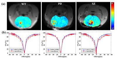 |
1 | Detecting dopamine in vivo using paramagnetic CEST-MRI at 11.7T
Ying Liu1, Xianfu Meng2,3, and He Wang1,4
1Institute of Science and Technology for Brain-Inspired Intelligence, Fudan University, Shanghai, China, 2Department of Materials Science and State Key Laboratory of Molecular Engineering of Polymers, Fudan University, Shanghai, China, 3Department of Medical Ultrasound, Shanghai Tenth People’s Hospital, School of Medicine, Tongji University, Tongji University Cancer Center, Shanghai, China, 4Human Phenome Institute, Fudan University, Shanghai, China Detecting dopamine in vivo is critical for understanding many neurologic and psychiatric disorders. In this work, we developed a paramagnetic CEST-MRI method for dopamine detection using NaHoF4@DBA nanoparticles. Our results showed that the dopamine in mouse brain can be selectively enriched in the surface of the nanoparticles and detected by CEST-MRI. Moreover, we found that the magnitude of ST% map reflecting dopamine levels in the brain of SZ mice was relatively higher than that of PD mice. |
||
3845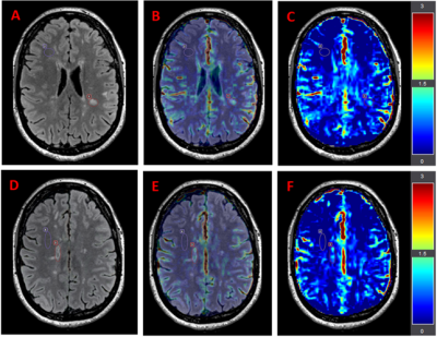 |
2 | Using 3D Amide Proton Transfer weighted imaging at 3T to investigate lesions and normal-appearing white matter in Multiple Sclerosis
Ibrahim Khormi1,2,3, Oun Al-iedani1,2, Bryan Paton2,4, Jeannette Lechner-Scott 2,5,6, Abdulaziz Alshehri1,2, Amir Fazlollahi7,8, Stefano Casagranda9, Christos Papageorgakis9, Margarita Arango-Lievano9, Anne-Louise Ponsonby10,11, Patrick Liebig12, and Saadallah Ramadan1,2
1School of Health Sciences, University of Newcastle, Newcastle, Australia, 2Hunter Medical Research Institute, Newcastle, Australia, 3College of Applied Medical Sciences, University of Jeddah, Jeddah, Saudi Arabia, 4School of Psychology, University of Newcastle, Newcastle, Australia, 5School of Medicine and Public Health, University of Newcastle, Newcastle, Australia, 6Department of Neurology, John Hunter Hospital, New Lambton Heights, Australia, 7CSIRO Health and Biosecurity, Brisbane, Australia, 8Queensland Brain Institute, The University of Queensland, Brisbane, Australia, 9Olea Medical, La Ciotat, France, 10The Florey Institute of Neuroscience and Mental Health, Melbourne, Australia, 11Murdoch Children's Research Institute, Royal Children's Hospital, University of Melbourne, Melbourne, Australia, 12Siemens Healthcare GmbH, Erlangen, Germany This novel study explores amide proton transfer weighted (APTw) imaging in people with relapsing-remitting multiple sclerosis (pw-RRMS). We evaluated the APTw signal intensity in selected MS lesions and normal-appearing white matter (NAMW) regions in 9 pw-RRMS. Compared to NAWM regions, a statistically significant increase in APTw signal intensity was observed in the MS lesions. Elevated APTw signal intensity could mark increased mobile myelin proteins decomposition and accumulation from the demyelination process. |
||
3846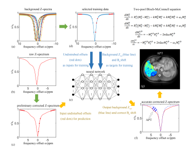 |
3 | A new approach for in-vivo liver CEST imaging
Zhichao Wang1, Jilei Zhang2, Yu Zhao3, Ting Hua4, Guangyu Tang4, Jianqi Li5, and Yu Zhang1
1Research Center for Healthcare Data Science, Zhejiang Lab, Hangzhou, Zhejiang, China, 2Philips Healthcare, Shanghai, China, 3Institute of Imaging Science, Vanderbilt University, Nashville, TN, United States, 4Renji Hospital affiliated to Shanghai Jiao Tong University Medical College, Shanghai, China, 5Shanghai Key Laboratory of Magnetic Resonance, School of Physics and Electronic Science, East China Normal University, Shanghai, China Chemical exchange saturation transfer (CEST) imaging shows great potentials in the diagnosis of brain diseases. However, there are still many challenges in the hepatic disease due to the fat suppression and large B0 inhomogeneity. In this study, we achieved CEST imaging with simultaneous B0 correction in one sequence, and conducted a pixel-by-pixel prediction on the background reference Z-spectra representing the magnetization transfer and direct saturation effects. Our approach also effectively separated the amide proton transfer (APT) effects from the mixing effect of fat and nuclear Overhauser enhancement (NOE), by subtracting the background reference Z-spectra from the predicted Z-spectra. |
||
3847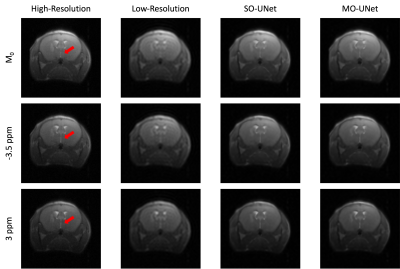 |
4 | Single-offset and multi-offset super-resolution for CEST MRI using deep transfer learning
Rohith Saai Pemmasani Prabakaran1, Zilin Chen2, Joseph H.C. Lai2, Se Weon Park1,2, Yang Liu1,2, Jianpan Huang2, and Kannie W.Y. Chan1,2,3,4
1Hong Kong Centre For Cerebro-Cardiovascular Health Engineering, Hong Kong, Hong Kong, 2Department of Biomedical Engineering, City University of Hong Kong, Hong Kong, Hong Kong, 3Russell H. Morgan Department of Radiology and Radiological Science, The Johns Hopkins University School of Medicine, Baltimore, MD, United States, 4City University of Hong Kong Shenzhen Research Institute, Shenzhen, China
CEST MRI is an unique molecular imaging approach to reveal the exchangeable proton information related to physiology and pathology. However, long scanning time has hindered its translation into clinics. While deep-learning based super-resolution methods have been explored to reduce scanning time in conventional MRI, adaptation of these methods to CEST MRI has been limited due to lack of large public CEST datasets. Therefore, this study proposes two transfer learning based super-resolution methods, Single-Offset UNet and Multi-Offset UNet, for accelerating CEST MRI acquisition by using public MRI databases for pretraining and a very small CEST dataset for training.
|
||
3848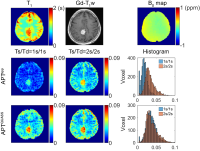 |
5 | Quasi-steady-state reconstruction provides fast equilibrium APT imaging of brain tumor patients at 3T
Yin Wu1, Zhou Liu1,2, Qian Yang2, Liyan Zou2, Fan Zhang2, Long Qian3, Xin Liu1, Hairong Zheng1, Dehong Luo2, and Phillip Zhe Sun4
1Shenzhen Institute of Advanced Technology, Chinese Academy of Sciences, Shenzhen, China, 2National Cancer Center/National Clinical Research Center for Cancer/Cancer Hospital & Shenzhen Hospital, Chinese Academy of Medical Sciences and Peking Union Medical College, Shenzhen, China, 3GE Healthcare, Beijing, China, 4Emory University School of Medicine, Atlanta, GA, United States APT MRI scans are often performed under non-equilibrium conditions, confounding imaging quantification and tissue characterization. This study proposed a quasi-steady-state (QUASS) algorithm for fast and accurate tumor APT imaging. Seven healthy volunteers and nineteen tumor patients were scanned at 3T, with two representative RF saturation times (Ts) and relaxation delays (Td). The routine APT measures significantly varied with Ts and Td (P<.001) and were significantly smaller than the corresponding QUASS indices (P<.001). In contrast, QUASS reconstruction results showed little dependence on scan protocols (P>.05). The QUASS algorithm enables robust APT imaging, promising to expedite and standardize clinical APT tumor MRI. |
||
3849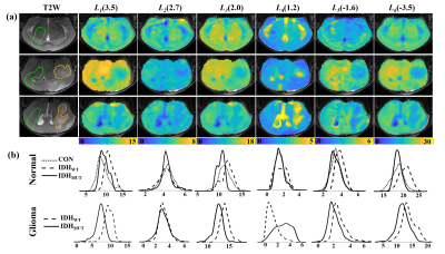 |
6 | Voxel-based quantitative mapping of glioma heterogeneity for the differentiation of IDH genotype using CEST-MRI
Ying Liu1, Botao Zhao1, and Xiao-Yong Zhang1
1Institute of Science and Technology for Brain-Inspired Intelligence, Fudan University, Shanghai, China The identification of the isocitrate dehydrogenase mutant (IDHMUT) glioma and IDH wild-type (IDHWT) glioma has significant diagnostic, prognostic, and therapeutic implications. In this work, we evaluated the non-invasive prediction of IDH mutation status in glioma mice by employing a combination of high spectral resolution CEST MRI at 11.7T together with data analysis using machine learning approaches. We demonstrated that in vivo CEST signals correspond very well with glioma genotypes. Among several CEST signals, the signals at Δω =3.5 ppm may serve as the major indicator to differentiate the IDH genotype glioma from other types. |
||
3850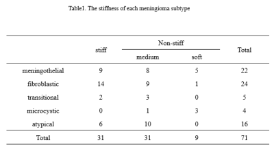 |
7 | Amide proton transfer weighted MR imaging in predicting the stiffness of meningiomas: a feasibility study Video Not Available
Hao Yu1, Yanting Wang1, Zhanguo Sun2, Weiwei Wang1, Zhe Zhou1, Weiqiang Dou3, Zhibo Wen4, and Yueqin Chen2
1Radiology, Affiliated Hospital of Jining Medical University, Jining, China, 2Affiliated Hospital of Jining Medical University, Jining, China, 3GE Healthcare MR Research, Beijing, China, 4Zhujiang Hospital, Guangzhou, China
Stiffness of meningioma is an important factor affecting the surgical resection. In this study we aimed to explore if amide proton transfer-weighted MR imaging has clinical potential of predicting meningioma stiffness
|
||
3851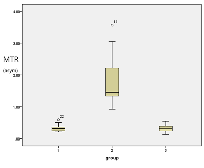 |
8 | Amide Proton Transfer Imaging of Bladder Cancer at 3 T: A Preliminary Study
Fang Wang1, Yun Xu1, Yong-Sheng Xiang1, Peng Wu2, Ai-Jun Shen1, and Pei-Jun Wang1
1Tongji Hospital, School of Medicine, Tongji University, Shanghai, China, 2Philips Healthcare, Shanghai, China
Amide proton transfer (APT) imaging is an emerging chemical exchange saturation transfer (CEST)-based MRI technique that is sensitive to mobile proteins and peptides in tissue and has drawn considerable attention in the field of cellular and molecular imaging. In the present work, our result demonstrates that it is feasible to use APT imaging for Bladder Cancer (BCa) and it has shown great potential for bladder cancer imaging, thus opening new research avenues in this field.
|
||
3852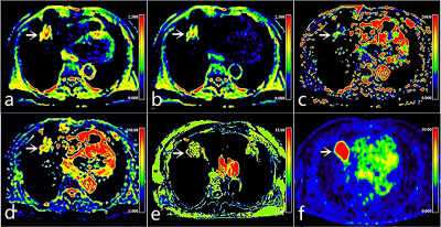 |
9 | Non-invasive assessment of PD-L1 expression status in non-small cell lung cancer using multi-parametric 18F-FDG PET/MRI
Nan Meng1, Fangfang Fu2, Zhun Huang3, Ziqiang Li4, Yu Luo1, Pengyang Feng3, Yaping Wu2, Jianmin Yuan5, Yang Yang6, and Meiyun Wang*1
1Department of Medical Imaging, Zhengzhou University People’s Hospital & Henan Provincial People’s Hospital, Zhengzhou, China, 2Department of Medical Imaging, Henan Provincial People’s Hospital, Zhengzhou, China, 3Department of Medical Imaging, Henan University People’s Hospital & Henan Provincial People’s Hospital, Zhengzhou, China, 4Department of Medical Imaging, Xinxiang Medical University Henan Provincial People’s Hospital, Zhengzhou, China, 5Central Research Institute, UIH Group, Shanghai, China, 6Beijing United Imaging Research Institute of Intelligent Imaging, UIH Group, Beijing, China
18F-Fluorodeoxyglucose positron-emission tomography/magnetic resonance imaging (18F-FDG PET/MRI) allows multimodal quantitative MRI sequences to be scanned in parallel with PET imaging, providing a more multidimensional reflection of lesion information. Our results showed that intravoxel incoherent motion (IVIM), amide proton transfer-weighted imaging (APTWI), and metabolism related parameters can be beneficial for the non-invasive assessment of PD-L1 expression in NSCLC patients, and the combination of SUVmax, D, and MTRasym(3.5ppm) may serve as a prognostic biomarker to guide immunotherapy.
|
||
3853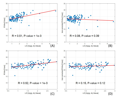 |
10 | Characterization and correction of the hepatic iron effects on T1rho relaxation in the liver at 3.0 Tesla
Yurui QIAN1, Jian Hou1, Baiyan Jiang1,2, Vincent Wai-Sun Wong3, Queenie Chan4, Yixiang Wang1, Winnie Chiu-Wing Chu1, and Weitian Chen1
1Department of Imaging and Interventional Radiology, The Chinese University of Hong Kong, Hong Kong, Hong Kong, 2Illuminatio Medical Technology Limited, Hong Kong, Hong Kong, 3Medicine & Therapeutics, The Chinese University of Hong Kong, Hong Kong, Hong Kong, 4Philips Healthcare, Hong Kong, Hong Kong
Spin-lock T1rho is a non-invasive MR imaging method. It has the potentials for the assessment of chronic liver diseases. However, liver iron content can potentially confound T1rho quantification of the liver. In this work, we reported a method to correct the influence of liver iron content on T1rho quantification of the liver at 3.0T.
|
||
3854 |
11 | Simultaneous macromolecular proton fraction and proton density fat fraction quantification using chemical shift-encoding based spin-lock MRI
Jian Hou1, Yurui Qian1, Baiyan Jiang1,2, Queenie Chan3, Zhigang Wu4, Vincent Wai-Sun Wong5, Dimitrios Karampinos6, Winnie Chiu-Wing Chu1, and Weitian Chen1
1Department of Imaging and Interventional Radiology, The Chinese University of Hong Kong, Hong Kong, Hong Kong, 2Illuminatio Medical Technology Limited, Hong Kong, Hong Kong, 3Philips Healthcare, Hong Kong, Hong Kong, 4Philips Healthcare, Shenzhen, China, 5Department of Medicine & Therapeutics, The Chinese University of Hong Kong, Hong Kong, Hong Kong, 6Department of Diagnostic and Interventional Radiology, Technical University of Munich, Munich, Germany
Chronic liver disease is a major healthcare problem worldwide. Liver fibrosis and fat fraction are two main features of chronic liver diseases. Recent work reported that macromolecular proton fraction (MPF) mapping has potential for diagnosis of liver fibrosis. In this work, we reported a novel technique to quantify MPF and fat fraction of the liver simultaneously in a brief breath hold.
|
||
3855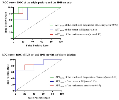 |
12 | Prediction of IDH, 1p/19q and TERTp Status: combination of APTw value of tumor solid and peritumoral area Video Not Available
Xinying Ren1, Diaohan Xiong1, Tiejun Gan1, Pengfei Wang1, Rui Wang1, Tao Wen1, Yujing Li1, Jing Zhang1, and Kai Ai2
1Lanzhou University Second Hospital, Lanzhou, China, 2Philips Healthcare, Xi'an, China
This study aims to analyze the metabolic information in both tumor solid and peritumoral area of gliomas to predict its genotype by using Amide Proton Transfer weighted (APTw) imaging. As a complementary method of pathological evaluation, APTw based MRI technique could provide a prediction of the gliomas genotype. Unlike other studies focus on tumor core region, our research investigated the APTmean value of tumor solid and the peritumoral area. Interestingly, the results showed that the AUC value of peritumoral area was higher than the tumor solid. The APTw images may serve as a potential marker for the genotyping of gliomas.
|
||
3856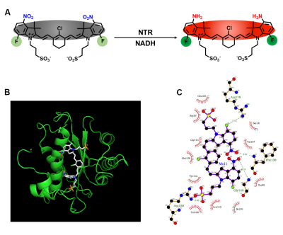 |
13 | A Nitroreductase Responsive Fluorescent/ 19F MRI Dual-Imaging Probe for Hypoxia Tumor Detection In Vivo Video Permission Withheld
Long Xiao1, Yu Li1, Sha Li1, Yaping Yuan1, Lei Zhang1, Shizhen Chen1, and Xin Zhou1
1Key Laboratory of Magnetic Resonance in Biological Systems, State Key Laboratory of Magnetic Resonance and Atomic and Molecular Physics, National Center for Magnetic Resonance in Wuhan, Wuhan Institute of Physics and Mathematics, Innovation Academy for Precision Measurement Science and Technology, Chinese Academy of Sciences–Wuhan National Laboratory for Optoelectronics, Wuhan, China
We designed and synthesized a molecular probe-based on fluorescence imaging and 19F MRI bimodal imaging to identify overexpressed nitroreductase in hypoxic tumor. The two imaging modes complement each other in sensitivity and imaging depth, providing more abundant information. More importantly, the bimodal molecular probe is enriched in the tumor region depending on the passive targeting ability and has been well verified by fluorescence imaging and 19F MRI in the metastatic tumor model. And we successfully identified the cancerous left lung and healthy right lung in lung cancer model mouse.
|
||
3857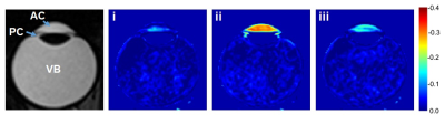 |
14 | In vivo measurement of human intraocular water movement using 1H-MRI with H217O saline eye drops
Moyoko Tomiyasu1, Yasuka Sahara1, Etsuko Mitsui1, Hiroki Tsuchiya2, Takamasa Maeda2, Nobuhiro Tomoyori3, Makoto Kawashima3, Tatsuya Higashi1, Atsushi Mizota3, Kohsuke Kudo4, and Takayuki Obata1
1Department of Molecular Imaging and Theranostics, National Institutes for Quantum Science and Technology, Ciba, Japan, 2Department of Medical Technology, National Institutes for Quantum Science and Technology, Ciba, Japan, 3Department of ophthalmology, Teikyo University, Tokyo, Japan, 4Department of Diagnostic and Interventional Radiology, Hokkaido University Hospital, Sapporo, Japan
We observed the movement of 17O-labelled water (H217O) in the eyes of three volunteers using dynamic T2W 1H-MRI (3T). After a drop of H217O saline in the right eye, the signal intensity in the right anterior chamber decreased, reaching a minimum at 7–9 min, and then gradually recovered to close to that seen before the eye drop, by about 40 min. Signal decrease and recovery was also observed in the posterior chamber, but not in the vitreous body. These results show that H217O drops flow smoothly into the human anterior chamber and flow out slowly.
|
||
3858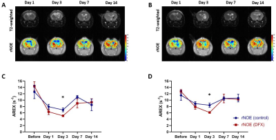 |
15 | rNOE imaging detects distinctive neuropathology in Intracerebral Hemorrhage (ICH) under deferoxamine treatment
Ho Chi Joseph Lai1, Tian Yang2, Jianpan Huang1, Yang Liu1, Youngjin Lee2, and Wai Yan Kannie Chan1,3,4,5
1Department of Biomedical Engineering, City University of Hong Kong, Hong Kong, Hong Kong, 2Department of Neuroscience, City University of Hong Kong, Hong Kong, Hong Kong, 3Russell H. Morgan Department of Radiology and Radiological Science, The Johns Hopkins University School of Medicine, Baltimore, MD, United States, 4City University of Hong Kong Shenzhen Research Institute, Shenzhen, China, 5Hong Kong Centre for Cerebro-Cardiovascular Health Engineering, Hong Kong, Hong Kong
We have shown that CEST (APTw and rNOE) contrast demonstrated distinctive changes of ICH mouse brains longitudinally1. As demonstrated by our published work2, the rNOE changes could be primarily associated with changes in myelin. Here, we studied the APTw and rNOE contrast after ICH up to 14 days, and under DFX treatment. We observed regional changes of APTw and rNOE contrast in the core and peri-hematoma regions, especially the significant difference (P<0.05) on day 3 with and without DFX treatment. Our immunohistochemistry data indicated that rNOE contrast correlated with myelin, which supports that rNOE could detect myelin pathology during ICH.
|
||
 Back to Meeting Home
 Back to the Program-at-a-Glance
The International Society for Magnetic Resonance in Medicine is accredited by the Accreditation Council for Continuing Medical Education to provide continuing medical education for physicians.
 Back to Meeting Home
 Back to the Program-at-a-Glance
 View the Presentation