Online Gather.town Pitches
Perfusion & Permeability IV
Joint Annual Meeting ISMRM-ESMRMB & ISMRT 31st Annual Meeting • 07-12 May 2022 • London, UK

Online Gather.town Pitches
Perfusion & Permeability IV
| Booth # | ||||
|---|---|---|---|---|
4900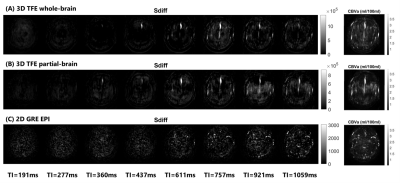 |
1 | On the optimization of 3D inflow-based vascular-space-occupancy (iVASO) MRI for the quantification of arteriolar cerebral blood volume (CBVa)
Chunming Gu1,2,3, Di Cao1,2,3, Xinyuan Miao2,3, Adrian G Paez2,3, Jitong Cai1, Yinghao Li1,2,3, Wenbo Li2,3, Jay J Pillai4,5, Peter C.M. van Zijl2,3, and Jun Hua2,3
1Biomedical Engineering, Johns Hopkins University, Baltimore, MD, United States, 2Neurosection, Division of MRI Research, Russell H. Morgan Department of Radiology and Radiological Science, Johns Hopkins University School of Medicine, Baltimore, MD, United States, 3F.M. Kirby Research Center for Functional Brain Imaging, Kennedy Krieger Institute, Baltimore, MD, United States, 4Division of Neuroradiology, Russell H. Morgan Department of Radiology and Radiological Science, Johns Hopkins University School of Medicine, Baltimore, MD, United States, 5Department of Neurosurgery, Johns Hopkins University School of Medicine, Baltimore, MD, United States
The iVASO MRI is a noninvasive approach for quantitative mapping of CBVa in the brain. It was originally developed in the single-slice mode. Recently, CBVa measured by single-slice iVASO has been validated using histological markers of arteriolar blood vessels in a mouse model. Here, we demonstrate an optimized 3D iVASO MRI protocol with a 3D turbo-field-echo (TFE) readout and a whole-brain coverage. It showed consistent CBVa measures when compared to the original single-slice iVASO. In 3D-TFE-iVASO, the imaging slab volume did not show significant effects on the measured CBVa values. CBVa measured with 3D-TFE-iVASO showed reasonable intra-subject reproducibility.
|
||
4901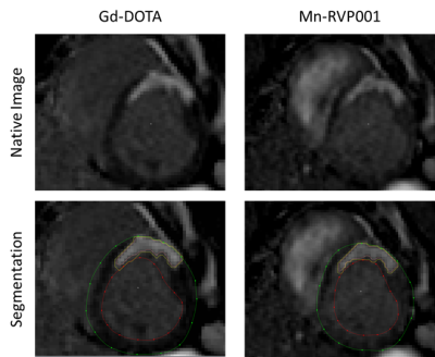 |
2 | Evaluation of a Novel Manganese-Based Contrast Agent for Cardiac MRI in the Setting of Myocardial Ischemia-Reperfusion
Benjamin Pryor Bonner1,2,3, Jaume Coll-Font1,2,4, Salva Yurista1,2,4, Anna Foster1,2, Robert Eder1,2, Shi Chen1,2, Peter Caravan2,4, Eric Gale2,4, and Christopher Nguyen1,2,4,5
1Cardiovascular Research Center, Massachusetts General Hospital, Boston, MA, United States, 2Athinoula A. Martinos Center for Biomedical Imaging, Massachusetts General Hospital, Boston, MA, United States, 3Louisiana State University Health Sciences Center, New Orleans, New Orleans, LA, United States, 4Harvard Medical School, Boston, MA, United States, 5Division of Health Science Technology, Harvard-Massachusetts Institute of Technology, Cambridge, MA, United States
Cardiac MRI a powerful imaging modality to assess cardiac anatomy and function, and its utility can be greatly enhanced with the use of contrast agents. Conventional contrast agents are nephrotoxic which limits their utility. We evaluate a novel contrast agent which is safe for use in renal dysfunction. We compare its performance against that of a conventional agent a porcine model of ischemia-reperfusion. This novel contrast agent demonstrates equivalent performance in assessing myocardial fibrotic scars, indicating its potential clinical use in the workout of cardiac patients with renal impairment.
|
||
4902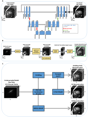 |
3 | Radial perfusion cardiac magnetic resonance imaging using deep learning image reconstruction.
Salah Assana1, Manuel Morales1, Kei Nakata1, Eiryu Sai1, Amine Amyar1, Hassan Haji-Valizadeh1, and Reza Nezafat1
1Department of Medicine, Cardiovascular Division, Beth Israel Deaconess Medical Center, Boston, MA, United States
Myocardial perfusion assessment using cardiac MRI allows non-invasive assessment of myocardial ischemia. In myocardial perfusion sequence, imaging is collected after a saturation pulse. An alternative approach based on steady-state imaging with radial sampling has been recently proposed. However, image reconstruction using compressed sensing in steady-state myocardial perfusion remains long and clinically not feasible. In this study, we sought to develop a deep learning-based image reconstruction platform for myocardial perfusion imaging.
|
||
4903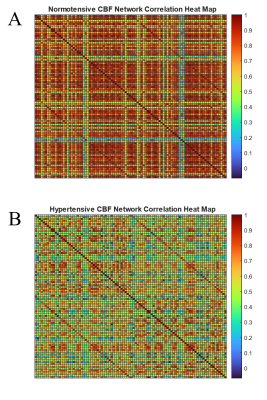 |
4 | Analyzing CBF Connectivity Differences Affected by Hypertension in Arterial Spin Labelled Data Using Graph Theory
William D Reeves1, Ishfaque Ahmed1, Wenwu Sun1, Michelle Brown2, Celestine Williams2, Catherine L Davis2, Jennifer E McDowell3, Nathan Yanasak4, Shaoyong Su2, and Qun Zhao1
1Department of Physics, University of Georgia, Athens, GA, United States, 2Georgia Prevention Institute, Augusta University, Augusta, GA, United States, 3Department of Psychology, University of Georgia, Athens, GA, United States, 4Department of Radiology and Imaging, Augusta University, Augusta, GA, United States In a cross-sectional study of hypertension and its effects on brain connectivity, graph theory can be used to show cerebral blood flow connectivity differences between normotensive (n=32) and hypertensive (n=37) subjects. We obtain adjacency matrices from processed arterial spin labelling scans which can be used to determine several quantifications based on graph theory analysis. We find that normotensive subjects exhibit higher small-worldness and global efficiency (σ = 3.30 and Eglob = 0.73) compared to hypertensive subjects (σ = 1.82 and Eglob = 0.46). This trend continues for nearly all thresholding values and implies a measurable perfusion difference between the groups. |
||
4904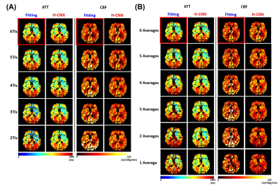 |
5 | Parametric ATT and CBF Mapping Using a Three-Dimensional Convolutional Neural Network
Donghoon Kim1,2, Megan Lipford3, Hongjian He4, Qiuping Ding4, Vladimir Ivanovic2, Samuel Lockhart5, Suzanne Craft5, Christopher T. Whitlow3, and Youngkyoo Jung1,2,3
1Biomedical Engineering, University of California, Davis, CA, United States, 2Radiology, University of California, Davis, CA, United States, 3Radiology, Wake Forest University, Winston-Salem, NC, United States, 4Center for Brain Imaging Science and Technology, College of Biomedical Engineering and Instrumental Science, Zhejiang University, Zhejiang, China, 5Internal Medicine, Wake Forest University, Winston-Salem, NC, United States
A CNN algorithm was proposed to estimate ATT and CBF simultaneously, using multi-TI ASL, acquiring ASL images at multiple PLDs. Hierarchical structure of CNN was used to reduce the estimation error of ATT and CBF. The proposed method successfully estimated ATT and CBF maps using reduced numbers of TIs or averages with a higher accuracy than a conventional non-linear model fitting. The successful estimation of ATT and CBF using our H-CNN may allow a total scan time reduction with a smaller number of TIs or averages and improvements of image quality when a part of acquisition is corrupted.
|
||
4905 |
6 | Resting-state based Cerebrovascular Reactivity (CVR) mapping at high spatial resolutions
Lincoln Kartchner1, Dengrong Jiang2, Parimal Joshi1, Cuimei Xu2, Hanzhang Lu2, and Peiying Liu1
1Department of Diagnostic Radiology & Nuclear Medicine, University of Maryland School of Medicine, Baltimore, MD, United States, 2Department of Radiology, Johns Hopkins University School of Medicine, Baltimore, MD, United States Cerebrovascular reactivity (CVR) is typically assessed with a carbon dioxide (CO2) stimulus combined with BOLD fMRI. Recently, resting-state BOLD fMRI has been shown capable of generating CVR maps, providing a potential for broader CVR applications in neuroimaging studies. However, prior RS-CVR studies have primarily been performed at a spatial resolution of 3-4mm3 voxel sizes. It remains unknown whether RS-CVR can also be obtained at high-resolution without major degradation in image quality. In this study, we investigated the feasibility of high-resolution CVR mapping based on resting-state fMRI data. Our results showed good CVR quality can be obtained at 2-3mm3 voxel sizes. |
||
4906 |
7 | Normalization for Relative Cerebrovascular Reactivity in Multifocal Vascular Disease Patients Using Calibrated Perfusion and Temporal Shift
Xiuyuan Wang1, Siddhant Dogra2, Koto Ishida3, Alejandro Gupta2, Deqiang Qiu4, and Seena Dehkharghani2,3
1Department of Radiology, Weill Cornell Medicine, New York, NY, United States, 2Department of Radiology, New York University Langone Health, New York, NY, United States, 3Department of Neurology, New York University Langone Health, New York, NY, United States, 4Department of Radiology and Imaging Sciences, Emory University, Atlanta, GA, United States
Cerebrovascular reactivity (CVR) is a widely used estimation of hemodynamic stress and ischemic stroke risk but its semi-quantitative nature relegates it primarily to estimations of relative change by comparison to putatively normal territories. Diagnostic and prognostic utility is thus attenuated in commonly encountered patients with bihemispheric disease, thus we have tested approaches for identification of candidate healthy voxels accompanying perfusion imaging or inline calibration of BOLD temporal shift, optimized in a cohort of subjects with strictly unilateral macrovascular disease. Excellent agreement with normative CVR values was observed, suggesting its applicability in patients with multi-focal or ambiguous vascular disease patterns.
|
||
4907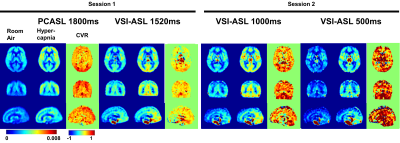 |
8 | Comparison of Velocity Selective ASL and PCASL with Phase-Contrast MRI for Measuring CO2-induced Cerebrovascular Reactivity
Feng Xu1,2, Cuimei Xu1, Dapeng Liu1,2, Dan Zhu2, Hanzhang Lu1,2, and Qin Qin1,2
1The Russell H. Morgan Department of Radiology and Radiological Science, Johns Hopkins University, Baltimore, MD, United States, 2F.M. Kirby Research Center for Functional Brain Imaging, Kennedy Krieger Institute, Baltimore, MD, United States
Saturation based velocity-selective ASL (VSASL) was recently investigated for acetazolamide challenge and notably lower cerebrovascular reactivity (CVR) was observed when comparing to PET results. Here we compared the performance of velocity-selective inversion (VSI) prepared ASL and pseudo-continuous ASL in response to the hypercapnia challenge for CO2-induced CVR quantification with phase-contrast MRI as a global reference. VSI-ASL could underestimate the regional CVR when the trailing edge of the labeled bolus arrive before the PLD as the CBF during hypercapnia would be underestimated. In contrast, PCASL could overestimate CVR in areas with long ATT as the baseline CBF would be underestimated.
|
||
4908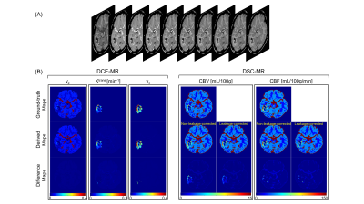 |
9 | MR Multitasking-based Dynamic Imaging for Cerebrovascular Evaluation (MT-DICE): A Simulation Study for Accuracy Validation
Zhehao Hu1,2, Nan Wang3, Anthony Christodoulou2,3, Mark Shiroishi1, Gabriel Zada4, Debiao Li2,3, and Zhaoyang Fan1,5,6
1Department of Radiology, Keck School of Medicine, University of Southern California, Los Angeles, CA, United States, 2Department of Bioengineering, University of California, Los Angeles, Los Angeles, CA, United States, 3Biomedical Imaging Research Institute, Cedars-Sinai Medical Center, Los Angeles, CA, United States, 4Department of Neurosurgery, Keck School of Medicine, University of Southern California, Los Angeles, CA, United States, 5Department of Radiation Oncology, Keck School of Medicine, University of Southern California, Los Angeles, CA, United States, 6Department of Biomedical Engineering, University of Southern California, Los Angeles, CA, United States
MR Multitasking-based Dynamic Imaging for Cerebrovascular Evaluation (MT-DICE) technique is recently developed and has the potential to provide DCE- and leakage-corrected DSC-MRI parameters simultaneously with one 7.6-minute scan and a single-dose contrast injection. However, it remains a practical challenge to validate the technique against their respective references with repeated contrast injections in separate days. In this work, we perform a numerical simulation study to validate the accuracy of MT-DICE in the estimation of permeability and leakage-corrected perfusion metrics.
|
||
4909 |
10 | Cerebral Blood Flow Patterns in Term and Premature Neonates measured at Term-Equivalent-Age: a PCASL study
Antonio Maria Chiarelli1, Eleonora Piccirilli1, Carlo Sestieri1, Daniele Mascali1, Emma Biondetti1, Antonio Ferretti1, Richard Wise1, and Massimo Caulo1,2
1Department of Neuroscience, Imaging and Clinical Sciences, University G. D'Annunzio of Chieti Pescara, Chieti, Italy, 2Department of Radiology, SS. Annunziata University Hospital, Chieti, Italy
During the perinatal period the brain undergoes extensive development, including increasing brain perfusion and metabolism. More rapidly modifying brain areas may be more sensitive to insults and transient hypoxic states. We performed PCASL on 115 infants at term equivalent age (TEA) with variable gestational age at birth. Insular-subcortical and somato-motor regions exhibited high grey matter CBF(GM) in both term and preterm newborns. However, premature neonates showed a redistribution of perfusion compared to term infants, with somato-motor regions showing even higher CBFGM. These results suggest that insular-subcortical and somato-motor regions may reflect brain health and development at TEA.
|
||
4910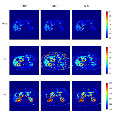 |
11 | Deep learning DCE-MRI parameter estimation: Application in pancreatic cancer
Tim Ottens1, Sebastiano Barbieri2, Matthew Orton3, Remy Klaassen4, Hanneke van Laarhoven4, Hans Crezee5, Aart Nederveen1, and Oliver Gurney-Champion1
1Radiology and Nuclear Medicine, Amsterdam UMC, Amsterdam, Netherlands, 2Centre for Big Data Research in Health, Sydney, Australia, 3Radiology, The Royal Marsden NHS Foundation Trust and The Institute for Cancer Research, London, United Kingdom, 4Medical Oncology, Amsterdam UMC, Amsterdam, Netherlands, 5Radiation Oncology, Amsterdam UMC, Amsterdam, Netherlands
Quantitative physiological perfusion parameters can be obtained from dynamic contrast-enhanced (DCE)-MRI. Conventionally, fitting is done with the non-linear least squares (NLLS) approach. However, the NLLS-fit suffers from long processing times and results in noisy parameter maps. In this work, we implemented a physics-informed gated recurrent unit (GRU) network with attention layers for estimating physiological parameters using the extended Tofts model. In simulations, we show it outperforms NLLS with more accurate and precise parameter maps. We show our method produced substantially less noisy parameter maps than NLLS in a fraction of the time in pancreatic cancer patients.
|
||
4911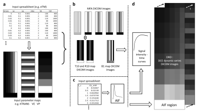 |
12 | Open source MATLAB code for the generation of digital reference objects for DCE-MRI analysis software validation
Andrew B. Gill1 and Martin J. Graves1
1Radiology, University of Cambridge, Cambridge, United Kingdom Extending work by the QIBA initiative and others, a method is presented here for the generation of configurable digital reference objects (DROs) for the validation of dynamic-contrast enhanced (DCE-) MRI analysis software packages. Five commonly used kinetic models are supported and two software analysis packages were independently tested, one open source and one commercial. A sample set of DROs yielded excellent concordance with ‘ground-truth’ when analyzed with each package, indicating the software’s correctness and providing a benchmark for testing newly written software. Our MATLAB code is provided on an open-source basis to help researchers evaluate and test DCE-MRI analysis software. |
||
4912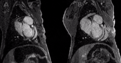 |
13 | 3D Inversion recovery-FLASH and myocardial perfusion MRI imaging in a sheep model of myocardial infarction
Steven KS Cho1, Jack RT Darby2, Georgia K Williams3, Catherine G Dimasi2, Stacey L Holman2, Joseph B Selvanayagam4, Christopher K Macgowan5, Janna L Morrison2, and Mike Seed6
1Physiology, University of Toronto, North York, ON, Canada, 2Early Origins of Adult Health Research Group, Health and Biomedical Innovation, University of South Australia, Adelaide, Australia, 3South Australian Health & Medical Research Institute, Preclinical, Imaging & Research Laboratories, Adelaide, Australia, 4Cardiac Imaging Research Group, Department of Heart Health, South Australian Health and Medical Research Institute, Flinders University, Adelaide, Australia, 5Department of Medical Biophysics, University of Toronto, Toronto, ON, Canada, 6Division of Cardiology, The Hospital for Sick Children, Toronto, ON, Canada
We investigated the accuracy of cardiac MRI for detecting myocardial infarction (MI) induced by coronary artery ligation surgery in a sheep model. 3D-inversion recovery (IR) FLASH sequence and first-pass perfusion imaging was feasible and provided accurate information about the size and location of MI immediately after surgery and at 15-day follow-up. The IR-FLASH sequence detected infarcts (91.2% sensitivity, 99.9% specificity) compared with gold-standard pathology staining findings. This may prove useful for preclinical research in which it is important to document the natural history of myocardial injury, such as in studies investigating interventions aimed at mitigating the long-term impact of MI.
|
||
 Back to Meeting Home
 Back to the Program-at-a-Glance
The International Society for Magnetic Resonance in Medicine is accredited by the Accreditation Council for Continuing Medical Education to provide continuing medical education for physicians.
 Back to Meeting Home
 Back to the Program-at-a-Glance
 View the Presentation