Online Gather.town Pitches
Advances in Data Acquisition II
Joint Annual Meeting ISMRM-ESMRMB & ISMRT 31st Annual Meeting • 07-12 May 2022 • London, UK

Online Gather.town Pitches
Advances in Data Acquisition II
| Booth # | ||||
|---|---|---|---|---|
4058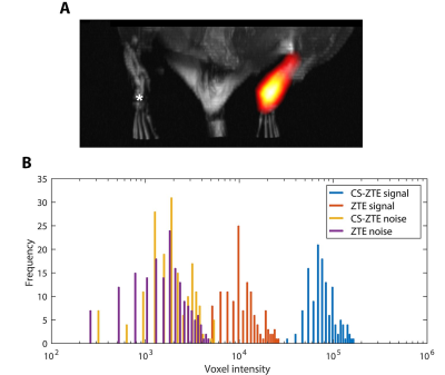 |
1 | Enhanced detection of paramagnetic fluorine-19 MRI agents using compressed sensing zero echo time sequence
Jiawen Chen1, Piya Pal1, and Eric Ahrens2
1Department of Electrical and Computer Engineering, University of California San Diego, La Jolla, CA, United States, 2Department of Radiology, University of California San Diego, La Jolla, CA, United States To accelerate the imaging speed and improve the signal-to-noise ratio of three-dimensional, whole body 19F MRI, we describe an ultrafast compressed sensing data acquisition scheme adapted for a zero echo time (ZTE) pulse sequence, with data reconstruction via a sparsity-promoting algorithm using a nonuniform fast Fourier transform (NUFFT) sensing operator. These methods are well suited for MRI of labeled cells using 19F tracer agents, particularly paramagnetic metallo-perfluorocarbon nanoemulsions. In phantom data and an in vivo immunotherapy mouse model, we show the feasibility of boosting MRI detection sensitivity by greater than an order of magnitude without increasing total scan time. |
||
4059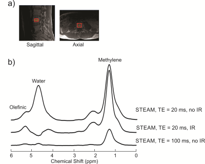 |
2 | Inversion Recovery for Resolving the Olefinic Resonance in Spinal Bone Marrow at 3 T
Clara J. Fallone1 and Atiyah Yahya1,2
1Department of Oncology, University of Alberta, Edmonton, AB, Canada, 2Department of Medical Physics, Cross Cancer Institute, Edmonton, AB, Canada
Magnetic resonance spectroscopy measures of the olefinic resonance (≈ 5.3 ppm) enables estimation of fat unsaturation levels. In spinal bone marrow, the olefinic peak is significantly overlapped by that of water (≈ 4.7 ppm). Previously, STEAM with a TE of 100 ms was demonstrated to resolve the olefinic resonance from that of water in spinal bone marrow at 3 T. In this work, we show that an inversion recovery with short-TE STEAM (20 ms) suppresses the water signal, resolving the olefinic resonance and yielding a signal to noise ratio 3.3x larger than that obtained with long-TE STEAM.
|
||
4060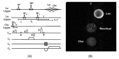 |
3 | In vivo phase-incrementing SEE-HSelMQC method to resolve tumor biomarker images by synchronizing RF phase increments and phase-encoding steps
Qiuhong He1,2, Hong Yuan2,3, and Yen-Yu Ian Shih1,2,4
1Center for Animal MRI, University of North Carolina at Chapel Hill, Chapel Hill, NC, United States, 2Biomedical Research Imaging Center (BRIC), University of North Carolina at Chapel Hill, Chapel Hill, NC, United States, 3Department of Radiology, University of North Carolina at Chapel Hill, Chapel Hill, NC, United States, 4Department of Neurology, University of North Carolina at Chapel Hill, Chapel Hill, NC, United States
We have developed the phase-incrementing MRSI (pi-MRSI) method to resolve overlapping biomarker images in the presence of a frequency encoding gradient during acquisition time. We report here the pi-SEE-HSelMQC experiments on a pre-clinical Bruker 9.4T spectrometer. The choline-selective and lactate CH-selective RF pulses in the pulse sequence were phase incremented by 10° in opposite signs in synchronization with the phase-encoding steps. In vivo two-dimensional pi-SEE-HSelMQC imaging of lactate and choline acquired from the PC3 human prostate cancer xenograft in a nude mouse showed opposite image offsets, shifted away from spurious signals. The pi-SEE-HSelMQC method completely suppresses lipid and water.
|
||
4061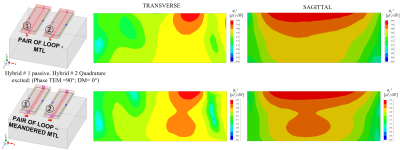 |
4 | Quadrature RF array using High Impedance concept for improved transmit RF field B1 efficiency at 7 Tesla
Komlan Payne1 and Xiaoliang Zhang1
1Jacobs School of Medicine & Biomedical Sciences, University at Buffalo, The State University of New York, Buffalo, NY, United States
Decoupling performance between coil elements is one of the factors that limits the transmit B1 field efficiency and SNR. However, mutual coupling between coil elements is an inevitable electromagnetic interaction that alters the isolated current distribution and impedance of individual elements. In contrast to linear arrays, decoupling challenge of quadrature arrays is more pronounced. In the pursuit of low inter-coupling, a high-impedance technique is adopted. A quadrature hybrid coil composed of loop and microstrip is used in a planar configuration. By employing the high-impedance method, sufficient decoupling between quadrature coils is obtained without the use of additional decoupling network.
|
||
4062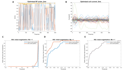 |
5 | Selective Refocusing Pulse Design via Time-varying Nonlinear Shim Array Fields and RF Pulse with Decomposition Property
Molin Zhang1, Nicolas Arango1, Jason Stockmann2, Jacob White1, and Elfar Adalsteinsson1,3,4
1EECS, MIT, Cambridge, MA, United States, 2Athinoula A. Martinos Center for Biomedical Imaging, Charlestown, MA, United States, 3Harvard-MIT Health Sciences and Technology, MIT, Cambridge, MA, United States, 4Institute for Medical Engineering and Science, MIT, Cambridge, MA, United States
Shim arrays provide nonlinear, time-varying fields that can extend the possibility of linear gradient field for RF excitations. In this work, we explore the possibility of designing a selective refocusing 180° pulse for a restricted slice pattern of 1.2cm thickness, aimed at application for the fetal brain. We use an auto-differentiable Bloch simulator framework for the design, but without the presentence of crusher gradients. Decomposition property is employed to overcome the undetermined initial magnetization. The selective refocusing profile achieves selective refocusing for 85% of voxels in the ROI (My< -0.9).
|
||
4063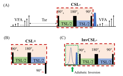 |
6 | Improved Phase-cycling Preparations in Quantitative T1rho Mapping
Gregory Peng1, Can Wu2, and Qi Peng1
1Department of Radiology, Albert Einstein College of Medicine, Bronx, NY, United States, 2Department of Medical Physics, Memorial Sloan Kettering Cancer Center, New York, NY, United States
Phase-cycling (PC) has been proposed as an effective method to minimize the impact of B0 and B1 field inhomogeneities in quantitative T1rho mapping, which needs both positive and negative longitudinal magnetization T1rho preparations. We propose here a simple magnetization inversion with an adiabatic inversion RF pulse before an established T1ρ preparation module to achieve an opposite PC. This approach has been shown to be much less sensitive to B0/B1 field imperfections compared with a traditional approach of reversing the phase of the last 90° RF pulse.
|
||
4064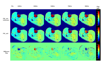 |
7 | Fast 3D T1rho Dispersion MRI with Interleaved Phase Cycling MAPSS
Qi Peng1 and Can Wu2
1Department of Radiology, Albert Einstein College of Medicine, Bronx, NY, United States, 2Department of Medical Physics, Memorial Sloan Kettering Cancer Center, New York, NY, United States
T1ρ dispersion imaging based on repeated T1ρ measurements at multiple spin-lock frequencies allows characterization of different dynamic processes of tissues. The clinical potential of this techniques is however limited by the total scan time needed to obtain reliable and consistent quantitative measurements. In this work, we propose an unpaired, interleaved phase-cycling scheme for T1ρ dispersion imaging, which almost reduces total scan time by a half compared with the traditional paired PC approach. This method, when combined with other advanced imaging and reconstruction techniques, could potentially enable high-resolution T1rho dispersion imaging acquired within clinically acceptable scan duration.
|
||
4065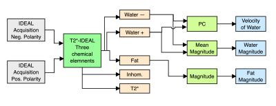 |
8 | Simultaneous acquisition of water-fat concentrations and velocity images using a Phase-contrast T2*-IDEAL method
Esteban Denecken1,2,3, Cristobal Arrieta1,3, Hernán Mella1,3, and Sergio Uribe1,3,4
1Biomedical Imaging Center, Pontificia Universidad Católica de Chile, Santiago de Chile, Chile, 2Department of Electrical Engineering, School of Engineering, Pontificia Universidad Católica de Chile, Santiago de Chile, Chile, 3ANID – Millennium Science Initiative Program – Millennium Nucleus for Cardiovascular Magnetic Resonance, Santiago de Chile, Chile, 4Department of Radiology, School of Medicine, Pontificia Universidad Católica de Chile, Santiago de Chile, Chile Phase-contrast in areas with a significant fat signal is subject to chemical shift artifacts where the fat signal interferes with the neighboring water signal. The purpose of this work is to present a novel method that combines phase-contrast with T2*-IDEAL water-fat separation to obtain water-fat concentrations and velocity images from a single acquisition. We validate our method using a numerical phantom with different combinations of water-fat concentrations, velocity, and noise, and MRI of a 2D axial section of the neck acquired in volunteers. PDFF comparisons showed a significant agreement between phase-contrast T2*-IDEAL and standard methods.
|
||
4066 |
9 | General Analytical Solution for Phase-Cycled bSSFP Imaging with Three Acquisitions
Qing-San Xiang1
1Radiology, University of British Columbia, Vancouver, BC, Canada
Elliptical signal model for bSSFP imaging has found various applications such as banding artifact removal and quantitative mapping of physical parameters. Typically, four or more phase-cycled acquisitions are needed and thus impose a limiting factor in term of total scan time. In this work, it is found that the phase-cycled bSSFP system can be unlocked using a general analytical solution with only three acquisitions, leading to a significant reduction of total scan time. Mathematical frame work is presented, followed by validation with simulated data. Future work includes further demonstration, optimization, and applications with phantoms and in vivo subjects.
|
||
4067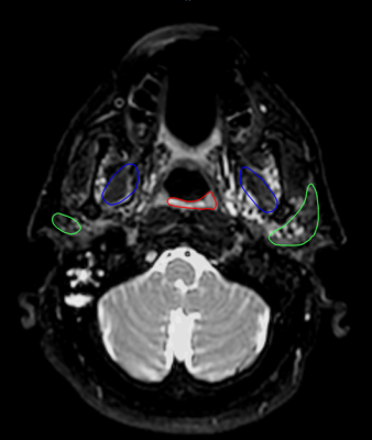 |
10 | Development and robust analysis of 3D fat-suppressed T2 MRI for head and neck radiotherapy treatment planning
Travis Salzillo1, Alex Dresner2, Brigid McDonald1, Ashley Way1, Sara Ahmed 1, Lauren Andring1, Kelsey Corrigan1, Gohar Manzar1, Alison Yoder1, Abdallah Mohamed1, Chelsea Pinnix1, Jason Stafford3, Jihong Wang4, and Clifton Fuller1
1Radiation Oncology, MD Anderson Cancer Center, Houston, TX, United States, 2Philips Healthcare, Best, Netherlands, 3Imaging Physics, MD Anderson Cancer Center, Houston, TX, United States, 4Radiation Physics, MD Anderson Cancer Center, Houston, TX, United States
In order to improve head and neck radiotherapy treatment planning, a series of 3D fat-suppressed T2-weighted sequences were developed with varying pulse sequence parameters. One non-fat-suppressed and five fat-suppressed sequences were acquired on five patients, and on each image, four structures were segmented by six radiation oncology physicians (observers). Robust and comprehensive analysis was performed to assess qualitative and quantitative image quality metrics. This included geometric distortion, SNR and CNR measurements, structure conspicuity, interobserver segmentation variability, and qualitative image quality rankings. The results of these were used to determine the optimal fat-suppressed sequence for head and neck radiation treatment planning.
|
||
4068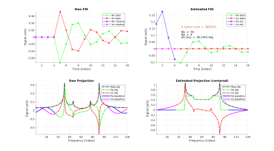 |
11 | Robust and Computationally Efficient Missing Point and Phase Estimation for Multi-Channel Acquisition with ZTE Sequences
Curtis A Corum1,2, Stanley J Kruger2, Mathews Jacob3, Vincent A Magnotta2, and James H Holmes2
1Champaign Imaging LLC, Shoreview, MN, United States, 2Radiology, University of Iowa, Iowa City, IA, United States, 3Electrical and Computer Engineering, University of Iowa, Iowa City, IA, United States
Missing point estimation has been a part of Fourier Transform NMR and MRI since the beginning. FID based imaging sequences, such as the ZTE (zero echo time) sequence, are missing needed data points corresponding to the region around the center of K-space. In multi-channel ZTE acquisitions, this estimate must be made for each channel. Missing data can be estimated by separate reference scans (WASPI and PETRA) but require extra time. In dynamic acquisitions missing points are optimally estimated from the data itself. Here we investigate applying a method borrowed from solid state NMR to estimate the missing points.
|
||
 Back to Meeting Home
 Back to the Program-at-a-Glance
The International Society for Magnetic Resonance in Medicine is accredited by the Accreditation Council for Continuing Medical Education to provide continuing medical education for physicians.
 Back to Meeting Home
 Back to the Program-at-a-Glance
 View the Presentation