Online Gather.town Pitches
New Systems & Devices III
Joint Annual Meeting ISMRM-ESMRMB & ISMRT 31st Annual Meeting • 07-12 May 2022 • London, UK

Online Gather.town Pitches
New Systems & Devices III
| Booth # | ||||
|---|---|---|---|---|
3947 |
1 | Characterization and Correction of Diffusion Gradient-Induced Eddy Currents in Echo-Planar and Spiral Cardiac Diffusion Tensor Imaging
Robbert J.H. van Gorkum1, Christian Guenthner1, Andreas Koethe1,2, Christian T. Stoeck1,3, and Sebastian Kozerke1
1Institute for Biomedical Engineering, University and ETH Zurich, Zurich, Switzerland, 2Center for Proton Therapy, Paul Scherrer Institute, Villigen, Switzerland, 3Division of Surgical Research, University Hospital Zurich, University Zurich, Zurich, Switzerland
Second-order motion-compensated diffusion encoding gradient waveforms cause substantial eddy currents (ECs), which can violate the linear-system assumption of a gradient impulse response function and thus require a dedicated measurement and reconstruction approach. Using a 3-dimensional spectroscopic imaging method, diffusion encoding gradient-induced ECs were characterized and non-linear effects were identified. When accounting for zeroth- and first-order diffusion encoding gradient-induced ECs besides off-resonance and trajectory-induced concomitant fields and ECs, image distortions could be adequately mitigated in echo-planar and spiral in vivo second-order motion-compensated cardiac diffusion tensor imaging sequences.
|
||
3948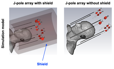 |
2 | Simulation of a shield effect on a J-pole antenna array for ultra-high field MR-PET
Chang-Hoon Choi1, Suk-Min Hong1, Jörg Felder1,2, Christoph Lerche1, and N. Jon Shah1,3,4,5
1Institute of Neuroscience and Medicine - 4, Forschungszentrum Juelich, Juelich, Germany, 2RWTH Aachen University, Aachen, Germany, 3Institute of Neuroscience and Medicine - 11, Forschungszentrum Juelich, Juelich, Germany, 4JARA - BRAIN - Translational Medicine, Aachen, Germany, 5Department of Neurology, RWTH Aachen University, Aachen, Germany
Interest in simultaneously operating MR-PET systems as a means of accessing multiple biological parameters with high spatial and temporal resolution is increasing. In a hybrid acquisition, the RF antenna is used inside the PET FOV generating potential photon loss and artefacts. A multi-channel J-pole antenna array, which does not include any high-density parts in the imaging FOV, has been recently introduced, and its outstanding performance has been demonstrated. To prevent interference between MR and PET, including an RF shield is necessary. Here, we conducted a simulation to compare the characteristics of the J-shape antenna array with and without the shield.
|
||
3949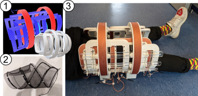 |
3 | iMPI – first real-time imaging with a human-sized interventional Magnetic Particle Imaging scanner
Patrick Vogel1, Martin A Rückert1, Teresa Reichl1, Johanna Günther1, Christoph Greiner1, Liana Mirzojan1, Alexander von Böhn1, Thomas Kampf2, Thorsten A. Bley3, Volker C. Behr1, and Stefan Herz3
1Experimental Physics 5 (Biophysics), University of Würzburg, Würzburg, Germany, 2Diagnostic and Interventional Neuroradiology, University Hospital Würzburg, Würzburg, Germany, 3Diagnostic and Interventional Radiology, University Hospital Würzburg, Würzburg, Germany In this work a first human-sized Magnetic Particle Imaging (MPI) scanner designed and specifically engineered for experimental (cardio)vascular interventions is presented. Based on a novel open design implementing the so called traveling wave MPI approach, the open iMPI system provides imaging of Tracers based on superparamagnetic iron oxide nanoparticles (SPIONs) with high sensitivity, optimal patient handling and would even allow hybrid imaging of magnetic tracers within gold standard x-ray based interventional angiography systems. In initial experiments, the feasibility of a human-sized interventional MPI scanner with real-time data reconstruction and image visualization is demonstrated. |
||
3950 |
4 | 8ch multi transmit with ultrasonic dual axis gradient and 32ch receive setup for fast spatial encoding at 7T Video Not Available
Mark Gosselink1, Aris van Ieperen1, Wout Schuth2, Martino Borgo2, Dimitri Welting1, Edwin Versteeg1, and Dennis Klomp1
1Department of Radiology, University Medical Center Utrecht, Utrecht, Netherlands, 2Futura composites, Heerhugowaard, Netherlands
We propose a setup to boost spatiotemporal encoding for MRI in the human brain using a conventional receiver array (32ch) and fast gradients (6100T/m/s). Moreover, we demonstrate that the operation frequency of a high-end gradient-amplifier can be increased to ultrasonic frequencies so to avoid unpleasant acoustic noise. By using this amplifier with 8 RF transmitters, 32 receivers and a 2-channel gradient coil, a light-weight setup is constructed for operation in a 7 Tesla MRI system. The setup is demonstrated for operation in the human brain showing good FLAIR data and no peripheral nerve stimulation at the slew rate of 6100T/m/s.
|
||
3951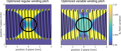 |
5 | Optimized variable winding pitch coil designs for miniature B0 field modification. Video Permission Withheld
Niklas Wehkamp1, Philipp Rovedo2, and Maxim Zaitsev3
1Department of Radiology, Medical Physics, Medical Center – University of Freiburg, Freiburg, Germany, 2Neurozentrum, Medical Center – University of Freiburg, Freiburg, Germany, 3High Field MR Center, Center for Medical Physics and Biomedical Engineering, Medical University of Vienna, Vienna, Austria We present numerical optimization of variable winding pitch coil designs for miniature coils with respect to target field homogeneity. The optimization is based on a continuous coil winding layout that was used to simulate field homogeneity in the target volume via the Biot-Savart law. The example coil designs expand the range of applications for frequency adjustable field probes, as they increase the B0 modification field’s homogeneity by factor of 7. Due to the specific requirements of miniature B0 modification coils, the dimensions of the coil are restricted in length, width and wire diameter. |
||
3952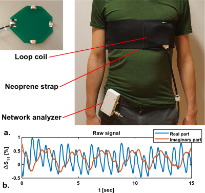 |
6 | Measuring stroke volume with wearable RF antennas: a validation study with EM simulations and MRI
Bart Romke Steensma1, Christina Anna Louka1,2, Alexander Jurriaan Eberhardt Raaijmakers3, and Cornelis Antonius Theodorus van den Berg1
1Center for Image Sciences, Computational Imaging Group, University Medical Center Utrecht, Utrecht, Netherlands, 2School of Applied Mathematical and Physical Sciences, Department of Physics, National Technical University of Athens, Athens, Greece, 3Biomedical Engineering, Medical Imaging Analysis, Eindhoven University of Technology, Eindhoven, Netherlands
We developed a wearable setup to detect heart motion and stroke volume with an RF antenna connected to a miniature network analyzer. EM simulations were used to demonstrate the possibility of measuring changes in stroke volume with an RF antenna and to investigate the spatial sensitivity of the antenna. A Valsalva manoeuver was used to provoke changes in stroke volume, which were observed with the RF antenna and in cine MRI acquisitions.
|
||
 |
3953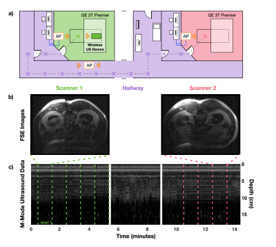 |
7 | Wireless Physiological Motion Monitoring with an Integrated RF/Wireless Coil Array and MR-Compatible Ultrasound-Based System
Devin Willey1,2, Olivia Jo Dickinson1,2, Devon Karl Overson1,2, Trong-Kha Truong1,2, Fraser Robb3, Allen W Song1,2, Bruno Madore4, and Dean Darnell1,2
1Brain Imaging And Analysis Center, Duke University, Durham, NC, United States, 2Medical Physics Graduate Program, Duke University, Durham, NC, United States, 3Brain Imaging And Analysis Center, GE Healthcare, Aurora, OH, United States, 4Radiology, Brigham and Women's, Harvard Medical School, Boston, MA, United States
A wireless MR-compatible ultrasound-based device consisting of an integrated RF/wireless coil, organ-configuration motion sensor, Raspberry Pi, pulser/receiver, and analog-to-digital converters was developed to acquire, digitize, and wirelessly transmit ultrasound data while simultaneously acquiring MR-images. Experiments were performed to demonstrate the system's 1) mobility through continuously acquiring, digitizing, and wirelessly transmitting ultrasound data at multiple locations, and 2) ability to wirelessly monitor physiological motion during coughing/gasping. This integrated system enables the OCM sensor to travel with patients throughout the hospital while also demonstrating the ability of the iRFW coil and backend electronics to process and wirelessly transmit a high-fidelity signal.
|
|
 Back to Meeting Home
 Back to the Program-at-a-Glance
The International Society for Magnetic Resonance in Medicine is accredited by the Accreditation Council for Continuing Medical Education to provide continuing medical education for physicians.
 Back to Meeting Home
 Back to the Program-at-a-Glance
 View the Presentation