Online Gather.town Pitches
Gray Matter & Neurofluids II
Joint Annual Meeting ISMRM-ESMRMB & ISMRT 31st Annual Meeting • 07-12 May 2022 • London, UK

Online Gather.town Pitches
Gray Matter & Neurofluids II
| Booth # | ||||
|---|---|---|---|---|
4846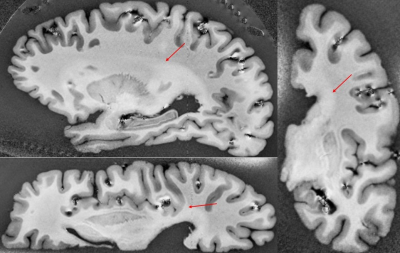 |
1 | Addressing ultra highfield MRI challenges in ex-vivo
Nadim Farhat1, Heching Jin2, Tales Santini1, Tiago Martins2, Jacob Berardinelli1, Noah Schweitzer1, Salem AlKhateeb 1, Milos Ikonomovic1, Julia Ann Kofler3, Joseph Mettenburg3, Howard J. Aizenstein1, and Tamer S. Ibrahim1
1University of Pittsburgh, pittsburgh, PA, United States, 2Bioengineering, University of Pittsburgh, pittsburgh, PA, United States, 3UPMC Presbyterian Hospital, Pittsburgh, PA, United States |
||
4847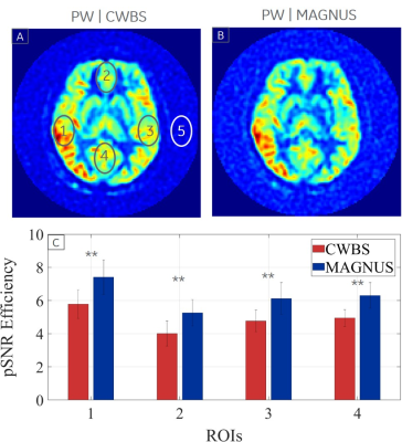 |
2 | 3D Pseudo-Continuous Arterial Spin Labeling Acquisition using a High-Performance Gradient System: A Scan Time and Image Quality Assessment
Afis Ajala1, Seung-Kyun Lee1, Daehun Kang2, Luca Marinelli1, Desmond Teck Beng Yeo1, and Thomas Foo1
1Magnetic Resonance Imaging, GE Global Research, Niskayuna, NY, United States, 2Mayo Clinic, Rochester, MN, United States
The maximum gradient amplitude (Gmax) and slew rate (SRmax) determine the minimum number of interleaves (and total scan time) in a 3D pseudo-continuous arterial spin-labeling (3DpCASL) acquisition with a stack-of-spirals readout module for a given spatial resolution. The higher Gmax and SRmax of a recently developed head-only gradient system (MAGNUS) was utilized to reduce the total scan time of a clinical 3DpCASL acquisition by a factor of 2 compared to a similar acquisition carried out at a lower Gmax and SRmax of a conventional whole-body system (CWBS) while maintaining comparable image quality.
|
||
4848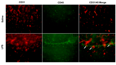 |
3 | Systemic inflammation alters brain blood flow and R2*: An in-vivo 9.4T MRI animal study
Qandeel Shafqat1, Ying Wu1, A. Max Hamilton1, Mada Hashem1, and Jeff F Dunn1
1Department of Radiology, University of Calgary, Calgary, AB, Canada
Systemic inflammation is linked to a range of neurological diseases. Reductions in cerebral blood flow (CBF) and the presence of brain hypoxia have been detected in animal models of inflammation and in multiple sclerosis, a disease with significant inflammation. Reduced CBF combined with hypoxia could exacerbate damage in neuroinflammatory conditions. To study this link, we used in-vivo 9.4T MRI to quantify CBF and R2*, a marker of deoxyhemoglobin, following systemic inflammation induced by bacterial lipopolysaccharide. We found reduced CBF and increased R2* in 4 regions, including the cortex and hippocampus—indicating that inflammation is accompanied by hypoxia and reduced CBF.
|
||
4849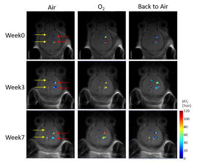 |
4 | Quantitative Mapping of Brain Tissue Oxygenation around chronically implanted neural interfaces
Yuka Sugamura1, Arati Sridharan1, Umu Jalloh1, Babak Moghadas1, Livia De Mesquita Teixeira1, Jitendran Muthuswamy1, and Vikram Kodibagkar1
1Arizona State University, Tempe, AZ, United States
Chronic implantation of neural interfaces often experiences decrease in signal reliability over time. Tissue response, including the alteration of the oxygen kinetics, is thought to be a major cause. Here, we report the spatial mapping of brain oxygenation around chronically implanted electrodes using a previously developed quantitative MRI-based oxygen sensing technique, PISTOL. Seven 200 um diameter electrodes implanted in four mice allowed quantitative pO2 mapping throughout the 7-week study. No changes were observed in the baseline brain tissue oxygenation as well as the response to respiratory challenge. Future studies will combine this approach with electrophysiology, and ultimately perform simultaneous assessment.
|
||
4850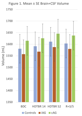 |
5 | INTRACRANIAL EFFECTS OF ARTIFICIAL GRAVITY: A 3T MRI STUDY Video Permission Withheld
Larry Allen Kramer1, Khader H. Hasan1, Xu Zhang2, Brandon R. Macias3, Karina Marshall-Goebel4, Steven S. Laurie4, and Eric M. Bershad5
1Diagnostic Imaging, UTSHC-Houston, Houston, TX, United States, 2UTSHC-Houston, Houston, TX, United States, 3NASA, Houston, TX, United States, 4KBR, Houston, TX, United States, 5BCM, Houston, TX, United States
Headward fluid shift is a natural consequence of working in microgravity. It is theorized to be the cause of changes in brain morphology and the development of optic nerve pathology in astronauts. This study looked at the application of artificial gravity in the form of short-arm centrifugation at 0.3g as a potential countermeasure in 24 healthy volunteers over a 60 day period. Headward fluid shift was simulated using a head down tilt bed rest model. Both continuous and intermittent artificial gravity for 30 minutes per day failed to show any benefit on intracranial changes associated with chronic headward fluid shift.
|
||
4851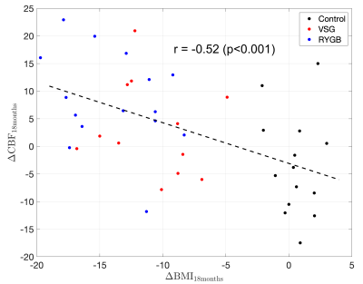 |
6 | Bariatric Surgery increases Cerebral Blood Flow in Women with Obesity
Sudipto Dolui1, Ariana M. Chao1, Noel N. Williams1, Thomas A. Wadden1, and John A. Detre1
1Perelman School of Medicine, University of Pennsylvania, Philadelphia, PA, United States
Obesity is a significant risk factor for cerebrovascular disease including dementia. Cerebral blood flow (CBF) provides a biomarker of cerebrovascular health and can be measured non-invasively using arterial spin labeled (ASL) perfusion MRI. Here we examined longitudinal ASL data from adult women with obesity who underwent bariatric surgery. We demonstrated that weight loss was associated with significant increases in CBF from baseline to 6 and 18 months after surgery. Change in glucose and insulin level did not mediate the relationship, suggesting a more direct association between obesity and CBF.
|
||
4852 |
7 | Is T2 a biomarker of inflammation in normal appearing gray matter: Maybe!
Qandeel Shafqat1, Rehman Tariq1, Rania Muhammed1, Ying Wu1, and Jeff F. Dunn1
1Department of Radiology, University of Calgary, Calgary, AB, Canada
With the reduction in use of gadolinium enhanced MRI for assessment of neuroinflammation, it is important to identify new non-invasive biomarkers of brain inflammation, as well as to understand the link between inflammation induced pathology and MRI. We used quantitative T2, behaviour and histology to study gray matter in an LPS mouse model of inflammation. There was a significant reduction in T2, along with behaviour and histological markers indicating inflammation. Although T2 appears useful as biomarker for inflammation, it may be that the precision needed will make it difficult to implement in patients.
|
||
4853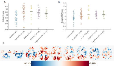 |
8 | Dynamic Changes in the Pituitary Gland and Brain Stiffness with Treatment of Idiopathic Intracranial Hypertension
Petrice M Cogswell1, Matthew C Murphy1, Muhammad T Bhatti2, Jeremy K Cutsforth-Gregory3, John Huston III1, and John J Chen2
1Radiology, Mayo Clinic, Rochester, MN, United States, 2Opthalmology, Mayo Clinic, Rochester, MN, United States, 3Neurology, Mayo Clinic, Rochester, MN, United States
Morphological changes in the pituitary gland and brain stiffness in idiopathic intracranial hypertension (IIH) are not well understood. We evaluated the difference in pituitary height and regional brain stiffness between IIH patients and controls and how these metrics change following acute and chronic treatment. The pituitary gland is smaller in IIH patients than controls and increases in size after chronic, but not acute, treatment of raised intracranial pressure. IIH patients have a different pattern of brain stiffness than controls, including stiffer occipital lobes. Following chronic treatment, the stiffness pattern becomes more like controls, though occipital stiffness does not significantly change.
|
||
4854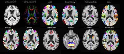 |
9 | Development and evaluation of a comprehensive array of gray matter labels for the MIITRA atlas: Interoperability with complementary atlases
Mohammad Rakeen Niaz1, Abdur Raquib Ridwan1, Yingjuan Wu1, Shengwei Zhang2, David A. Bennett2, and Konstantinos Arfanakis1,2
1Biomedical Engineering, Illinois Institute of Technology, Chicago, IL, United States, 2Rush Alzheimer's Disease Center, Rush University Medical Center, Chicago, IL, United States
The Multichannel Illinois Institute of Technology & Rush university Aging (MIITRA) atlas constructed using high quality MRI data on a large (N=400), diverse, community cohort of non-demented older adults, contains high resolution (0.5mm isotropic) structural and diffusion tensor imaging templates. The purpose of this work was to build and evaluate a comprehensive array of gyral-based, cytoarchitecture-based, and functional connectivity-based gray matter labels in MIITRA space in order to enhance the functionality of the MIITRA atlas and its interoperability with complementary atlases.
|
||
4855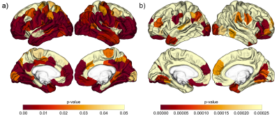 |
10 | Correlates of Perivascular Spaces Volume Based on Automated Segmentation of T1-weighted MRI
Sichen Ludwig Zhao1, William S Tackett2, Banafsheh Shakibajahromi2, Sudipto Dolui3, Ilya M Nasrallah3, David A Wolk2, Corey T McMillan2, Farshid Sepehrband4, and John A Detre2
1Department of Bioengineering, School of Engineering and Applied Science, University of Pennsylvania, Philadelphia, PA, United States, 2Department of Neurology, Perelman School of Medicine, University of Pennsylvania, Philadelphia, PA, United States, 3Department of Radiology, Perelman School of Medicine, University of Pennsylvania, Philadelphia, PA, United States, 4Laboratory of Neuroimaging, Keck School of Medicine of USC, University of Southern California, Los Angeles, CA, United States
Perivascular spaces (PVS) are a component of the glymphatic system, the brain-wide waste drainage pathway, but the pathophysiological significance of enlarged perivascular space volume is incompletely characterized. We obtained PVS segmentations from 167 cognitively intact or mildly symptomatic older subjects from high-resolution T1-weighted MRI using an automated approach and correlated PVS volume with both amyloid status, as determined by amyloid PET, and cortical thickness changes from normative values. Automated PVS segmentations required additional masking of CSF spaces and manual exclusion of some lacunar infarcts. PVS volume was correlated with brain atrophy and not amyloid status.
|
||
4856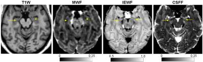 |
11 | Increased hippocampal cerebrospinal fluid fraction is associated with episodic verbal learning and memory deficits in multiple sclerosis
Thanh D Nguyen1, Liangdong Zhou1, Elizabeth M Sweeney1, Melanie Marcille1, Susan A Gauthier1, Yi Wang1, and Yi Li1
1Weill Cornell Medicine, New York, NY, United States
We applied FAST-T2 multi-component T2 relaxometry to 145 MS patients and found that the mean hippocampal CSF fraction, a measure of glymphatic clearance dysfunction in the hippocampus, was negatively associated with verbal learning and memory performance measured by the California Verbal Learning Test after adjusting for age, sex, MS phenotype, disease duration, and log total lesion volume.
|
||
4857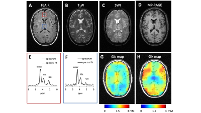 |
12 | A Multi-Sequence, Interleaved MRI-DMI Protocol for Human Brain
Yanning Liu1, Henk M De Feyter2, Robert K Fulbright2, Scott McIntyre2, Terence W Nixon2, and Robin A de Graaf1,2
1Department of Biomedical Engneering, Yale University, New Haven, CT, United States, 2Department of Radiology and Biomedical Imaging, Yale University, New Haven, CT, United States Deuterium Metabolic Imaging (DMI) is an emerging method to spatially map metabolism in vivo. To enhance the time efficiency of a combined MRI-DMI protocol, interleaved MRI-DMI acquisitions were developed for multiple clinical MRI sequences. The protocol includes four MRIs that are commonly used in neurological MRI exams, namely FLAIR, T2W, SWI and T1W MP-RAGE, with DMI acquisition in parallel, acquired in 28 minutes. We demonstrate the performance of the interleaved MRI-DMI protocol on healthy human brain and discuss the guiding principles as well as limitations of extending other MRI sequences with DMI using the presented interleaving strategy. |
||
4858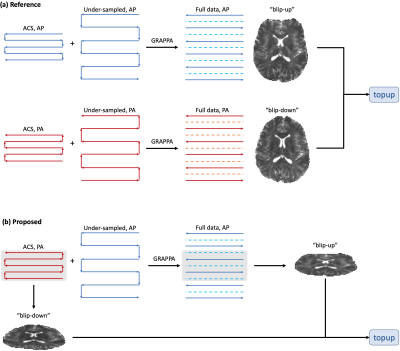 |
13 | EPI with Parallel Imaging (eπ2) self-calibrates the image distortion due to B0 field inhomogeneity
Hong Hsi Lee1, Berkin Bilgic1, and Susie Y Huang1
1Athinoula A. Martinos Center for Biomedical Imaging, Massachusetts General Hospital, Charlestown, MA, United States
Correcting image distortions in echo-planar-imaging (EPI) due to B0 field inhomogeneity usually requires additional acquisitions, such as a B0 field map or blip-up-blip-down image pair. Interestingly, susceptibility distortions in EPI with parallel imaging can be corrected by leveraging the mismatch in phase-encoding polarity and/or effective echo-spacing between the auto-calibration-signal and under-sampled data, obviating the need for additional image acquisitions. Here we demonstrate the proposed pipeline of susceptibility distortion correction in in vivo diffusion MRI data of a healthy subject, and the results and estimated field values are comparable with the conventional method.
|
||
 Back to Meeting Home
 Back to the Program-at-a-Glance
The International Society for Magnetic Resonance in Medicine is accredited by the Accreditation Council for Continuing Medical Education to provide continuing medical education for physicians.
 Back to Meeting Home
 Back to the Program-at-a-Glance
 View the Presentation