Online Gather.town Pitches
Cardiac I
Joint Annual Meeting ISMRM-ESMRMB & ISMRT 31st Annual Meeting • 07-12 May 2022 • London, UK

| Booth # | ||||
|---|---|---|---|---|
4156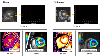 |
1 | Assessment of myocardial involvement in patients with Fabry disease using metabolic imaging by 1H-MR Spectroscopy (1H-MRS)
Masaru Shiotani1, Yoshiaki Morita1, Yoshito Ichiba2, Yasuhiro Nagai1, Wataru Ueki1, Tatsuhiro Yamamoto1, Yasutoshi Ohta1, Keizo Murakawa1, and Tetsuya Fukuda1
1Department of Radiology, National Cerebral and Cardiovascular Center, Suita, Osaka, Japan, 2MR Research & CollaborationDpt. Diagnostic Imaging Division, Siemens Healthcare K.K., Shinagawa-ku, Tokyo, Japan
The peak ratio of 3.5ppm to water in 1H-MRS of myocardium showed the higher values in Fabry disease and the association with native T1 values and regional function. 1H-MRS has a potential as disease-specific imaging biomarker for direct quantification of myocardial sphingolipid accumulation of Fabry disease.
|
||
4157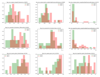 |
2 | The feasibility of radiomic analysis based on T1 mapping to distinguish between hypertensive heart disease and hypertrophic cardiomyopathy Video Permission Withheld
Shengliang Liu1, Jianxiu Lian2, Haixia Li2, Guokun Wang1, Yunling Li1, Yanming Zhao1, Bing Xu1, and Bo Yu1
1Department of Cardiology, the Second Affiliated Hospital of Harbin Medical University, Harbin, China, 2Philips Healthcare, Beijing, China
Hypertrophic cardiomyopathy (HCM) and hypertensive heart disease (HHD) share similar features, such as thickened left ventricular (LV) wall and reduced compliance, which make it difficult to distinguish HCM from HHD clinically. In this retrospective study, patients with HCM and HHD were enrolled to evaluate the feasibility of radiomic analysis to differentiate between these two diseases. For all calculated texture analysis (TA) features, AUCs of nine parameters had the values above 0.65, Logarithm_glszm_ZoneEntropy reached the best performance than other parameters (AUC: 0.790, sensitivity: 71.4 %, specificity: 81.8 %, P < 0.01).
|
||
4158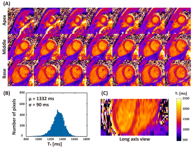 |
3 | 3D free-breathing simultaneous whole heart T1 and T2 mapping based on SAturation Recovery and Variable flip Angle (SAVA)
Dongyue Si1, Rui Guo2, Bowei Liu1, Daniel A. Herzka3, and Haiyan Ding1
1Center for Biomedical Imaging Research, Department of Biomedical Engineering, Tsinghua University, Beijing, China, 2Department of Medicine, Beth Israel Deaconess Medical Center and Harvard Medical School, Boston, MA, United States, 3National Heart, Lung, and Blood Institute, National Institutes of Health, Bethesda, MD, United States
Myocardial T1 and T2 mapping enable quantitative detection of various cardiac diseases. Here we propose a fast 3D free-breathing simultaneous T1 and T2 mapping sequence, which acquires four volumes for joint estimation of T1 and T2. A very small flip angle is used for efficient sampling of the equilibrium longitudinal magnetization. Whole-heart T1 and T2 maps were acquired with high resolution of 1.5×1.5×4mm3 within 5 min in normal human subjects with image quality comparable to conventional 2D methods.
|
||
4159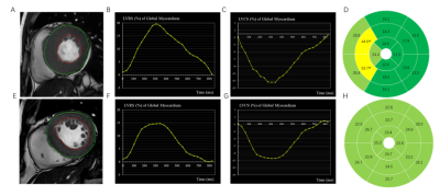 |
4 | Combination of strain with LGE can improve diagnostic ability for differentiation hypertrophic cardiomyopathy from hypertensive heart disease Video Permission Withheld
Shengliang Liu1, Jianxiu Lian2, Yunling Li1, Guokun Wang1, Haixia Li2, Xueying Wang1, Yanming Zhao1, Bing Xu1, and Bo Yu1
1Department of Cardiology, the Second Affiliated Hospital of Harbin Medical University, Harbin, China, 2Philips Healthcare, Beijing, China
To evaluate the value of left ventricular radial strain (LVRS) and left ventricular circumferential strain (LVCS) in discriminating between hypertrophic cardiomyopathy (HCM) and hypertensive heart disease (HHD) by using cardiac magnetic resonance feature tracking (CMR-FT). HCM patients presented higher values of LVRS and LVCS when compared with HHD patients. And both LVRS and LVCS correlated obviously with left ventricular ejection fraction (LVEF) in HCM patients. Furthermore, the combination of strain with LGE can significantly improve the diagnostic ability for differentiation HCM from HHD (AUC: 0.86; sensitivity: 86.9 %; specificity: 76.3 %).
|
||
4160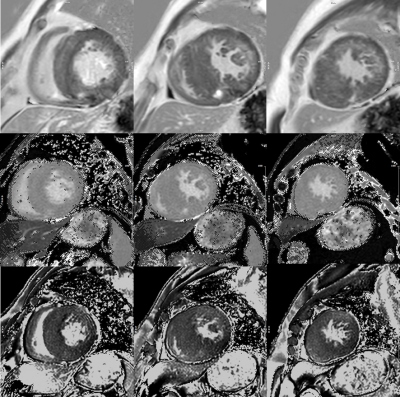 |
5 | T1 rho mapping and native T1 mapping for endogenous assessment of myocardial fibrosis in hypertrophic cardiomyopathy
Gang Yin1, Zhixiang Dong1, Shihua Zhao1, Xiuyu Chen1, Kai Yang1, Ke Jiang2, and Zhigang Wu2
1Fuwai Hospital, Beijing, China, 2Philips Healthcare, Beijing, China
This study is to examine the feasibility of T1 rho and native T1 mapping for endogenous detection of diffuse and focal myocardial fibrosis in patients with hypertrophic cardiomyopathy (HCM). Results showed that absolute T1 rho increased progressively and significantly from healthy controls to HCM without LGE and then to HCM with LGE, whereas T1 native increased significantly only from healthy controls to HCM with LGE. Besides, both T1 rho and native T1 had moderate correlation with LGE ratio in severe segments.
|
||
4161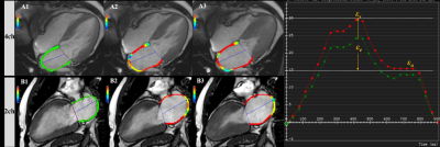 |
6 | Characteristics of left atrial strain in hypertrophic cardiomyopathy comorbid HF with preserved EF: evaluation by CMR-feature tracking
Shi Rui1, Gao Yue1, Shen Li-ting1, and Yang Zhi-gang1
1West China hospital of Sichuan University, Chengdu, China
The majority of heart failure (HF) in hypertrophic cardiomyopathy (HCM) manifests as a phenotype with preserved left ventricular (LV) ejection fraction, however, the exact contribution of left atrial (LA) phasic function to HF with preserved ejection fraction (HFpEF) in HCM remains unresolved. We designed the study to define the association between LA function and HFpEF in HCM patients using cardiac MRI feature tracking. The result revealed that LA phasic function was severely impaired in HCM patients with HFpEF, whereas LV function was not further impaired compared with non-HF patients.
|
||
4162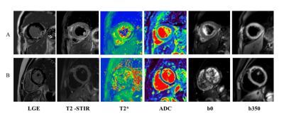 |
7 | The feasibility of evaluating acute ST-segment elevation myocardial infarction by using diffusion weighted imaging and T2* mapping Video Permission Withheld
Shengliang Liu1, Jianxiu Lian2, Ke Jiang2, Guokun Wang1, Yunling Li1, Xueying Wang1, Yanming Zhao1, Bing Xu1, and Bo Yu1
1Department of Cardiology, the Second Affiliated Hospital of Harbin Medical University, Harbin, China, 2Philips Healthcare, Beijing, China
Acute myocardial infarction is the result of occlusion of cardiac arteries, which leads to myocardial ischemic injury and even necrosis. Diffusion weighted imaging (DWI) and T2* mapping were performed to investigate the correlation between apparent diffusion coefficient (ADC) and T2* values in ST-segment elevation myocardial infarction (STEMI) patients here. ADC values of infarct regions were higher than those in border regions, remote regions in patients with STEMI, and also higher when compared with control group. Conversely, T2* values showed opposite trend. What’s more, there was a negative correlation between T2* and ADC values (r = 0.-734; P < .001).
|
||
4163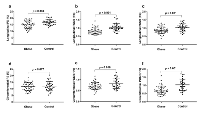 |
8 | Regional fat distributions are associated with subclinical right ventricular dysfunction in adults with uncomplicated obesity Video Not Available
Jing Liu1, Jing Li2, Huaxia Pu2, Wenzhang He1, Xue Li1, Xiaoyue Zhou3, Nanwei Tong2, and Liqing Peng1
1Radiology, West China Hospital, Sichuan University, Chengdu, China, 2Endocrinology and Metabolism, West China Hospital, Sichuan University, Chengdu, China, 3MR Collaboration, Siemens Healthineers Ltd., Shanghai, China This study evaluated right ventricular (RV) functional changes using cardiac magnetic resonance (CMR) tissue tracking and the association of these changes with fat distributions in obese adults with no clinical signs or comorbidities. The results showed that CMR tissue tracking can detect subclinical RV dysfunction with preserved RV ejection fraction in obese adults. Central obesity, represented by android fat, trunk fat, and the android/gynoid fat mass ratio, had a deleterious effect on RV subclinical dysfunction, whereas peripheral obesity (gynoid fat) might have had a protective effect. These findings could contribute to more precise obesity management in clinical practice. |
||
4164 |
9 | Left ventricular diastolic dysfunction in uncomplicated obesity: evaluated by CMR tissue tracking and volume-time curve Video Not Available
Jing Liu1, Jing Li2, Huaxia Pu1, Wenzhang He1, Xue Li1, Xiaoyue Zhou3, Nanwei Tong2, and Liqing Peng1
1Radiology, West China Hospital, Sichuan University, Chengdu, China, 2Endocrinology and Metabolism, West China Hospital, Sichuan University, Chengdu, China, 3MR Collaboration, Siemens Healthineers Ltd., Shanghai, China This study evaluated left ventricular (LV) diastolic functional changes and left atrial (LA) functional indices using cardiac magnetic resonance (CMR) tissue tracking and volume-time curve in obese adults with no clinical signs or comorbidities. The association of LV diastolic function with fat distributions was also assessed. The results showed that CMR detected subclinical LV diastolic dysfunction, impaired LA reservoir and conduit function with preserved LV ejection fractions in adults with obesity. LV diastolic function was associated with LA reservoir and conduit function. Visceral fat was deleterious for LV diastolic function, while peripheral obesity might have had a protective effect. |
||
4165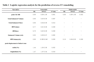 |
10 | Predictive value of cardiac magnetic resonance for reverse left ventricular remodeling after acute ST-segment elevation myocardial infarction
Jianing Cui1, Tao Li1, Yanan Zhao1, and Xiuzheng Yue2
1Department of Radiology, the First Medical center, Chinese People's Liberation Army Hospital, Beijing, China., Beijing, China, 2Philips Healthcare, Beijing, China, Beijing, China
Many ST-segment elevation myocardial infarction (STEMI) patients treated with primary percutaneous coronary intervention (PCI) are still exposed to a consequence known as left ventricular (LV) remodeling. Most studies have reported that reverse LV remodeling is associated with improved patient outcomes. Cardiac magnetic resonance (CMR) imaging has become a beneficial imaging modality to assess myocardial morphology, LV function and infarct characteristics simultaneously. This study assesses the predictive role of LV volume, LV function and infarct characteristics by CMR on reverse LV remodeling after STEMI. Our data showed that peak CK-MB, extent of MVO volume of LV were independent predictors of reverse LV remodeling.
|
||
4166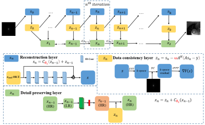 |
11 | Detail-preserving multi-scale deep learning reconstruction for cardiac magnetic resonance imaging
Juan Zou1, Cheng Li1, Ruoyou Wu1, Zhenzhen Xue1, Xin Liu1, Hairong Zheng1, and Shanshan Wang1
1Paul C. Lauterbur Research Center for Biomedical Imaging, Shenzhen Institute of Advanced Technology, Chinese Academy of Sciences, shenzhen city, China
Fast data acquisition and high-quality image reconstruction are vital for dynamic MRI, which can capture both anatomical and temporal information. High-resolution acquisition approaches in k-space and super-resolution approaches after reconstruction have been frequently reported. However, these methods may get details lost at high acceleration factors. To address this issue, we propose a multi-scale detail preserving reconstruction method for dynamic MR images. The residuals of multi-scale intermediate images in the iterative procedure are explored and the temporal and spatial dependencies between frames are considered. Promising results are achieved by the proposed method at the high acceleration factor of 11.
|
||
4167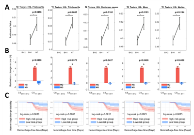 |
12 | Early MR radiomic biomarkers for re-hemorrhage after Gamma Knife Radiosurgery in Cavernous Malformation
Pei-Hsuan Kuo1, Cheng-Chia Lee2,3,4, Huai-Che Yang2,3, Hsiu-Mei Wu3,5, and Chia-Feng Lu1
1Department of Biomedical Imaging and Radiological Sciences, National Yang Ming Chiao Tung University, Taipei, Taiwan, 2Department of Neurosurgery, Neurological Institute, Taipei Veteran General Hospital, Taipei, Taiwan, 3School of Medicine, National Yang Ming Chiao Tung University, Taipei, Taiwan, 4Brain Research Center, National Yang Ming Chiao Tung University, Taipei, Taiwan, 5Department of Radiology, Taipei Veteran General Hospital, Taipei, Taiwan
Cavernous malformation (CM) is one of the common cerebral vascular diseases. Hemorrhage is a common and dangerous symptom of CMs, and re-hemorrhage may still occur in 30% of patients after the treatment of Gamma Knife radiosurgery (GKRS). Till now, the prediction of occurrences of future re-hemorrhage in CMs are still less explored. In this study, we used statistical analyses to observe longitudinal changes in MRI radiomic features before re-hemorrhage after GKRS. We aimed to identify the reliable image biomarkers using the quantitative and non-invasive MRI technique as early predictors to guide the clinical management for CM patients.
|
||
 Back to Meeting Home
 Back to the Program-at-a-Glance
The International Society for Magnetic Resonance in Medicine is accredited by the Accreditation Council for Continuing Medical Education to provide continuing medical education for physicians.
 Back to Meeting Home
 Back to the Program-at-a-Glance
 View the Presentation