Online Gather.town Pitches
MSK III
Joint Annual Meeting ISMRM-ESMRMB & ISMRT 31st Annual Meeting • 07-12 May 2022 • London, UK

| Booth # | ||||
|---|---|---|---|---|
4630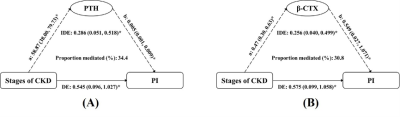 |
1 | Associations and mediation effects analysis of cortical porosity index, CKD stages, and bone metabolism markers
Yan Xiong1, Shuang Hu1, Yao Zhang 1, Donglin Wen1, Weiyin Vivian Liu2, and Xiaoming Li 1
1Radiology, Tongji Hospital, Tongji Medical College, Huazhong University of Science and Technology, Wuhan, China, 2GE Healthcare, Beijing, China
Chronic kidney disease (CKD) has a significant negative impact on bone health. However, the mechanisms of cortical bone deterioration and cortical porosity enlargement caused by CKD have not been fully described. Double-echo ultrashort echo-time magnetic resonance imaging (UTE MRI) provides the possibility of quantifying cortical porosity in vivo. The increasing CKD stages were associated with a higher PI value (Ptrend < 0.001). The association of CKD stages and PI mediated 34.4% and 30.8% of the total effect by increased PTH and β-CTX, respectively. Our study revealed the internal mechanism of bone deterioration caused by CKD to some extent.
|
||
4631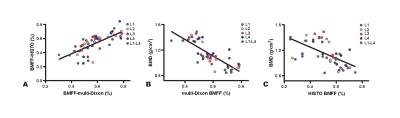 |
2 | Application of Multi-echo DIXON and HISTO to evaluate bone marrow fat/iron content in lumbar osteoporosis patients
Yang Wang1, Fengyu Sun1, Chen Zhang2, Haoran Sun1, Dong Li1, and Yuezeng Cai1
1Department of Radiology, Tianjin Medical University General Hospital, Tianjin, China, 2MR Scientific Marketing, Siemens Healthineers, Beijing, China
Our study investigated the feasibility of LiverLab technique, consisted of multi-echo DIXON and HISTO, to evaluate the vertebral BMFF and iron content for the diagnosis of osteoporosis. BMFF measured by multi-echo DIXON and HISTO was significantly increased in osteoporosis patients and strong negative correlation with BMD. Vertebral iron content had no difference in osteoporosis patients and no correlation with BMD. LiverLab technique, composed of multi-echo DIXON and HISTO, can quantitatively evaluate the changes in bone marrow fat content, and can be used as a potential biomarker for evaluating abnormal bone density and severity of osteoporosis.
|
||
4632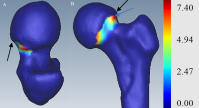 |
3 | Focused shape model assessment of cam-type femoroacetabular impingement: 3D MR cam morphology visualizations with associated surgical reviews Video Permission Withheld
Jessica Marie Bugeja1,2, Ying Xia1, Shekhar Chandra2, Nicholas Murphy3,4, Stuart Crozier2, David Hunter3,5, Jurgen Fripp1, and Craig Engstrom6
1AEHRC, CSIRO, Brisbane, Australia, 2School of Information Technology and Electrical Engineering, University of Queensland, Brisbane, Australia, 3Institute of Bone and Joint Research, University of Sydney, Sydney, Australia, 4Department of Orthopaedic Surgery, John Hunter Hospital, Newcastle, Australia, 5Department of Rheumatology, Royal North Shore Hospital, St Leonards, Australia, 6School of Human Movement and Nutrition Sciences, University of Queensland, Brisbane, Australia
Evaluating hip shape change with statistical shape models (SSMs) has been reported as a biomarker for OA incidence, progression and end-stage outcomes. However, there have been few MR studies using 3D SSMs for assessing proximal femur shape, encompassing the femoral head-neck region, in patients with conditions such as FAI. To the best of our knowledge, we present the first study to use focused shape models from 3D MR hip images to assess a focal region of (clinical) interest for cam morphology evaluation in patients classified as having an ‘inadequate’, ‘borderline’ or ‘satisfactory’ surgical outcome as determined from expert surgical review.
|
||
4633 |
4 | Denoising of 3D Fast Spin Echo Magnetic Resonance Images Using Convolutional Neural Networks with Rapid 2-NEX Acquisitions
Shutian ZHAO1, Fan XIAO1, Siyue LI1, Dόnal G. Cahill1, James F. Griffith1, and Weitian CHEN1
1Imaging and Interventional Radiology, CUHK lab of AI in radiology (CLAIR), Department of imaging and interventional radiology, The Chinese university of Hong Kong, Hong Kong SAR, China
We propose a deep learning denoising network with rapid 2-NEX acquisitions that can improve structure detail on rapid acquisition 3D FSE knee MR images. We investigated and compared this method to both conventional and deep learning methods. We demonstrated that this new method has the potential to suppress noise while improving structural detail on 3D FSE knee MR images.
|
||
4634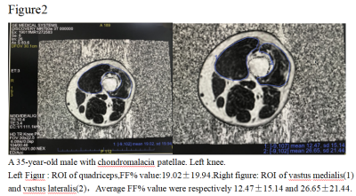 |
5 | Study on the relationship between accurate quantitative fat fraction of quadriceps femoris and patellofemoral arthritis by IDEAL-IQ
Binbin Yang1, Xiao cheng Wei2, and Xiao wen Ma1
1Honghui Hospital,Xi'an jiaotong University, Xi'an, China, 2GE Healthcare, Beijing, China
we aim to to explore the relationship between fat infiltration in quadriceps femoris with patellofemoral arthritis,It was concluded that IDEAL-IQ canbe a tool for quantification and prediction of patellar cartilage defect progression
|
||
4635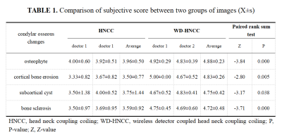 |
6 | Evaluation of condylar osseous changes using wireless detector combined with PDWI sequence
Chong Tian1, Xinge Cheng2, Rongpin Wang1, Chunqi Qian3, Xianchun Zeng1, and Lisha Nie4
1Guizhou Provincial People's Hospital, Guiyang,Guizhou, China, 2Zunyi Medical University, Guiyang,Guizhou, China, 3Michigan State University, East Lansing, MI, USA, MI, United States, 4GE Healthcare, MR Research China, Beijing, Beijing,China, China
MRI is considered to be the reference method for the imaging and understanding of temporomandibular disease (TMD) , but it provides little or no detectable signal from cortical bone. A disc-shaped wireless detector (WD), which is more suitable for body superficial tissue or organ examination, is used to overcome the sensitivity limitation of conventional MRI in condyle imaging. This study aims to observe the capability of WD in showing the bone changes of TMD condyle on PDWI. Compared with the traditional MRI, WD is suitable for imaging the condyle bone changes of TMD patients, and significantly improves the image quality.
|
||
4636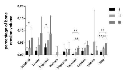 |
7 | Quantitative assessment of bone erosion in patients with rheumatoid arthritis using ZTE-MRI
Xingyao Yu1, Weiyin Vivian Liu2, Chao Liu3, Hu Chen3, Jingyu Jiang1, Qianqian Feng3, and Lin Xu3
1Biomedical Engineering College, Hubei University of Medicine, Shiyan Hubei, China, 2GE Healthcare, Beijing, China, 3Department of Radiology, Taihe Hospital, Hubei University of Medicine, Shiyan Hubei, China
MRI is reckoned as the most sensitive tool for monitoring changes such as synovitis and bone marrow edema at early stage of rheumatoid arthritis (RA)[1, 2]. Our results showed that ZTE-MRI can better precisely detect bone erosion compared with fs PD as well as DR and reflect RA degree via the volume percentage of bone erosion over carpal bone. Overall, ZTE-MRI could be an alternative to routine PDWI in clinic practice to help clinicians make better diagnosis without influenced by bone marrow edema, joint effusion and thickening of the synovial membrane and also offer articular function assessment as radiography.
|
||
4637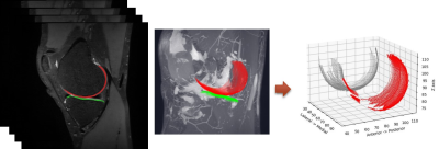 |
8 | Longitudinal Multiparametric Assessment of MRI in Symptomatic Knee Osteoarthritis and Correlation with Clinical Severity over 1 year Video Permission Withheld
Woo Young Kang1, Suk-joo Hong1, Hyeonbin Lee1, Ji-Hoon Bae1, Zepa Yang1, and In-Seong Kim2
1Korea University Guro Hospital, Seoul, Korea, Republic of, 2Siemens Healthcare, Seoul, Korea, Republic of
MRI is a valuable tool for to assess osteoarthritis (OA) progression. Although OA is a progressive degenerative disease involving the entire joint structure, cartilage degeneration is the central hallmark of the disease. MRI enables quantitative and semiquantitative assessment of longitudinal changes in articular cartilage morphology and composition in knee OA. We investigated the association between MRI biomarkers and knee OA progression over 12 months.
|
||
4638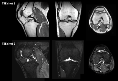 |
9 | MIXTURE-DOSMA: A comprehensive and multi-parametric quantitative 3D knee MR exam in 10 minutes
Jihun Kwon1, Takayuki Sakai2, Masami Yoneyama1, Daichi Murayama2, Quin Lu3, Arjun D Desai4, Akshay S Chaudhari5, and Marc Van Cauteren6
1Philips Japan, Tokyo, Japan, 2Department of Radiology, Eastern Chiba Medical Center, Chiba, Japan, 3Philips Healthcare NA, San Francisco, CA, United States, 4Electrical Engineering, Stanford University, Stanford, CA, United States, 5Radiology, Stanford University, Stanford, CA, United States, 6Philips Healthcare, Best, Netherlands
Multi-Interleaved X-prepared TSE with inTUitive RElaxometry (MIXTURE) is a recently developed 3D magnetization-prepared turbo spin-echo sequence. MIXTURE provides 3D high-resolution isotropic knee morphometry and T1ρ relaxometry in a clinically reasonable scan time. In this study, we integrated MIXTURE with the Deep Open-Source Medical Analysis (DOSMA) framework for deep learning-powered musculoskeletal MR image analysis to expand its clinical usage. By exploiting the advantage of both techniques, we show that the combination of MIXTURE and DOSMA enables a comprehensive whole knee MR exam including 3D proton density-weighted isotropic submillimeter imaging, T2- and T1ρ-mapping, and standardized image analysis techniques.
|
||
 Back to Meeting Home
 Back to the Program-at-a-Glance
The International Society for Magnetic Resonance in Medicine is accredited by the Accreditation Council for Continuing Medical Education to provide continuing medical education for physicians.
 Back to Meeting Home
 Back to the Program-at-a-Glance
 View the Presentation