Online Gather.town Pitches
MSK II
Joint Annual Meeting ISMRM-ESMRMB & ISMRT 31st Annual Meeting • 07-12 May 2022 • London, UK

| Booth # | ||||
|---|---|---|---|---|
4429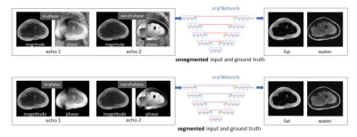 |
1 | Water-Fat Separation from Dual-Echo Dixon Imaging Using Deep Learning
Yan Wu1, Marc Alley 1, Keshav Datta1, Zhitao Li 1, Christopher Sandino 1, Zhifei Wen2, Michael Lustig3, John Pauly1, and Shreyas Vasanawala1
1Stanford University, Stanford, CA, United States, 2Hoag Hospital, Newport Beach, CA, United States, 3University of California, Berkeley, Berkeley, CA, United States
We design a data-driven method to generate water/fat images from dual-echo complex Dixon images, aimed at near-instant water-fat separation with high robustness. A hierarchical convolutional neural network is employed, where ground truth images are obtained using a binary quadratic optimization approach. With IRB approval and informed consent, 9281 image sets are collected from 30 pediatric patients to train and test networks, with the application of six-fold cross validation. In addition to high fidelity and significantly reduced processing time, the predicted images are superior to the ground truth in mitigation of water/fat swaps and correction of artifacts introduced by metallic implants.
|
||
4430 |
2 | AI-based prediction of patient mobility using MRI data from the Osteoarthritis Initiative
Bragi Sveinsson1,2 and Matthew S Rosen1,2,3
1Radiology, A. A. Martinos Center, Massachusetts General Hospital, Boston, MA, United States, 2Radiology, Harvard Medical School, Boston, MA, United States, 3Physics, Harvard University, Cambridge, MA, United States
With an aging population, decreased muscle mass from conditions such as sarcopenia can be expected to lead to frequently decreased mobility, lowering patient quality of life. To develop effective treatments to slow down mobility loss, it is essential to obtain robust, objective mobility measurements that ideally do not require patient tasks. In this work, we explore the feasibility of predicting patient mobility by applying a neural network on sagittal knee MR images and accelerometry data from the Osteoarthritis Initiative.
|
||
4431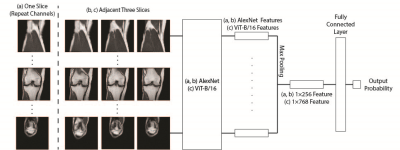 |
3 | Prediction of Total Knee Replacement using Vision Transformers
Chaojie Zhang1, Haresh Rengaraj Rajamohan2, Kyunghyun Cho2, Gregory Chang1, and Cem M. Deniz1
1Department of Radiology, New York University Langone Health, New York, NY, United States, 2Center for Data Science, New York University, New York, NY, United States
Vision transformers were used to predict total knee replacement within 9 years from magnetic resonance images. Inspired by MRNet, 2D slices of an MR image were encoded by a vision transformer and these encodings were aggregated to provide a single prediction outcome from a 3D MR volume. Our results suggest that the prediction performance of vision transformers was comparable with the models based on convolutional neural networks for the outcome prediction task. Moreover, training models with stochastic gradient descent optimizer provided a better performance compared with the Adam optimizer.
|
||
4432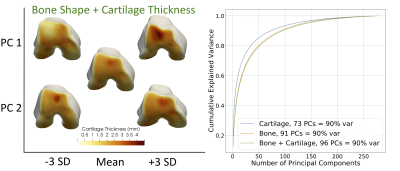 |
4 | Statistical Shape Models of Bone and Cartilage for Predicting Demographics: Data from the Osteoarthritis Initiative
Anthony A Gatti1, Kuan-Chieh Wang2, Garry E. Gold1, Scott L. Delp3, and Akshay C. Chaudhari1
1Radiology, Stanford University, Stanford, CA, United States, 2Computer Science, Stanford University, Stanford, CA, United States, 3Bioengineering, Stanford University, Stanford, CA, United States Three-dimensional statistical shape models built from MRI data can predict future disease and distinguish between groups. However, these models do not leverage the major advantage of MRI – the ability to visualize and quantify soft tissues such as cartilage. This study built three MRI-based statistical shape models, 1) bone shape, 2) cartilage thickness, 3) both bone shape and cartilage thickness. We showed that bone shape and bone shape + cartilage thickness models predicted sex with an R2 of ~0.9, significantly outperforming cartilage thickness alone (0.8). However, cartilage thickness alone was significantly better at predicting radiographic knee osteoarthritis. |
||
4433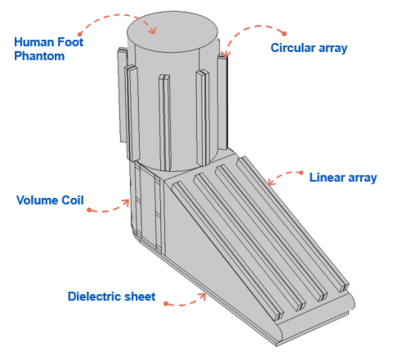 |
5 | Design of a 13-Channel hybrid RF array with field rectification of dielectric material for foot/ankle imaging at 7T
Aditya Ashok Bhosale1, Leslie L Ying1, and Xiaoliang Zhang1
1Biomedical Engineering, University at Buffalo, Buffalo, NY, United States
This study proposes a 13-channel hybrid array system consisting of Microstrip transmission line resonators and half-birdcage coil and demonstrates the high-permittivity dielectric material’s positive effect on the B1 field distribution.
|
||
4434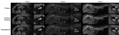 |
6 | Deblurring 3D FSE Images via Regularized Iterative Demodulation of T2 Decay
Yan Wen1, Ek Tsoon Tan2, Maggie Fung1, and Darryl B. Sneag2
1GE Healthcare, New York, NY, United States, 2Department of Radiology and Imaging, Hospital for Special Surgery, New York, NY, United States The 3D FSE sequence is widely used in clinical MRI for its capability to collect many phase encoding lines in each RF excitation. However, the signals during the long echo train readout are modulated by the T2 decay, resulting in blurring. An algorithm is presented to retrospectively demodulate estimated T2 decay from the actual signal. In the preliminary data, the proposed algorithm demonstrated success in deblurring the images. |
||
4435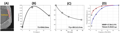 |
7 | Quantitative evaluation of tendon enthesis using ultrashort echo time magnetic resonance imaging (UTE-MRI) techniques
Saeed Jerban1, Alecio Lombardi1, Yajun Ma1, Amir M Afsahi1, Dina Moazamian1, Jiyo Athertya1, Hyungseok Jang1, Christine B Chung1, Jiang Du1, and Eric Y Chang1,2
1Department of Radiology, University of California, San Diego, San Diego, CA, United States, 2Radiology Service, VA San Diego Healthcare System, San Diego, CA, United States
Entheses are transition zones connecting flexible tissues such as tendons and ligaments to bone. Conventional MRI sequences detect little or no signal from entheses as a result of their short T2* values. Alternatively, ultrashort echo time MRI (UTE-MRI) with TE<50 μs can be used to image tissues with short T2 and T2* for quantitative assessment. The feasibility of using quantitative UTE-MRI techniques for the evaluation of entheses regions of Achilles tendons in cadaveric ankle specimens was investigated. Using 20 cadaveric ankle specimens, high quality images were obtained with significant entheses signal which enabled excellent UTE-T1, UTE-Adiab-T1r, and UTE-MT model fittings.
|
||
 Back to Meeting Home
 Back to the Program-at-a-Glance
The International Society for Magnetic Resonance in Medicine is accredited by the Accreditation Council for Continuing Medical Education to provide continuing medical education for physicians.
 Back to Meeting Home
 Back to the Program-at-a-Glance
 View the Presentation