Online Gather.town Pitches
Gray Matter & Neurofluids I
Joint Annual Meeting ISMRM-ESMRMB & ISMRT 31st Annual Meeting • 07-12 May 2022 • London, UK

Online Gather.town Pitches
Gray Matter & Neurofluids I
| Booth # | ||||
|---|---|---|---|---|
3633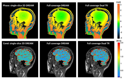 |
1 | Towards fast and precise tissue conductivity imaging in brain
Jun Cao1,2, Iain Ball3, and Caroline Rae1,2
1Neuroscience Research Australia, Sydney, Australia, 2School of Medical Sciences, University of New South Wales, Sydney, Australia, 3Philips Australia & New Zealand, Sydney, Australia
Here we tested the boundaries of the speed and precision of phase MREPT in brain using balanced FFE. Baseline maps with conductivity variance of < 5% could be safely accelerated with compressed SENSE 6 without sacrifice of precision, and scans acquired as quickly as 1.89 s. Precision could be further improved by whole head B1 mapping. MREPT mapping should be conducted task free for best results.
|
||
3634 |
2 | Brain structure and volume reveal neurodevelopmental abnormalities in preterm infants with low-grade intraventricular hemorrhage
Chunxiang Zhang1, Xin Zhao1, Jinxia Guo2, Kaiyu Wang2, Meiying Cheng1, Honglei Shang1, Xueyuan Wang1, Desheng Xuan1, Lingjie Zhang1, and Xiaoan Zhang1
1Department of Radiology, The Third Affiliated Hospital of Zhengzhou University, Zhengzhou, China, 2GE Healthcare, MR Research China, Beijing, China
Premature infants with low-grade intraventricular hemorrhage (IVH) are accompanied by neurodevelopmental abnormalities. Synthetic MRI is an emerging technology that can provide brain volume quantification. Diffusion kurtosis imaging (DKI) can be sensitive to assess brain microstructure changes. Smaller myelin volume, brain parenchymal fraction, lower radial kurtosis value in cerebellum were associated with lower neonatal behavioral neurological assessment. DKI and Synthetic MRI are able to quantitatively evaluate the abnormality of the brain development in preterm infants with low-grade IVH.
|
||
3635 |
3 | Postnatal age-related change of brain volume and its association with neurobehavior outcome in term neonates
Yuying Feng1, Linlin Zhu1, Peng xuan Bai1, Yao Ge1, Congcong Liu1, Na Zhang1, Yichu He2, Feng Shi2, Xiaocheng Wei3, Jian Yang1, and Chao Jin1
1The First Affiliated Hospital of Xi’an Jiaotong University, Xi'an, China, 2Department of Research and Develpoment, Shanghai United Imaging Intelligence Co., Shanghai, China, 3Xiaocheng Wei, GEHealthcare, Beijing, China
Neonatal period is an important stage of brain development, exploring the development and evolution of brain region at this stage is significant for studying brain development and disease mechanisms. We analyzed the relationship between segmented brain region volume and postnatal age in term neonates, the results showed that, after controlling for gestational age, birth weight, body length, head circumference and gender factors, the volume of most brain regions of neonates was positively correlated with postnatal age. In addition, there is no correlation between the volume of most brain regions and neurobehavioral scores.
|
||
3636 |
4 | Hippocampal subfield volumes exhibit differences in ME/CFS and are associated with clinical measures
Kiran Thapaliya1,2, Donald Staines1, Sonya Marshall-Gradisnik11, Jiasheng Su1, and Leighton Barnden1
1Menzies Health Queensland, NCNED, Griffith University, Southport, Australia, 2Centre for Advanced Imaging, The University of Queensland, St Lucia, Australia
Myalgic Encephalomyelitis/chronic fatigue syndrome (ME/CFS) patients suffer from a variety of physical and neurological complaints indicating involvement of the central nervous system including cognitive and memory dysfunction along with a variety of disabling physical symptoms. The hippocampus plays a key role in cognitive function and is altered in neurodegenerative diseases. In this study, we evaluated the volumetric changes in the subfields of the hippocampus in ME/CFS and performed a correlation between hippocampal subfield volumes and clinical measures. Our study showed that hippocampal subfield volumes were lower/higher in ME/CFS patients compared with healthy controls and are also associated with clinical measures
|
||
3637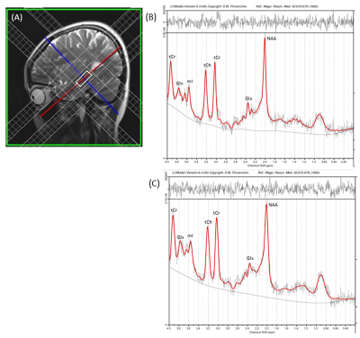 |
5 | Regional Hippocampal Changes in Patients with Hyperthyroidism Video Not Available
Sadhana Singh1,2, Poonam Rana1, Pawan Kumar1, Prabhjot Kaur1, L Ravi Shankar3, and Subash Khushu1,2
1NMR Research Centre, Institute of Nuclear Medicine and Allied Sciences (INMAS), Delhi, India, 2The University of Trans-disciplinary Health Sciences and Technology, Bangalore, India, 3Thyroid Research Centre, Institute of Nuclear Medicine and Allied Sciences (INMAS), Delhi, India
Hyperthyroid patients showed structural and functional abnormalities in the hippocampus region of the brain as suggested by neuroimaging studies. However, the extent of structural, functional, and metabolic changes in hyperthyroid patients is still unclear. We examined the regional hippocampal volume and neuro-metabolite changes in hyperthyroid patients using high resolution T1-weighed imaging and 1H MRS techniques. Hyperthyroid patients showed reduced bilateral hippocampal volume and altered metabolic changes, i.e., Glu/tCr, mI/tCr, and NAA/tCr ratios in the hippocampus, compared to healthy controls. These findings provide evidence that hyperthyroidism results in structural and metabolic alterations in the hippocampus region of the brain.
|
||
3638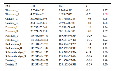 |
6 | Iron deposition decreased in the thalamus and functional connectivity increased between the thalamus and the hippocampus in T2DM patient
Minhua Ni1,2, Linfeng Yan1, Xiaocheng Wei3, and Guangbin Cui1
1Tangdu Hospital, XI an, China, 2Shaanxi University of Chinese Medicine, Xian yang, China, 3MR Research, GE Healthcare, Beijing, China
In this study, change of susceptibility level in the gray matter nucleus of T2DM patients and their functional connectivity were investigated using quantitative susceptibility mapping (QSM) and fMRI techniques. We found iron deposition decreased in the thalamus and functional connectivity increased between the thalamus and the hippocampus. Susceptibility reduction suggested an involvement of chronic microglia activation in the depletion of iron from oligodendrocytes in this central and integrative brain region. The functional connectivity may indicate a compensatory enhancement. These results remind that the thalamus may play an important role in patients with T2DM.
|
||
3639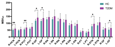 |
7 | Global and Regional Iron Content Changes of Gray Nucleus Using Quantitative Susceptibility Mapping in Type 2 Diabetes Mellitus Patients Video Not Available
Rui Hu1, Yangyingqiu Liu1, Bingbing Gao1, Wanyao Li1, Weiwei Wang1, Qingwei Song1, Ailian Liu1, and Yanwei Miao1
1Radiology, First Affiliated Hospital of Dalian Medical University, Dalian, China Assessing whole-structural and regional high iron content in deep gray matter nuclei using STAGE-based QSM in T2DM Patients. Excessive deposition and uneven distribution of iron protein occur in all nuclei in T2DM patients. The MSVwh of significant differences (P < 0.05) between groups were present in right HCN, right PUT, bilateral GP, right STN and left GF, with the largest AUC of 0.723 in the right GP. The MSVhi in right HCN, right PUT, bilateral GP and left GF were significant differences (P < 0.05) With a largest AUC of 0.714 in right GP. |
||
3640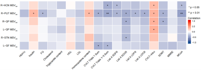 |
8 | Correlation between Iron Burden and Cognitive Status in Type 2 Diabetes Mellitus Patients Using Quantitative Susceptibility Mapping Video Not Available
Rui Hu1, Yangyingqiu Liu1, Bingbing Gao1, Wei Du1, Weiwei Wang1, Qingwei Song1, Ailian Liu1, and Yanwei Miao1
1Radiology, First Affiliated Hospital of Dalian Medical University, Dalian, China The relationship between iron accumulation and cognitive decline in T2DM has not been fully revealed and publicized. This research focuses on assessing the global iron content and high iron content levels of the gray nucleus using QSM, and to analyze the correlation between MSV with significant differences and clinical laboratory indicators and cognitive scores in T2DM Patients. Excessive deposition and uneven distribution of iron protein occur in all nuclei in T2DM patients. Iron is important in the pathophysiology of T2DM, especially the MSVhi in the right PUT was significantly negatively correlated with MoCA (r = -0.485, P = 0.004). |
||
3641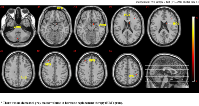 |
9 | Gray matter difference associated with hormone replacement therapy in menopausal women: a voxel-based morphometry (VBM) study Video Permission Withheld
Tae-Hoon Kim1, Chang-Won Jeong1, ByoungRyun Kim2, Youe Ree Kim3, Chungsub Lee1, Young Hwan Lee3, and Kwon-Ha Yoon3
1Medical Convergence Research Center, Wonkwang University, Iksan, Korea, Republic of, 2Obstetrics and Gynecology, Wonkwang University School of Medicine and Hospital, Iksan, Korea, Republic of, 3Radiology, Wonkwang University School of Medicine and Hospital, Iksan, Korea, Republic of
Hormone replacement therapy (HRT) in menopausal women can reduce troublesome menopause symptoms and prevent cognitive decline. Several structural and volumetric researches reported that localized morphologic changes associated with estrogen meditation. However, it is still unclear how to minimize menopausal symptoms and to protect a woman’s brain from decline. This study investigated the potential effects of HRT on sex hormone level and brain morphology using an optimized voxel-based morphometry (VBM) method.
|
||
3642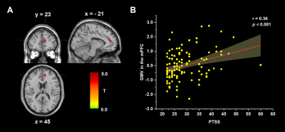 |
10 | Distinct brain structures prospectively predict posttraumatic stress symptoms and posttraumatic growth related to COVID-19 Video Permission Withheld
Huan Lan1, Xueling Suo1, Chao Zuo1, Nanfang Pan1, Song Wang1, and Qiyong Gong1
1Huaxi MR Research Center (HMRRC), Department of Radiology, West China Hospital of Sichuan University, Chengdu, China
Post-traumatic stress symptoms (PTSS) and post-traumatic growth (PTG) might develop after a major trauma, but their neurological bases are largely unknown. Here, we used structural magnetic resonance imaging acquired prepandemic to explore the neuroanatomical correlates in 115 college students. The PTSS and PTG scores were collected during the epidemic. We found that PTSS was positively associated with gray matter volume (GMV) in the medial prefrontal cortex, and PTG was negatively correlated with the GMV in the dorsolateral prefrontal cortex. Our research revealed the neural underpinnings of post-traumatic consequences in healthy students, suggesting PTSS and PTG are two distinct psychological changes.
|
||
3643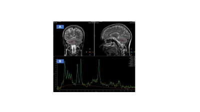 |
11 | Altered neurochemical cerebral metabolism in patients with gluten ataxia: A pilot in-vivo proton MRS study
Uma Sharma1, Vishwa Rawat1, Prasenjit Das2, Achal Kumar Srivastava3, and Govind Makharia4
1Department of NMR, All India Institute of Medical Sciences (AIIMS), New Delhi, India, 2Department of Pathology, All India Institute of Medical Sciences (AIIMS), New Delhi, India, 3Department of Neurology, All India Institute of Medical Sciences (AIIMS), New Delhi, India, 4Department of Gastroenterology and Human Nutrition, All India Institute of Medical Sciences (AIIMS), New Delhi, India
Present study evaluated the neurochemical profile of vermis and right cerebellum of GA patients using 1H MRS. The concentration of N-acetyl aspartate (NAA), glycerophosphocholine (GPC)+phosphocholine (PC), creatine (Cr) + phosphocreatine (PCr) and glutamine (Gln)+glutamate (Glu) were significantly lower in the right cerebellum of patients with GA compared to HC. The concentration of NAA, Cr and Glu were significantly lower in vermis region of patients with GA compared to HC indicating altered cerebral metabolism in GA patients that would have contributed to cerebral damage. The metabolites NAA, GPC+PC, Glu+Gln may have the potential to serve as early indicators of neuronal damage.
|
||
3644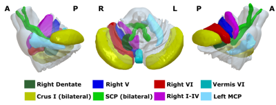 |
12 | Posterior fossa radiomics based diagnosis of Essential tremor from T1-weighted MRI
Archith Rajan1, Shweta Prasad2, Priyanka Tupe Waghmare1,3, Jitender Saini4, Pramod Kumar Pal5, and Madhura Ingalhalikar1
1Symbiosis Centre for Medical Image Analysis, Symbiosis International University, Lavale, Mulshi, Pune, India, 2Department of Clinical Neurosciences and Neurology, National Institute of Mental Health & Neurosciences, Bangalore, India, Bengaluru, India, 3Symbiosis Institute of Technology, Symbiosis International University, Lavale, Mulshi, Pune, India, 4Department of Neuroimaging & Interventional Radiology, National Institute of Mental Health & Neurosciences, Bangalore, India, Bengaluru, India, 5Department of Neurology, National Institute of Mental Health & Neurosciences, Bangalore, India, Bengaluru, India
The cerebellum and its connections (cerebellar peduncles) have been implicated to play a significant role in the pathogenesis of essential tremor (ET). However, these abnormalities may not be grossly evident on basic structural imaging. To this end, we employ radiomics on 3D-T1 weighted images to capture subtle features of pathology and use it in a machine learning framework to deliniate patients with ET. We demonstrate a test accuracy of 83% with radiomics features from dentate nucleus contributing the most, followed by the right V. It thus suggests the potential utility of radiomics features from these structures for diagnosis of ET.
|
||
3645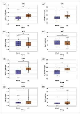 |
13 | Decreased brain GABA levels in patients with migraine without aura: an exploratory 1H-MRS study
Xiaojuan Wu1, Shuting Han1, Yang Yang1, Hui Dai1, Peng Wu2, Hongru Zhao3, and Yonggang Li1
1Department of Radiology, The First Affiliated Hospital of Soochow University, Suzhou, China, 2Philips Healthcare, Shanghai, China, 3Department of Neurology, The First Affiliated Hospital of Soochow University, Suzhou, China Increasing neurophysiological studies had revealed that regional excitation-inhibition imbalance in brain played a key role in the pathogenesis of migraine. This study explored the alterations in GABA and Glx levels in the anterior cingulate gyrus (ACC) and medial prefrontal lobe (mPFC) of patients with migraine without aura (MWoA) using MEGA-PRESS at 3 Tesla. Compared with healthy controls, the GABA+ levels in the ACC and mPFC of patients were lower and correlated with attack frequency, and Glx levels had no difference, suggesting a relationship of the abnormalities of GABAergic system in the ACC and mPFC with the pathophysiological mechanism of MWoA. |
||
3646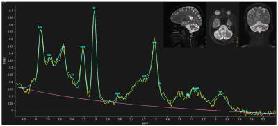 |
14 | Neuronal metabolic biomarkers in cerebellum of Spinocerebellar ataxia type 2 and 12 patients
Pankaj Pankaj1, S Senthil Kumaran1, Achal Kumar Srivastava2, and Ramesh Kumar Agrawal3
1Department of NMR & MRI Facility, All India Institute of Medical Sciences, New Delhi, India, 2Department of Neurology, All India Institute of Medical Sciences, New Delhi, India, 3School of Computer & Systems Sciences, Jawaharlal Nehru University, New Delhi, India
Spinocerebellar Ataxia (SCA) is a genetic and hereditary disorder that leads to severe disability in balance, speech, posture, etc. SCA2 and SCA12 are two sub-types, which have different CAG repeat expansion on chromosome with different pathologies. In vivo MR spectroscopy was acquired using single voxel Point Resolved Spectroscopy Sequence (PRESS) in the left cerebellum. SCA2 showed a decrease in NAA and Cho and increased mI concentration as compared to controls and an increase in mI values as compared to that in SCA12, which may be attributed to the motor and cognitive dysfunctions in SCA, reflecting more atrophy in SCA2.
|
||
3647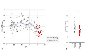 |
15 | Comparative analysis of diffusion-weighted image analysis along the perivascular space (DWI-ALPS) for evaluating interstitial fluid status
Toshiaki Taoka1, Rintaro Ito1, Rei Nakamichi2, Toshiki Nakane2, Kazushige Ichikawa3, Mayuko Sakai4, and Shinji Naganawa2
1Department of Innovative Biomedical Visualization (iBMV), Nagoya University, Nagoya, Japan, 2Department of Radiology, Nagoya University, Nagoya, Japan, 3Devision of Radiology, Nagoya University Hospital, Nagoya, Japan, 4Canon Medical Systems Corporation, Otawara, Japan
The diffusion tensor image analysis along the perivascular space (DTI-ALPS) method has been developed to evaluate glymphatic function. In order to evaluate wider clinical cases, we developed a diffusion weighted image ALPS (DWI-ALPS) technique. The ALPS-index was retrospectively calculated by clinical DWI of normal subjects and those with pathologies. In normal subjects, the ALPS-index was highest in those in their forties and lowest in those in their seventies. Patients with Alzheimer’s disease and subdural hematoma showed a lower ALPS-index than age-matched normal subjects. The DWI-ALPS method seems to be feasible for evaluation of glymphatic function in the clinical practice.
|
||
 Back to Meeting Home
 Back to the Program-at-a-Glance
The International Society for Magnetic Resonance in Medicine is accredited by the Accreditation Council for Continuing Medical Education to provide continuing medical education for physicians.
 Back to Meeting Home
 Back to the Program-at-a-Glance
 View the Presentation