Online Gather.town Pitches
White Matter & Nervous System II
Joint Annual Meeting ISMRM-ESMRMB & ISMRT 31st Annual Meeting • 07-12 May 2022 • London, UK

Online Gather.town Pitches
White Matter & Nervous System II
| Booth # | ||||
|---|---|---|---|---|
3040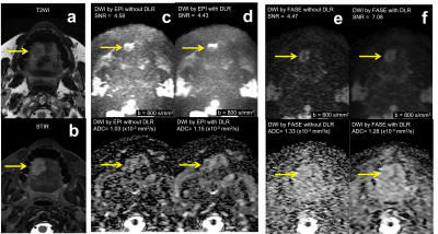 |
1 | Head and Neck DWI: Comparison of Image Quality and Diagnostic Performance among FASE and EPI Sequences with and without DLR Video Permission Withheld
Hirotaka Ikeda1, Yoshiharu Ohno1, Kaori Yamamoto2, Kazuhiro Murayama3, Masato Ikedo2, Masao Yui2, Satomu Hanamatsu1, Saki Takeda4, Akiyoshi Iwase4, Yuki Obama1, Takahiro Ueda1, Hiroyuki Nagata1, and Hiroshi Toyama1
1Radiology, Fujita Health University School of Medicine, Toyoake, Japan, 2Canon Medical Systems Corporation, Otawara, Japan, 3Joint Research Laboratory of Advanced Medical Imaging, Fujita Health University School of Medicine, Toyoake, Japan, 4Radiology, Fujita Health University Hospital, Toyoake, Japan
No major studies have been reported for assessing the utility of DWI obtained by Fast Advanced Spin-Echo (FASE) with Deep Learning Reconstruction (DLR) for head and neck MR. We hypothesized that DWI obtained by FASE with DLR could improve the image quality with less deformation and better diagnostic performance as compared with DWI obtained by Echo-Planar Imaging (EPI) in head and neck MR. The purpose of this study was to compare the capability of DWI obtained by FASE and EPI sequences with and without DLR for image quality and diagnostic performance on DWI in patients suspected head and neck tumors.
|
||
3041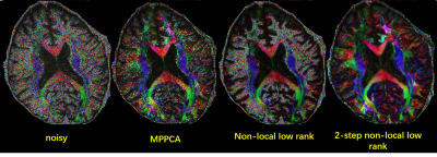 |
2 | Non-local low rank denoising method for complex-valued DWI
Xinyu Ye1, Xiaodong Ma2, Ziyi Pan1, Steen Moller2, Eddie Auerbach2, Kamil Ugurbil2, Xiaoping Wu2, and Hua Guo1
1Center for Biomedical Imaging Research, Department of Biomedical Engineering, School of Medicine, Tsinghua University, Beijing, China, 2Center for Magnetic Resonance Research, Radiology, Medical School, University of Minnesota, Minneapolis, MN, United States
Diffusion-weighted MRI (DWI) suffers from the intrinsic low SNR, especially for high-resolution and/or high b-value acquisitions. Thus, denoising methods are critical for diffusion images. Recently, increasing attention has been paid to complex DWI image denoising. Here, we propose a 2-step non-local low-rank joint denoising method to process complex-valued DWI images. Simulation data and in-vivo brain data were used to test the proposed method. The results showed that the proposed method may further improve the image quality.
|
||
3042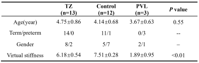 |
3 | Quantifying the Stiffness of Peritrigonal Terminal Zones in Preschool Children by Diffusion Imaging-based Virtual MRI Elastography
Na Zhang1, Xianjun Li1, Congcong Liu1, Yao Ge1, Yuying Feng1, Pengxuan Bai1, Miaomiao Wang1, and Jian Yang1
1Department of Radiology, The First Affiliated Hospital of Xi’an Jiaotong University, Xi'an, China
The high signal intensity on T2-weighted images in terminal zones (TZ) mainly associated with lower maturational degree. We hypothesized that the stiffness in these regions may be different from typical developing children without TZ. Virtual stiffness of brain can be measured safely with virtual magnetic resonance elastography (vMRE). Therefore, this study tried to use vMRE to investigate differences between children with and without TZ. Results demonstrated that virtual stiffness of TZ was lower than controls, while higher than periventricular leukomalacia. These suggest that vMRE may provide additional information for evaluating brain maturation and injury.
|
||
3043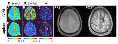 |
4 | Mathematically constrained intravoxel incoherent motion (IVIM)
Jiaqiu Wang1, Thomas Barrick2, Matt Hall3, Muge Karaman4, Richard Magin4, David Reiter5, Qianqian Yang6, Qiang Yu1, Fatima Nasrallah 7, Weeyao Koh8, and Viktor Vegh1,9
1Centre for Advanced Imaging, University of Queensland, Brisbane, Australia, 2Neuroscience Research Centre, St George's, University of London, London, United Kingdom, 3National Physical Laboratory, Teddington, United Kingdom, 4Department of Bioengineering, University of Illinois at Chicago, Chicago, IL, United States, 5Department of Radiology and Imaging Sciences, Emory University, Atlanta, GA, United States, 6School of Mathematical Sciences, Queensland University of Technology, Brisbane, Australia, 7Queensland Brain Institute, University of Queensland, Brisbane, Australia, 8National University Cancer Institute Singapore, National University Hospital, Singapore, Singapore, 9ARC Training Centre for Innovation in Biomedical Imaging Technology, Brisbane, Australia Intravoxel incoherent motion (IVIM) provides a method of mapping slow and fast diffusion using a two-exponential model. Whilst this method has been applied in acute stroke, brain cancer and muscle, for example, it does have challenges. These include how to best fit the three model parameters which on top of the two diffusion coefficients includes the fast diffusion volume fraction (i.e., perfusion fraction), and how to best sample b-values which lead to robust model parameter fits. Here we propose an approach based on quasi-diffusion which helps constrain the model fit and show reproducibility under different b-value sampling regimes. |
||
3044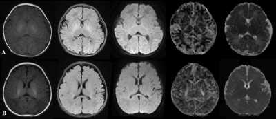 |
5 | The value of diffusion kurtosis imaging in detecting delayed brain development of premature infants
Xin Zhao1, Chunxiang Zhang1, Bohao Zhang2, Jiayue Yan3, Jinxia Guo4, Kaiyu Wang4, Zitao Zhu5, and Xiaoan Zhang6
1Department of Radiology, The Third Affiliated Hospital of Zhengzhou University, Zhengzhou, China, 2Henan Key Laboratory of Child Brain Injury, Institute of Neuroscience and Third Affiliated Hospital of Zhengzhou University, Zhengzhou, China, 3McGill university Montreal, Zhengzhou, China, 4GE Healthcare, MR Research China, Beijing, China, 5Wuhan University, Wuhan, China, 6The Third Affiliated Hospital of Zhengzhou University, Zhengzhou, China
Preterm infants are at high risk of adverse neurodevelopmental outcome. Therefore, it is necessary to find a tool to detect brain developmental abnormalities earlier. Diffusion kurtosis imaging (DKI) is considered as a more accurate technology based on a kurtosis model accounting for the variation of Gaussian distribution caused by complex cellular environments in compare with diffusion tensor imaging. The mean kurtosis (MK) in posterior limbs of the internal capsule (PLIC), anterior limb of internal capsule (ALIC), the mean diffusion (MD) in parietal white matter (PWM) were found with good diagnostic performance for abnormal brain microstructural change.
|
||
3045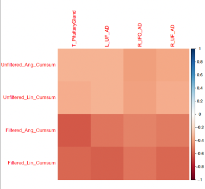 |
6 | What happens to the brain over a single season of playing high school rugby: structural and white matter fibre tract changes related to impact
Maryam Tayebi1,2, Eryn Kwon1,2, William Schierding3, Joshua McGeown2, Matthew McDonald2,4, Paul Condron2, Leigh Potter2, Miao Qiao5, Jerome Maller6, Samantha Holdsworth2,7, Justin Fernandez1, Alan Wang1, and Vickie Shim1,2
1Auckland Bioengineering Institute, University of Auckland, Auckland, New Zealand, 2Mātai Medical Research Institute, Gisborne, New Zealand, 3Liggins Institute, University of Auckland, Auckland, New Zealand, 4Department of Ophthalmology, University of Auckland, Auckland, New Zealand, 5School of Computer Science, University of Auckland, Auckland, New Zealand, 6General Electric Healthcare, Victoria, Australia, 7Faculty of Medical and Health Sciences & Centre for Brain Research, University of Auckland, Auckland, New Zealand
We conducted a multimodal MRI study on high school rugby players with bespoke kinematic mouthguard sensors to investigate the correlation between the cumulative effects of subconcussive head impact exposure and any change in the structure of the brain. Despite being underpowered, the study found that the volume of the corpus callosum changed the most over a season of rugby (PCA); Axial Diffusivity of the UF and ILF tracts negatively correlated with the cumulative impact sustained by the athletes. Further investigation of the cumulative effect of playing high-contact sports over an extended period is required, especially for children with developing brains.
|
||
3046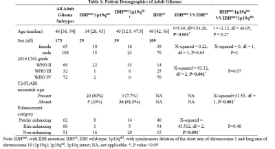 |
7 | Diagnostic performance of ADC and CBV imaging intersecting MRS for identifying molecular status in adult gliomas Video Permission Withheld
Shuang Li1, Xiaorui Su1, and Qiang Yue2
1radiology, Huaxi MR Research Center (HMRRC), Department of Radiology, West China Hospital of Sichuan University, Chengdu, China, Chengdu, China, 2radiology, Department of Radiology, West China Hospital of Sichuan University, Chengdu, China, Chengdu, China
Preoperative identification of IDH and 1p/19q codeletion could help clinical doctors make the optimal therapy plan. Although, there were few diagnostic studies could provide quantitative cutoff values of ADC and/or CBVfor identifying genogroup of gliomas. In this study, we calculated histogram metrics of ADC and CBV based on core tumor and used these metrics intersection MRS to establish diagnostic models to identify IDH mutant station and 1p/19q codeletion in turn. The main results showed that histogram metrics of ADC and CBV were powerful for identifying molecular status in adult gliomas. And MRS concentrations could improve diagnostic performance in both models.
|
||
3047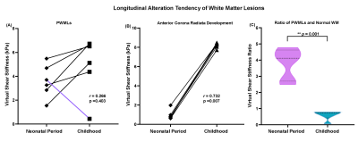 |
8 | Quantitatively Assessing Longitudinal Alteration of Shear Stiffness in Neonatal Punctate White Matter Lesions based on Virtual Elastography
Congcong Liu1, Miaomiao Wang1, Xianjun Li1, Yao Ge1, and Jian Yang1
1the Department of Radiology, the First Affiliated Hospital of Xi'an Jiaotong University, Xi'an, China
Punctate white matter lesions (PWMLs) were currently common white matter injuries, with incidence of 20-30% and associated with adverse neurological development. Longitudinal alteration of PWMLs was less researched which might be related to pathology. Therefore, this study aimed to assess alterations of PWMLs virtual shear stiffness in neonates via DKI-based virtual elastography. We found that virtual shear stiffness of PWMLs was higher than contemporaneous white matter at neonatal periods and did not change obviously with age, while virtual shear stiffness of normal white matter was increased with age. Virtual elastography might be a potential method to identify differently nosological PWMLs.
|
||
3048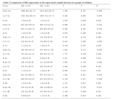 |
9 | An evaluation of the caput nuclei caudati in children with autism spectrum disorder and language deficits with DKI and Synthetic MR
Yongbing Sun1, Xin Zhao1, Jinxia Guo2, Lingsong Meng1, Desheng Xuan1, Xiongpeng He1, and Xiaoan Zhang1
1the Third Affiliated Hospital of Zhengzhou University, Zhengzhou, China, 2GE Healthcare, Beijing, China
In this study, DKI combined with synthetic MRI of magnetic resonance imaging compilation (MAGiC) was used to assess the developmental integrity of the caput nuclei caudati of children with ASD and language deficits, and the relationship between DKI parameters and language test scores were explored.
|
||
3049 |
10 | Assessment of relationship between chemotherapy-induced brain structural alteration and neuropsychological scale revealed by GQI
Wei Chuang1, Vincent Chin-Hung Chen2,3, Yuan-Hsiung Tsai2,4, and Jun-Cheng Weng1,3,5
1Department of Medical Imaging and Radiological Sciences, Graduate Institute of Artificial Intelligence, Chang Gung University, Taoyuan, Taiwan, 2School of Medicine, Chang Gung University, Taoyuan, Taiwan, 3Department of Psychiatry, Chang Gung Memorial Hospital, Chiayi, Taiwan, 4Department of Diagnostic Radiology, Chang Gung Memorial Hospital, Chiayi, Taiwan, 5Medical Imaging Research Center, Institute for Radiological Research, Chang Gung University and Chang Gung Memorial Hospital at Linkou, Taoyuan, Taiwan
Breast cancer is the most prevalent cancer among women, and most of them suffered from chemotherapy-related cognitive impairment (CRCI). In this study, we aimed to use generalized q-sampling imaging (GQI) to detect the relationship between GQI indices and mood symptoms caused by chemotherapy, post-traumatic stress disorder (PTSD), or post-traumatic growth (PTG). Using statistical multiple regression, we found the relationship change between GQI indices and depression, and a consistent relationship was obtained between GQI indices and anxiety with or without chemotherapy. The results indicated that neurotoxicity or traumatic stressors may affect brain structures.
|
||
3050 |
11 | Fixel-Based Analysis of White Matter Degeneration in Patients with Huntington Disease
Chih-Chien Tsai1,2, Sher Li Oh3, Chiung-Mei Chen4, Yih-Ru Wu4, Maria Valdes Hernandez5,6, Jur-Shan Cheng7, Yao-Liang Chen8, Yi-Ming Wu8, Yu-Chun Lin8, and Jiun-Jie Wang1,2,9,10
1Department of Medical imaging and Radiological Science, Chang-Gung University, Taoyuan City, Taiwan, 2Healthy Aging Research Center, Chang Gung University, Taoyuan City, Taiwan, 3Nanyang Technological University, Singapore, Singapore, 4Department of Neurology, Chang Gung Memorial Hospital, Taoyuan City, Taiwan, 5Centre for Cognitive Ageing and Cognitive Epidemiology, Department of Neuroimaging Sciences, University of Edinburgh, Edinburgh, United Kingdom, 6Centre for Clinical Brain Sciences, University of Edinburgh, Edinburgh, United Kingdom, 7Clinical Informatics and Medical Statistics Research Center, Chang-Gung University, Taoyuan City, Taiwan, 8Department of f Medical Imaging and Intervention, Chang Gung Memorial Hospital, Taoyuan City, Taiwan, 9Medical Imaging Research Center, Institute for Radiological Research, Chang Gung University/Chang Gung Memorial Hospital, Taoyuan City, Taiwan, 10Department of Diagnostic Radiology, Chang Gung Memorial Hospital, Taoyuan City, Taiwan
Microstructure damage in white matter might be linked to regional and global atrophy in Huntington Disease. Fixel-based analysis (FBA) has recently emerged as a useful fiber-specific tool for examining white matter structure, which could be an important contributor to the pathogenesis of Huntington’s disease. Therefore, we investigate the utility of FBA as a biomarker for disease progression. Our findings indicated the reductions in FBA occurs in major white matter tracts, which was closely co-localized with the regions of increased diffusivity in basal ganglia. FBA analysis is effective in studying white matter tractography and fiber changes in Huntington’s Disease.
|
||
3051 |
12 | Non-neuropsychiatric Childhood-onset Systemic Lupus Erythematosus: Changes in Brain Network Organization and White Matter Microstructure
Laleh Eskandarian1,2, Müşerref Kasap Cüceoğlu3, Selcan Demir3, Tuna Cak4, Ebru Cengel Kultur4, Yelda Bilginer3, Seza Ozen3, and Kader K Oguz2,5
1Neuroscience Department, Bilkent University, ANKARA, Turkey, 2National Magnetic Resonance Research Center (UMRAM), Bilkent University, ANKARA, Turkey, 3Faculty of Medicine, Department of Pediatrics, Division of Rheumatology, Hacettepe University, ANKARA, Turkey, 4Faculty of Medicine, Department of Pediatric Psychiatry, Hacettepe University, ANKARA, Turkey, 5Faculty of Medicine, Department of Radiology, Hacettepe University, ANKARA, Turkey
Central nervous system in childhood-onset SLE shows an increased risk of damage due to onset age, accumulating disease effect and other possible pathophysiologic mechanisms. Thus, an understanding of the timing and extent of changes occurring in the brains of these young patients with SLE is important for new therapeutic strategies. With this purpose, we investigated whether brain network and white matter microstructural changes occur in non-neuropsychiatric SLE patients with disease onset at childhood. TBSS and graph analysis of DTI data, showed significant alterations in white matter integrity and global and nodal connectivity, which revealed correlations with disease duration and severity.
|
||
3052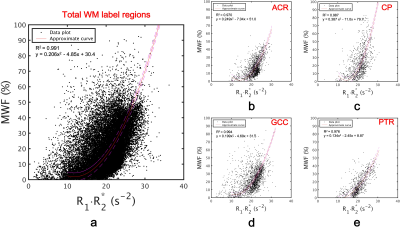 |
13 | Conversion map from quantitative parameter mapping to myelin water fraction
Shun Kitano1, Yuki Kanazawa2, Masafumi Harada2, Yo Taniguchi3, Yuki Matsumoto2, Hiroaki Hayashi4, Kosuke Ito3, Yoshitaka Bito3, and Akihiro Haga2
1Graduate of Health Science, Tokushima University, Tokushima, Japan, 2Graduate School of Biomedical Sciences, Tokushima University, Tokushima, Japan, 3FUJIFILM Healthcare Corporation, Tokyo, Japan, 4College of Medical, Pharmaceutical and Health Sciences, Kanazawa University, Kanazawa, Japan We determined MWF derived from QPM-relaxation parameters using quadratic polynomial approximation. Two types of myelin imaging were generated; MWF derived from multi-echo gradient-echo and R1·R2* map derived from QPM, which were standardized from twelve healthy volunteers to MNI. As a result, the relationship between MWF and R1·R2* values on the white matter region strongly correlated with a quadratic polynomial approximation (R2 = 0.991). Using acquired quadratic polynomial approximation, the R1·R2* map was converted to MWF. Our MWF map derived from QPM can dramatically improve image quality when compared to the general MWF. |
||
3053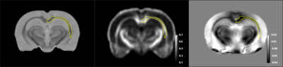 |
14 | Whole-brain assessment of age-related brain characteristics using Quantitative Susceptibility Mapping and Diffusion Tensor Imaging
Qiong Ye1
1High Magnetic Field Laboratory, Hefei Institutes of Physical Science, Chinese Academy of Sciences, Hefei, China
Quantitative Susceptibility Mapping (QSM) can reveal the pathophysiological changes such as myelin, iron deposition, and tissue oxygenation. Diffusion Tensor Imaging (DTI) is an effective tool to assess the integrity of white matter. In our study, we performed whole-brain analysis of rat QSM and DTI using the recently reported SIGMA rat brain template (Nature Communication, 2019). The whole-brain segmentation demonstrated excellent performance. The derived parameters show high repeatability and are sensitive to age-related changes.
|
||
 Back to Meeting Home
 Back to the Program-at-a-Glance
The International Society for Magnetic Resonance in Medicine is accredited by the Accreditation Council for Continuing Medical Education to provide continuing medical education for physicians.
 Back to Meeting Home
 Back to the Program-at-a-Glance
 View the Presentation