Online Gather.town Pitches
Gastrointestinal & Lungs I
Joint Annual Meeting ISMRM-ESMRMB & ISMRT 31st Annual Meeting • 07-12 May 2022 • London, UK

Online Gather.town Pitches
Gastrointestinal & Lungs I
| Booth # | ||||
|---|---|---|---|---|
3380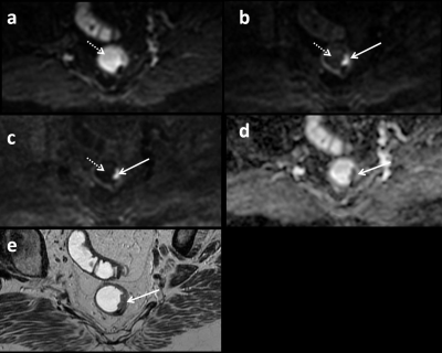 |
1 | Intra-rectal gel suppression effectiveness on rectal cancer MR diffusion-weighted images
Edward M Lawrence1,2 and David H Kim1
1Radiology, UW-Madison School of Medicine and Public Health, Madison, WI, United States, 2Radiology, WS Middleton VA Hospital, Madison, WI, United States
Retrospective evaluation of intra-rectal gel suppression on DWI from 60 rectal MRIs. The DWI, including b1000, computed synthetic b1500, and ADC maps were evaluated by 2 radiologists independently using a 5 point Likert scale with scores of 4-5 considered fully diagnostic. The median suppression score was significantly greater than 3 for both readers (p<0.05) with fully diagnostic suppression achieved in 57/60 (95.0%) and 53/60 (88.3%) cases for reader 1 and 2, respectively. No study was given a score of 1 or 2 by either reader. These results suggest that gel does not impede reader evaluation for tumor restriction at DWI.
|
||
3381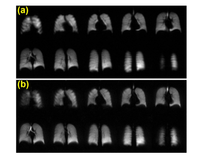 |
2 | Comparison of Spiral and Cartesian Acquisitions for Rapid Hyperpolarized 129Xe Ventilation Mapping in Pediatric Cystic Fibrosis Lung Disease
Brandon Zanette1, Samal Munidasa1,2, Yonni Friedlander1,2, Felix Ratjen1,3, and Giles Santyr1,2
1Translational Medicine, The Hospital for Sick Children, Toronto, ON, Canada, 2Medical Biophysics, University of Toronto, Toronto, ON, Canada, 3Division of Respiratory Medicine, The Hospital for Sick Children, Toronto, ON, Canada
A rapid 3D stack-of-spirals (3D-SoS) sequence was used for hyperpolarized 129Xe ventilation imaging in pediatric cystic fibrosis and healthy controls, allowing for 5x reduction in scan duration and breath-hold compared to a commonly used 2D gradient echo (2D-GRE) sequence. The ventilation defect percent (VDP) measured with 3D-SoS was not significantly different from VDP measured with 2D-GRE. Additionally, the transient drop in SpO2 associated with xenon breath-holds was reduced with the shorter breath-hold afforded by 3D-SoS. In future, the reduced acquisitions durations offered by 3D-SoS may allow for improved tolerability, or may be traded for improved resolutions.
|
||
3382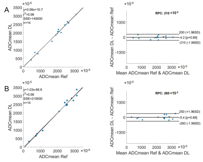 |
3 | A cross-modality deep learning model for esophageal cancer segmentation and quantitation on 18F-FDG PET/CT and diffusion weighted MRI
Zijian Zhou1, Bikash Panthi1, David E. Rauch1, Jong Bum Son1, Carol C. Wu1, Steven H. Lin1, Mark D. Pagel1, and Jingfei Ma1
1The University of Texas MD Anderson Cancer Center, Houston, TX, United States
We applied a deep learning (DL) model developed for 18F-FDG PET/CT of mantle cell lymphoma to esophageal cancers on 18F-FDG PET/CT and diffusion-weighted MRI. We compared the performance of the DL-based segmentation with the manual segmentation on PET and evaluated the quantitation on both PET and apparent diffusion coefficient (ADC). The model achieved promising results of detecting and segmenting esophageal cancers, and the DL-based imaging metrics were consistent with the reference standards.
|
||
3383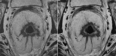 |
4 | 3D T2-Weighted Rectal Cancer Imaging using a 3D Fast Spin Echo Sequence with Deep Learning Reconstruction
Sarah Palmquist1, Usama Salem1, Nir Stanietzky1, Jia Sun2, Xinzeng Wang3, Ersin Bayram3, Ken-Pin Hwang4, Jong Bum Son4, Peng Wei2, Randy Ernst1, Harmeet Kaur1, and Jingfei Ma4
1Abdominal Imaging, M.D. Anderson Cancer Center, Houston, TX, United States, 2Biostatistics, M.D. Anderson Cancer Center, Houston, TX, United States, 3GE Healthcare, Houston, TX, United States, 4Imaging Physics, M.D. Anderson Cancer Center, Houston, TX, United States
Multiplanar high resolution T2-weighted (T2W) imaging with a 2D fast spin echo (FSE) sequence is currently an essential component of rectal cancer MRI. In this work, we performed T2W imaging of rectal cancer with a 3D FSE sequence and evaluated the quality and potential clinical value of the images after applying a postprocessing deep learning reconstruction (DLR) algorithm. We found that DLR images are non-inferior to conventional images and there are fair-moderate inter-reader agreements in a large majority of the categories evaluated for image quality and clinical usefulness.
|
||
3384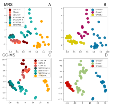 |
5 | A clustering method to classify stages of NAFLD based on the metabolites results obtained in two mouse models with MRS
Aline Xavier1,2,3, Constanza Gainza4,5, Flavia Zacconi6,7,8, Daniel Cabrera9,10, Marco Arese9,11, Carlos Sing-Long2,3,4,5,8, and Marcelo Andia2,3,12
1Instituto de Ciencias de la Ingeniería, Universidad de O’Higgins, Rancagua, Chile, 2Millennium Nucleus for Cardiovascular Magnetic Resonance, Santiago, Chile, 3Biomedical Imaging Center, Pontificia Universidad Católica de Chile, Santiago, Chile, 4Millennium Nucleus Center for the Discovery of Structures in Complex Data, Santiago, Chile, 5Institute for Mathematical and Computational Engineering, Pontificia Universidad Católica de Chile, Santiago, Chile, 6Faculty of Chemistry and Pharmacy, Pontificia Universidad Católica de Chile, Santiago, Chile, 7Research Center for Nanotechnology and Advanced Materials CIEN-UC, Pontificia Universidad Católica de Chile, Santiago, Chile, 8Institute for Biological and Medical Engineering, Pontificia Universidad Católica de Chile, Santiago, Chile, 9Gastroenterology Department, Pontificia Universidad Católica de Chile, Santiago, Chile, 10Faculty of Medical Sciences, Universidad Bernardo O’Higgins, Santiago, Chile, 11Center of Aging and Regeneration (CARE), Pontificia Universidad Católica de Chile, Santiago, Chile, 12Radiology Department, School of Medicine, Pontificia Universidad Católica de Chile, Santiago, Chile
In this study, we analyzed the liver fatty acids results obtained by magnetic resonance spectroscopy and gas chromatography in non-alcoholic fatty liver disease (NAFLD) mice with principal component analysis (PCA) and a clustering method. The study was made with two different mouse models (CDAA and Western diet), and a control group. Our results, as evidenced by PCA, showed that the liver fatty acid composition changed as NAFLD progressed and it was possible to identify the 3 most relevant NAFLD groups with MRS (healthy, steatosis, and steatohepatitis) with excellent agreement with the gold-standard histopathological scores.
|
||
3385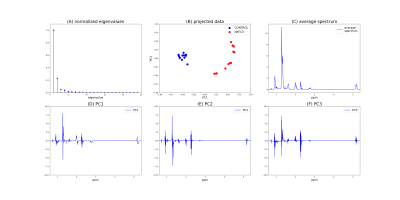 |
6 | Classifying healthy versus NAFLD mice from liver fatty acids’ 1H-MRS at 9.4 T using SVMs
Pedro Izquierdo Lehmann1,2, Aline Xavier2,3,4, Marcelo Andia2,4,5, and Carlos A Sing-Long1,2,4,6,7
1Institute for Mathematical and Computational Engineering, Pontificia Universidad Católica de Chile, Santiago, Chile, 2Millennium Nucleus for Cardiovascular Magnetic Resonance, Santiago, Chile, 3Instituto de Ciencias de la Ingeniería, Universidad de O’Higgins, Rancagua, Chile, 4Biomedical Imaging Center, Pontificia Universidad Católica de Chile, Santiago, Chile, 5Radiology Department, School of Medicine, Pontificia Universidad Católica de Chile, Santiago, Chile, 6Institute for Biological and Medical Engineering, Pontificia Universidad Católica de Chile, Santiago, Chile, 7Millennium Nucleus Center for the Discovery of Structure in Complex Data, Santiago, Chile
Nonalcoholic fatty liver disease (NAFLD) is a common liver disorder that is the first step in a cascade of liver damage. Hence, there is a clinical need for non-invasive, early identification of patients. We hypothesize that the intra-hepatocyte fatty acid composition provides information about NAFLD. To test this, the 1H-MRS spectra of fatty acids in the livers of healthy and NAFLD mice were measured at 9.4 T. Exploratory data analysis shows the spectra can be correctly classified with SVMs. Our results open an opportunity to develop a non-invasive tool for staging NAFLD patients without the need of liver biopsy.
|
||
3386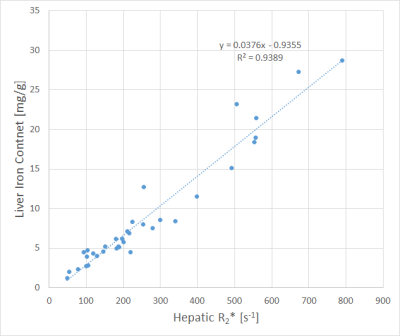 |
7 | MR-based Liver Iron Quantification using Gradient Echo: Co-Factors influencing the Calibration of R2* with Reference Liver Iron Content Values
Arthur Peter Wunderlich1,2, Holger Cario3, Stephan Kannengießer4, Lena Hering1, Michael Götz1,2, Meinrad Beer1, and Stefan Andreas Schmidt1
1Dept. for Diagnostic and Interventional Radiology, Ulm University, Medical Center, Ulm, Germany, 2Section for Experimental Radiology, Ulm University, Medical Center, Ulm, Germany, 3Clinic of Pediatrics and Adolescent Medicine, Ulm University, Medical Center, Ulm, Germany, 4MR Application Predevelopment, Siemens Healthcare GmbH, Erlangen, Germany Purpose: To analyze the Liver-Iron-Content-(LIC)-R2*-calibration with respect to co-factors. Methods: 116 patients were scanned with spin-echo (Ferriscan® protocol) and gradient-echo sequences. R2* was correlated to LIC reference values for all patients together and in subgroups divided by e.g. age, gender and underlying diseases. Linear correlations were analyzed with R2* only and including MR-PDFF as suspected co-factor. Results: Deviating slopes of regression lines were found e.g. in patients after splenectomy vs. patients with spleen, influence of MR-PDFF amongst others in females below 17 years. Conclusion: Co-factors like disease patterns should be considered in gradient echo based LIC quantification. |
||
3387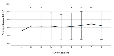 |
8 | 3D multi-echo GRE MRI of the whole liver: First experiences of a volumetric segment-by-segment R2* analysis
Arthur Peter Wunderlich1,2, Holger Cario3, Stephan Kannengießer4, Veronika Grunau1, Michael Götz1,2, Meinrad Beer1, and Stefan Andreas Schmidt1
1Dept. for Diagnostic and Interventional Radiology, Ulm University, Medical Center, Ulm, Germany, 2Section for Experimental Radiology, Ulm University, Medical Center, Ulm, Germany, 3Clinic of Pediatrics and Adolescent Medicine, Ulm University, Medical Center, Ulm, Germany, 4MR Application Predevelopment, Siemens Healthcare GmbH, Erlangen, Germany Purpose: To test liver segmentation of 3D-multi-gradient-echo MRI data and segmental R2* evaluation. Methods: 44 patients examined by multi-gradient-echo MRI underwent semiautomatic liver segmentation and subdivision into nine segments. Segmental R2* values were analyzed in all patients together and in patient subgroups. Results: R2* was lowest in segment 1 (S1), differences to S1 were significant for S3, S5 and S6, highly significant to S2 and S7. Patients with high average R2* showed also significant differences to S1 in some segments. Conclusions: Inhomogeneity of hepatic iron distribution was found. Low R2* in S1 may be explained by its special vascular supply. |
||
3388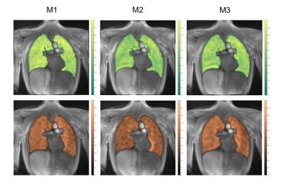 |
9 | Reproducibility of Functional Lung Parameters derived from non-contrast enhanced self-gated 2D Ultrashort Echo-Time (UTE) in Healthy Subjects Video Permission Withheld
Bingjie Yang1, Patrick Metze1, Anke Balasch1, Kilian Stumpf1, Meinrad Beer2, Wolfgang Rottbauer1, and Volker Rasche1
1Department of Internal Medicine II, Ulm University Medical Center, Ulm, Germany, 2Department of Radiology, Ulm University Medical Center, Ulm, Germany MRI lung imaging is challenging due to cardiac and respiratory motion and the short T2*. In this study the reproducibility of lung function parameters derived from data acquired either from a breath-hold or free-breathing tiny golden angle UTE (2D tyGA UTE) technique have been assessed. Inter-observer and inter-measurement reproducibility was assessed for different lung parameters including lung density and qualitative and quantitative ventilation and perfusion. In general, a very good inter-observer reproducibility and superior reproducibility of the lung function parameters was observed for the free-breathing approach. |
||
3389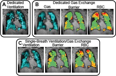 |
10 | Hyperpolarized 129Xe Ventilation and Gas Exchange Images Acquired in a Single 10 s Breath-hold.
Peter J Niedbalski1, Chase Hall1, and Mario Castro1
1Internal Medicine, University of Kansas Medical Center, Kansas City, KS, United States Hyperpolarized 129Xe MRI (Xe-MRI) can be used to acquire images of pulmonary ventilation and gas exchange. These ventilation and gas exchange images are typically acquired in separate breath-holds, which lengthens scan time, increases cost, and requires more of patients. We have combined 3D spiral (FLORET) ventilation imaging with radial 1-point Dixon gas exchange imaging within a single-breath-hold scan. This single-breath-hold scan produces both a gas exchange and a “high-resolution” (4x4x4 mm3 voxels) ventilation image that are qualitatively and quantitatively similar to images acquired in dedicated breath-holds. This increase in efficiency improves the clinical feasibility of hyperpolarized 129Xe MRI. |
||
 Back to Meeting Home
 Back to the Program-at-a-Glance
The International Society for Magnetic Resonance in Medicine is accredited by the Accreditation Council for Continuing Medical Education to provide continuing medical education for physicians.
 Back to Meeting Home
 Back to the Program-at-a-Glance
 View the Presentation