Online Gather.town Pitches
Machine Learning/Artificial Intelligence II
Joint Annual Meeting ISMRM-ESMRMB & ISMRT 31st Annual Meeting • 07-12 May 2022 • London, UK

Online Gather.town Pitches
Machine Learning/Artificial Intelligence II
| Booth # | ||||
|---|---|---|---|---|
3172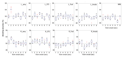 |
1 | MVPA using the hyperaligned 7T-BOLD signals revealed that the initial decrease contains finer information to decode facial expressions Video Permission Withheld
Toshiko Tanaka1, Naohiro Okamoto2, Ikuhiro Kida1,2, and Masahiko Haruno1,2
1National Institute of Information and Communications Technology, Suita Osaka, Japan, 2Osaka Universitiy, Suita Osaka, Japan
Previous studies suggested that the initial decrease in the BOLD signal reflects primary neuronal activity more than the later hemodynamic positive peak responses. We applied the hyper-alignment algorithm to 7T-BOLD timeseries during the facial expression discrimination task. and conducted the MVPA using the aligned data. We found decoding accuracies in the amygdala and superior temporal sulcus at 2 s after the face onset were significantly beyond baseline and the voxels contributing to the decoding accuracy displayed decreasing pattern in hemodynamics response, revealing that the initial decrease in 7T-BOLD signals contains finer information than thought previously.
|
||
3173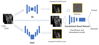 |
2 | Screening detection of abnormalities in knee joint MRI using deep machine learning Video Permission Withheld
Tsutomu Inaoka1, Akihiko Wada2, Rumiko Ishikawa1, Tomoya Nakatsuka1, Hisanori Tomobe1, Masaru Sonoda3, Akinori Yamamoto1, Ryousuke Sakai1, and Hitoshi Terada1
1Radiology, Toho University Sakura Medical Center, Sakura, Japan, 2Radiology, Juntendo University, Tokyo, Japan, 3Radiology, Seirei Sakura Citizen Hospital, Sakura, Japan
The DML model including fat-suppressed contrast generation, normal image restoration, and classification and determination models may make it possible to detect all abnormalities in knee joint MRI once.
|
||
3174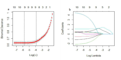 |
3 | MS or not MS: T2-weighted image (T2WI)-based radiomics findings distinguishes MS from its mimics
Ting He1, Yi Mao1, Yao Wang1, Lei Wang1, Qinmei Kuang1, Jie Xu1, Yuqi Ji1, Yujie He1, Meimei Zhu1, and Fuqing Zhou1
1The First Affiliated Hospital, Nanchang University, Nanchang, China
Multiple sclerosis (MS) is the most common immune-mediated disease of the central nervous system. Early identification of MS lesions and its mimics is very important to help alleviate the tension between the benefits of early diagnosis of MS and inaccurate diagnosis that can have serious health and economic consequences.The radiomics feature model based on T2-weighted images (T2WIs) has obvious clinical value and high specificity in differentiating patients with multiple sclerosis and ischemic demyelination. The high specificity of radiomic model could improve the accuracy of the 2017 McDonald diagnostic criteria for MS, by differentiating it from its mimics-- ischemic demyelination.
|
||
3175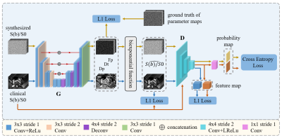 |
4 | Synthetic-to-real domain adaptation with deep learning for fitting IVIM-DWI parameters
Haoyuan Huang1, Baoer Liu2, Yikai Xu2, and Wu Zhou1
1School of Medical Information Engineering, Guangzhou University of Chinese Medicine, Guangzhou, China, 2Department of Medical Imaging Center, Nanfang Hospital, Southern Medical University, Guangzhou, China
The intravoxel incoherent motion (IVIM) model of DWI with IVIM parameters has been widely used in characterization. However, the optimal method to obtain the IVIM parameters is still being explored. In this work, we propose a synthetic-to-real domain adaptation method for fitting the IVIM parameters. Specifically, we use synthesized data to train the network to learn the accurate mapping of the b-value images to the parameter map, and design a discriminator to help the network gradually adapt the learned mapping to the real data. Experimental results demonstrate that the proposed method outperforms previously reported methods for fitting IVIM parameters.
|
||
3176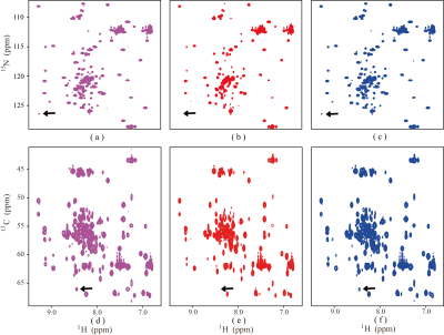 |
5 | XCloud-HyperLRF: Fast Hypercomplex NMR Spectroscopy with Cloud-based Low Rank Hankel Matrix Reconstruction
Di Guo1, Jiaying Zhan1, Zhangren Tu2, Yi Guo1, Yirong Zhou2, Jianfan Wu1, Qing Hong3, Vladislav Orekhov4, and Xiaobo Qu2
1School of Computer and Information Engineering, Xiamen University of Technology, Xiamen, China, 2Department of Electronic Science, Biomedical Intelligent Cloud R&D Center, Fujian Provincial Key Laboratory of Plasma and Magnetic Resonance, Xiamen University, Xiamen, China, 3China Mobile Group, Xiamen, China, 4Department of Chemistry and Molecular Biology, University of Gothenburg, Gothenburg, Sweden
Nuclear magnetic resonance (NMR) serves as an indispensable tool in revealing physical, chemical and structural information about molecules. We present a hypercomplex low rank approach to reconstruct hypercomplex NMR spectrum reconstruction. We first introduce an adjoint matrix operation to convert the hypercomplex signal into complex matrix and then propose a low-rank model and algorithm to reconstruct hypercomplex signal. The experiment results demonstrate that the proposed method provides a fast and high-fidelity reconstruction of hypercomplex NMR data. Furthermore, we made the method available at an open-access and easy-to-use cloud computing platform.
|
||
3177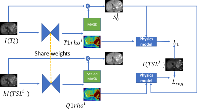 |
6 | Self-supervised Liver T1rho Mapping with Physics-constrained Regularization
Chaoxing Huang1, Yurui Qian1, Jian Hou1, Baiyan Jiang1,2, Queenie Chan3, Vincent Wong4, Winnie Chu1, and Weitian Chen1
1Department of Imaging and Interventional Radiology, The Chinese University of Hong Kong, Shatin, Hong Kong, 2Illuminatio Medical Technology Limited, Hong Kong, China, 3Philips Healthcare, Hong Kong, China, 4Department of Medicine and Therapeutics, The Chinese University of Hong Kong, Shatin, Hong Kong
Quantification of liver T1rho has gained interest in liver pathological study. Traditional fitting method requires acquisition of multiple T1rho-weighted images and it can be affected by respiratory motion. We propose a physics-informed self-supervised mapping method by taking only one T1rho-weighted image to do the mapping. Our preliminary experimental results show that our method has the potential to outperform the traditional multi-TSL acquisition method, particularly in the scenario of free-breathing MRI scan.
|
||
3178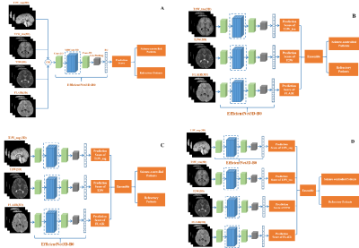 |
7 | Prediction of Drug Treatment Outcome among Epilepsy Children with Tuberous Sclerosis Complex based on Deep Neural Network and Multi-contrast MRI
Dian Jiang1,2, Zhanqi Hu3, Cailei Zhao4, Xia Zhao3, Jun Yang1,2, Yanjie Zhu2,5, Jianxiang Liao3, Dong Liang1,2,5, and Haifeng Wang2,5
1Research Centre for Medical AI, Shenzhen Institutes of Advanced Technology, Chinese Academy of Sciences, Shenzhen, China, 2University of Chinese Academy of Sciences, Beijing, China, 3Department of Neurology, Shenzhen Children’s Hospital, Shenzhen, China, 4Department of Radiology, Shenzhen Children’s Hospital, Shenzhen, China, 5Paul C. Lauterbur Research Center for Biomedical Imaging, Shenzhen Institutes of Advanced Technology, Chinese Academy of Sciences, Shenzhen, China
Distinguishing epilepsy drug treatment outcomes is crucial for treating children with tuberous sclerosis complex (TSC). Here, a deep-learning framework named AE-net was proposed to analyze epilepsy drug treatment outcomes using multi-contrast MRI data. Firstly, multi-contrast image-based models were respectively generated using the EfficientNet3D-B0 networks. Then, an averaging ensemble network was created as the final model. The proposed AE-net achieved the best AUC performance of 0.800 and sub-optimal AUC performance of 0.763 in the testing cohort, better than others. And the proposed method can predict epilepsy drug treatment outcomes to help clinical radiologists formulate more targeted treatments in the future.
|
||
3179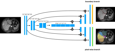 |
8 | Deep Learning Algorithm for Automated Liver Segmentation Using Portal Venous Phase Magnetic Resonance Images
Xinjun Han1, Niange Yu2, Qianjiang Xiao2, Mingyang Gao2, Dandan Zheng2, and Zhenghan Yang1
1Department of Radiology, Beijing Friendship Hospital, Capital Medical University, Beijing, China, 2Shukun (Beijing) Technology Co., Ltd, Beijing, China
Accurate segmentation of liver not only facilitates the subsequent quantitative assessment of the regions of interest but also benefits precise diagnosis, and surgical planning. These tasks are usually performed by radiologists via visual inspection and manual delineations, which are tedious, labor-intensive, time-consuming. Convolutional neural networks (CNNs) have shown promise for performing automated liver segmentation for CT examinations, but there is less research on MR images. In this study, we provide a 3D U-Net based model for robust whole-liver and Couinaud segment measurements to support the treatment decision-making process on MR images.
|
||
3180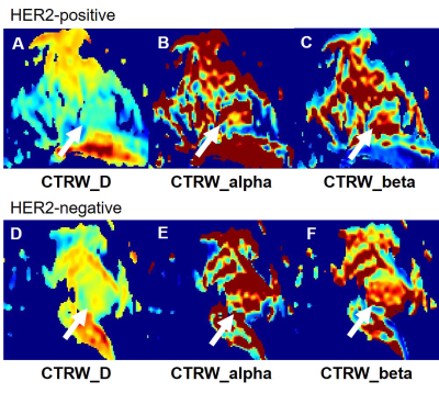 |
9 | Applying Continuous-Time-Random-Walk (CTRW) diffusion model based Radiomics in Predicting HER-2 Expression in Breast Invasive Ductal Cancer Video Not Available
Siyao Du1, Mengfan Wang1, Shasha Liu1, Xiaoqian Bian1, Xinyue Chen1, Liangcun Guo1, Guoliang Huang1, Ruimeng Zhao1, Can Peng1, Wenhong Jiang1, Qinglei Shi2, Xu Yan2, Guang Yang3, and Lina Zhang1
1Department of Radiology, The First Affiliated Hospital of China Medical University, Shenyang, China, 2MR Scientific Marketing, Siemens Healthineers Ltd., Beijing, China, 3Shanghai Key Laboratory of Magnetic Resonance, East China Normal University, Shanghai, China
In this study, we built a support vector machine (SVM) model based on quantitative parameters of continuous-time random-walk (CTRW) diffusion model in predicting the human epidermal growth factor receptor-2 (HER-2) expression in breast invasive ductal carcinoma. An AUC of 0.753 was achieved, which may have a great potential in future clinical practice.
|
||
3181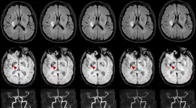 |
10 | Using the Artificial Intelligence Based Compressed SENSE Technology in Acute Cerebral Infarction
Cong Ning1, Yue Ma1, Di Yu1, Dan Tong1, Yi Zhu2, and Ke Jiang2
1Department of Radiology, The first hospital of jilin university, Changchun,Jilin, China, 2Philips Healthcare, Beijing, China
MRI has advantages in detecting acute ischemia and describing the core volume of the infarct without radiation, but the long scanning time affects clinical use. Compressed sense combined with artificial intelligence (CS-AI) technology accelerates the acquisition of imaging. This study aims to use the CS-AI technique to accelerate common sequences of Acute cerebral infarction and investigate the effect of different acceleration factors on image quality and diagnosis. The results show that CS-AI reconstruction scans reduce scanning time while maintaining image quality compared to conventional SENSE.
|
||
3182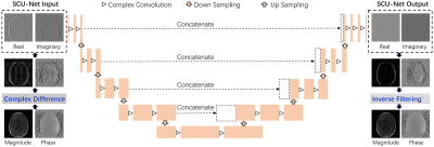 |
11 | Fast Magnetic Resonance Imaging by Deep Learning the Sparsified Complex Data
Zhaoyang Jin1 and Qing-San Xiang2
1Hangzhou Dianzi University, Hangzhou, China, 2University of British Columbia, Vancouver, BC, Canada
In this study, we exploited a sparsifying deep learning method and an inverse filtering reconstruction to obtain high quality complex MR images for under-sampled MRI data. This study allows much more flexible data representations for complex MRI data training, leading to significantly higher complex reconstruction quality for practical MRI applications.
|
||
3183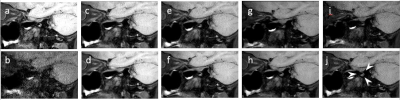 |
12 | 3D T1-weighted sequence of volumetric isotropic turbo spin-echo acquisition with a deep learning constrained Compressed SENSE reconstruction Video Not Available
Yue Ma1, Linna Li2, Yang Sun2, Chang Zhai2, Dan Tong2, Yi Zhu3, and Ke Jiang3
1Department of Radiology, The First Hospital of Jilin University, Changchun,Jilin, China, 2The First Hospital of Jilin University, Changchun,Jilin, China, 3Philips Healthcare,Beijing,China, Beijing, China
Compressed SENSE-AI reconstruction system was introduced with its sufficient noise removal to maintain high image quality while reducing scanning time. Intracranial and carotid vessel wall MR imaging (VW-MRI) are widely available in detecting and characterizing atherosclerosis occurring in the two vascular ranges, however, long scan time had so far limited high resolution VW-MR imaging. In this study, we investigated the impact of Compressed SENSE-AI acceleration factor on the diagnostic quality of magnetic resonance images and compared with standard vessel wall MR imaging protocol.
|
||
3184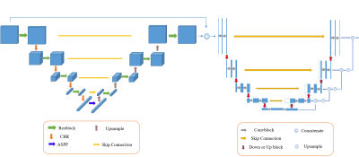 |
13 | 3D Automatic segmentation of breast lesion in dynamic contrast enhanced MRI using deep convolutional neural network Video Not Available
Fuliang Lin1,2, Zhou Liu3, Qinglei Zhou2, Pengyu Gao4, Jie Wen3, Meng Wang3, Ya Ren3, Dehong Luo3, Ye Li1, Dong Liang1, Xin Liu1, Hairong Zheng1, and Na Zhang1
1Paul C. Lauterbur Research Center for Biomedical Imaging, Shenzhen Institutes of Advanced Technology, Chinese Academy of Sciences, Shenzhen, China, 2Zhengzhou University, Zhengzhou, China, 3Department of Radiology, National Cancer Center/National Clinical Research Center for Cancer/Cancer Hospital & Shenzhen Hospital, Chinese Academy of Medical Sciences and Peking Union Medical College, Shenzhen, China, 4Henan University, Kaifeng, China
Breast cancer is the most common cancer in women with the highest incidence. Dynamic contrast enhanced MRI is one of the backbone sequences for breast cancer diagnosis. Accurate segmentation of breast lesions based on DCE-MRI images is helpful for clinically objective and quantitative evaluation of breast lesions. However, the commonly used manual segmentation method is subject to high inter-observer variability. In this study, a 3D automatic algorithm is proposed for segmentation of breast lesions in DCE-MRI. The results show that the proposed network can obtain accurate and automatic 3D segmentation of breast lesions and achieves better segmentation results than VNet.
|
||
3185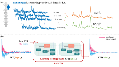 |
14 | MRS Denoising Model: ReLSTM-Net Trained by few In vivo Measured Data Video Permission Withheld
Dicheng Chen1, Wanqi Hu1, Huiting Liu1, Tianyu Qiu1, Yihui huang1, Liangjie Lin2, Di Guo3, Jianzhong Lin4, and Xiaobo Qu1
1Department of Electronic Science, National Institute for Data Science in Health and Medicine, Xiamen University, Xiamen, China, 2Healthcare, Philips, Beijing, China, 3School of Computer and Information Engineering, Xiamen University of Technology, Xiamen, China, 4Department of Radiology, The Zhongshan Hospital affiliated to Xiamen University, Xiamen, China 1H Magnetic Resonance Spectroscopy (MRS) suffers low Signal-Noise Ratio (SNR) due to low concentrations of metabolites. To improve the SNR, the current mainstream is to do Signal Averaging with repeated samplings but it is time-consuming. Therefore, we designed a novel denoising ReLSTM-Net to learn the mapping from the low SNR MRS to the high SNR one in the time-domain by a few in vivo measured data. Denoised spectra by the proposed method has higher accuracy and reliability in quantifying metabolites Glx, tCho and mI, compared with the state-of-art Low-Rank method. |
||
3186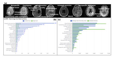 |
15 | Multi-class brain lesion detection: establishing a baseline for fastMRI+ dataset
Lifeng Mei1,2, Sixing Liu1,2, Guoxiong Deng1,2, Shaojun Liu1,2, Yali Zheng1,2, and Mengye Lyu1,2
1College of Health Science and Environmental Engineering, Shenzhen Technology University, Shenzhen, China, 2College of Applied Sciences, Shenzhen University, Shenzhen, China Computer aided diagnosis (CAD) is widely considered an important application of deep learning in healthcare. However, brain data with lesion location labels are rare in MRI domain. Recently, Microsoft Research has released a dataset of clinical pathology annotations based on the raw images from fastMRI and named it fastMRI+. Here, fastMRI+ brain dataset was analyzed and used to train deep learning-based models for lesion detection. Some well-known object detection architectures such as YOLOv3, Faster-RCNN and YOLOX were compared. Overall, this abstract established a baseline with improvement suggestions for future studies. |
||
 Back to Meeting Home
 Back to the Program-at-a-Glance
The International Society for Magnetic Resonance in Medicine is accredited by the Accreditation Council for Continuing Medical Education to provide continuing medical education for physicians.
 Back to Meeting Home
 Back to the Program-at-a-Glance
 View the Presentation