Online Gather.town Pitches
Neurological Conditions I
Joint Annual Meeting ISMRM-ESMRMB & ISMRT 31st Annual Meeting • 07-12 May 2022 • London, UK

Online Gather.town Pitches
Neurological Conditions I
| Booth # | ||||
|---|---|---|---|---|
3054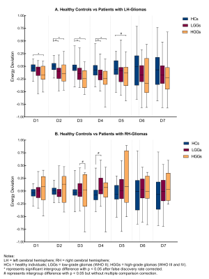 |
1 | Thalamic Plasticity in Brains with Glioma Investigated with Discrete Wavelet Transformation Analysis of Resting-state BOLD Fluctuations
Siqi Cai1,2, Zhifeng Shi3, Chunxiang Jiang1,2, Shihui Zhou1,2, and Lijuan Zhang*1
1Shenzhen Institutes of Advanced Technology, Chinese Academy of Sciences, Shenzhen, China, 2University of Chinese Academy of Sciences, Beijing, China, 3Huashan Hospital of Fudan University, Shanghai, China
Thalamic plasticity plays an important role in the functional remodeling in brains with glioma. This study investigated the frequency-specific functional alteration of the thalami in the context of gliomas with discrete wavelet transformation and wavelet-energy. Brains with glioma in the left hemisphere featured with amplified inter-thalamus energy deviation for BOLD signals at 15.63-250mHz irrespective of the malignant grades, as compared with healthy controls. Similar analysis revealed no energy changes for the right brain gliomas. These findings suggest a differential thalamic plasticity that is specific to the tumor laterality, and may leverage innovation in the treatment of glioma involving neuromodulation.
|
||
3055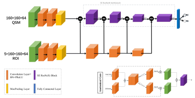 |
2 | Deep Learning with Anatomical Attention Mechanism for Distinguishing Parkinson’s Disease from Normal Controls in MR imaging
Yida Wang1, Naying He2, Chenglong Wang1, Yan Li2, Zhijia Jin2, Xiance Zhao3, Ewart Mark Haacke2,4, Fuhua Yan2, and Guang Yang1
1Shanghai Key Laboratory of Magnetic Resonance, East China Normal University, Shanghai, China, 2Department of Radiology, Ruijin Hospital, Shanghai Jiao Tong University School of Medicine, Shanghai, China, 3Philips Healthcare, Shanghai, China, 4Department of Biomedical Engineering, Wayne State University, Detroit, MI, United States
We proposed an automatic cascaded framework based on deep learning to segment deep brain nuclei and distinguish Parkinson’s disease from normal controls using quantitative susceptibility mapping (QSM) images. A 3D CA-Net model integrating channel attention, spatial attention and scale attention module was utilized to segment 5 brain nuclei from QSM and T1W data. Then, the QSM images and the segmented brain nuclei ROIs were fed into the SE-ResNeXt50 with anatomical attention mechanism to get the predicted PD probability. The proposed method provided good interpretability and achieved AUC values of 0.97 and 0.90 on training and testing cohort, respectively.
|
||
3056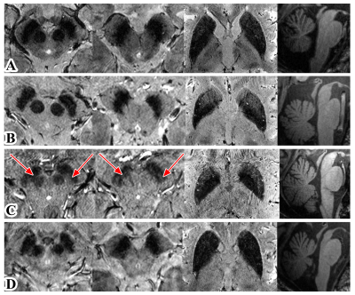 |
3 | Anatomical changes and iron deposition of Parkinson's disease, multiple system atrophy, and progressive supranuclear palsy on 7 Tesla MRI
Su Dongning1,2, Zhang Zhe2,3, Zhu Wanlin2,3, Sui Binbin2,3, Zhang Yingkui2,3, Bi Jingfeng2,3, Kong Qingle 4, Zhang Zhijin1,2, Gan Yawen1,2, Jin Jianing1,2, Wang Xuemei1,2, Wang Zhan1,2, Wang Yongjun 1,2,3, Wu Tao1,2,
Jing Jing1,2,3, and Feng Tao1,2
1Department of Neurology, Beijing Tiantan Hospital,Captital Medical University, Beijing, China, 2China National Clinical Research Center for Neurological Diseases, Beijing, China, 3Tiantan Neuroimaging Center of Excellence, Beijing Tiantan Hospital, Capital Medical University, Beijing, China, 4MR Collaboration, Siemens Healthineers Ltd., Beijing, China
Ultra-high field MRI enables visualization of anatomical alterations and quantification of iron deposition in the subcortical nucleus. To investigate whether 7T MRI could provide the diagnostic opportunity for Parkinson-plus syndromes, we recruited PD, MSA-P, MSA-C and PSP patients who underwent multi-modal scans including 3D MPRAGE and 3D multi-echo T2*-weighted MRI. All six patients with MSA-C showed visible nigrosome-1. The distances from SN to RN were significantly shorter in PSP than PD. The R2* values in SN, RN and putamen were significantly higher in MSA-P than PD. Our pilot study provided first-of-its-kind evidence that 7T MRI could help diagnose Parkinson-plus syndromes.
|
||
3057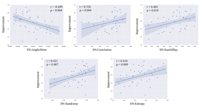 |
4 | Iron Spatial Distributions in Deep Gray Nuclei are Related to Motor Outcomes of Subthalamic Nuclei Deep Brain Stimulation for Parkinson's Disease
Weiwei Zhao1, Xi Wu2, Chunhui Yang2, Luguang Chen3, Yiqing Qiu2, Chao Ma3, Gaiying Li1, Yang Song4, Yi Wang5, and Jianqi Li1
1East Chian Normal University, Shanghai, China, 2Department of Neurosurgery, Changhai Hospital, Shanghai, China, 3Department of Radiology, Changhai Hospital, Shanghai, China, 4MR Scientific Marketing, Siemens Healthineers, Shanghai, China, 5Department of Radiology, Weill Medical College of Cornell University, New York, NY, United States
The objective of this study was to investigate the relationship between motor outcomes of subthalamic nuclei deep brain stimulation (STN-DBS) and spatial distribution of iron in the deep gray nuclei for patients with Parkinson's disease (PD). The first and second texture parameters were obtained from the preoperative QSM of 40 PD patients. Significant correlations were found between motor improvement after DBS and QSM texture parameters in substantia nigra (SN) and dentate nucleus (DN). Linear regression showed that second-order texture parameter angular second moment in SN (r = -0.449, p = 0.004) had the highest correlation with the STN-DBS motor outcomes.
|
||
3058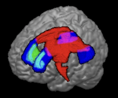 |
5 | Tract-specific association of myelin water fraction with cognitive decline and motor dysfunction in Parkinson’s disease
Septian Hartono1,2, Tiet Gan Ong3, Weiling Lee4, Zi Cheng Kuek4, Celeste Chen1, Kuan Jin Lee5, Jongho Lee6, Eng King Tan1,2, and Ling Ling Chan2,4
1National Neuroscience Institute, Singapore, Singapore, 2Duke-NUS Graduate Medical School, Singapore, Singapore, 3Nanyang Technological University, Singapore, Singapore, 4Singapore General Hospital, Singapore, Singapore, 5Institue of Bioengineering and Bioimaging, Singapore, Singapore, 6Seoul National University, Seoul, Korea, Republic of
We found associations between tract-specific myelin water fraction and cognitive decline, and motor dysfunction in patients with Parkinson’s disease (PD). Myelin water fractions (MWF) in the anterior corona radiata and posterior thalamic radiations were associated with cognitive decline in PD whilst those in the superior and posterior corona radiata, posterior limb of internal capsule and corticospinal tract were associated with moderate to severe motor dysfunction in PD. Our results suggest that MWF has potential utility as an objective imaging biomarker to monitor longitudinal changes in target WM tracts relevant to motor and cognitive dysfunction in PD neurodegeneration.
|
||
| 3059 | 6 | Altered intrinsic functional connectivity across nine brain networks in early stages of idiopathic Parkinson’s disease
Yao-Chia Shih1,2,3, Pohchoo Seow1, Hartono Septian2,4, Welton Thomas2,4, Weiling Lee1, Aeden Zi Cheng Kuek1, Say Lee Chong1, Samuel Yong Ern Ng4, Nicole Shuang Yu Chia4, Eng-King Tan4, Louis CS Tan4, and Ling-Ling Chan1,2
1Department of Diagnostic Radiology, Singapore General Hospital, Singapore, Singapore, 2Duke-NUS Medical School, Singapore, Singapore, 3Graduate Institute of Medicine, Yuan Ze University, Taoyuan, Taiwan, 4Department of Neurology, National Neuroscience Institute – SGH Campus, Singapore, Singapore
Parkinson’s disease (PD) is a common neurodegenerative disease. Besides motor deficits, autonomic and cognitive dysfunctions might be caused by global abnormalities beyond the dopaminergic system. We performed resting-state fMRI to measure functional connectivity between the central autonomic network and eight other critical resting networks in both early PD and healthy control groups. Using general linear models, we identified functional impairments in the inter- and intra-network connections in relation to various brain functions. These findings could shed light on the neural mechanisms behind the complex clinical manifestations evident even in the early stages of PD.
|
||
3060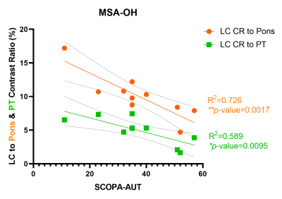 |
7 | Decreased LC MRI Contrast and Relationship with Orthostatic Hypotension in Multiple System Atrophy
Haiying Lv1, Qing Li2, and Yong Lu3
1Shanghai Jiao Tong University School of Medicine, Shanghai, China, 2MR Collaborations, Siemens Healthineers Ltd., Shanghai, China, 3Ruijin Hospital / Lu Wan Branch Affiliated to Shanghai Jiao Tong University School of Medicine, Shanghai, China
MRI imaging with Neuromelanin contrast has been thrived in recent years. We adopted this method again to explore the LC contrast differences in multiple system atrophy (MSA). Three methods for quantifying LC contrast were used, which all had the ability to reproduce the results that LC contrast decreases in MSA. We further did subgroup analyses of MSA with orthostatic hypotension (MSA-OH) and MSA without orthostatic hypotension (MSA-nonOH). Interestingly, although LC contrast of the two subgroups were not significantly different, adverse correlations between LC contrast and autonomic dysfunction severity were found in MSA-OH and MSA-nonOH subgroups.
|
||
3061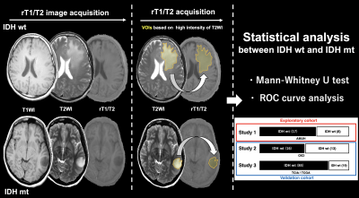 |
8 | The ratio of T1-Weighted to T2-Weighted Signal Intensity and IDH mutation in glioma
Takahiro Sanada1, Shota Yamamoto1, Hirotaka Sato1,2, Mio Sakai3, Masato Saito1, Nobuyuki Mitsui1, Satoru Hiroshima1, Ryogo Anei4, Yonehiro Kanemura5, Katsuyuki Nakanishi3, Haruhiko Kishima6, and Manabu Kinoshita1
1Neurosurgery, Asahikawa Medical University, Asahikawa, Japan, 2Neurosurgery, Japanese Red Cross Kitami Hospital, Asahikawa, Japan, 3Diagnostic and Interventional Radiology, Osaka International Cancer Institute, Osaka, Japan, 4Neurosurgery, Moriyama Hospital, Asahikawa, Japan, 5Biomedical Research and Innovation, National Hospital Organization Osaka National Hospital, Osaka, Japan, 6Neurosurgery, Osaka University Graduate School of Medicine, Suita, Japan
The authors investigated the correlation of the ratio of T1WI to T2WI signal intensity (rT1/T2) and IDH mutation status in lower-grade gliomas (LrGG). An exploratory study was conducted to determine an ideal cut-off of rT1/T2 for predicting IDH-mutation, followed by a validation study using two different cohorts. Mean rT1/T of IDH-wild type LrGG was higher than IDH-mutant and significantly predicted IDH mutation in the exploratory cohort. The domestic cohort validated this result, but the accuracy in the TCIA / TCGA cohort declined. The cause of this decline should be further investigated.
|
||
3062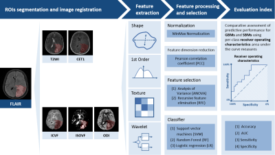 |
9 | Radiomics analysis of neurite orientation dispersion and density imaging: differentiation between glioblastoma and solitary brain metastasis
Guohua Zhao1, Huiting Zhang2, Eryuan Gao1, Ankang Gao1, Xiaoyue Ma1, Jie Bai1, Xu Yan2, Guang Yang3, Yusong Lin4, and Jingliang Cheng1
1The Department of Magnetic Resonance Imaging, The First Affiliated Hospital of Zhengzhou University, Zhengzhou, China, 2Siemens Healthineers, Shanghai, China, 3Shanghai Key Laboratory of Magnetic Resonance, East China Normal University, Shanghai, China, 4The School of Software, Zhengzhou University, Zhengzhou, China
Differentiation of glioblastomas and solitary brain metastases is clinically crucial for the prescription of patient management and assessment of prognosis. However, indistinguishable signs between two tumors on routine MRI leads to a high misdiagnosis rate. Neurite orientation dispersion and density imaging (NODDI) can not only estimate the intricacy of neurites in vivo but also provide data to illuminate pathology. We developed a series of radiomics models of NODDI parameter maps, routine MRI, combined routine MRI, and combined NODDI parameter maps to compare their performance in the identification of two tumors. Finally, the combined NODDI radiomics model obtained the best performance.
|
||
3063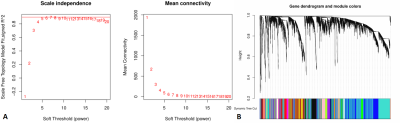 |
10 | Radiomics Molecular Mechanism and Candidate Prognostic Biomarker Investigation for Glioblastoma Multiforme Utilizing WGCNA
Jixin Luan1, Jun Chen1, Hua Fan1, Di Zhang1, Peng Wu2, and Chuanchen Zhang1
1Department of Radiology, Liaocheng People's Hospital, Shandong First Medical University & Shandong Academy of Medical Sciences, Liaocheng, China, 2Philips Healthcare, Shanghai, China
In this study, we aimed to explore the deeper molecular mechanism of radiomics features, and uncover new GBM prognostic biomarker. We extracted radiomics features out of 57 patients with MRI data obtained from TCIA database and corresponding clinical and transcription information from TCGA database. WGCNA revealed that the radiomics features are correlated with numerous cancer-associated biological processes, including regulation of vasculature development and so on. HMGA2 as the hub gene was screened by Cytoscape software. There was significant difference in survival analysis between the high and low expression groups, indicating that HMGA2 may be a new prognostic biomarker for GBM.
|
||
3064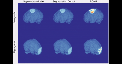 |
11 | Interpretable Meningioma Grading and Segmentation with Multiparametric Deep Learning
Yohan Jun*1, Yae Won Park*2, Hyungseob Shin1, Yejee Shin1, Jeong Ryong Lee1, Kyunghwa Han2, Soo Mee Lim3, Seung-Koo Lee2, Sung Soo Ahn**2, and Dosik Hwang**1
1Electrical and Electronic Engineering, Yonsei University, Seoul, Korea, Republic of, 2Department of Radiology and Research Institute of Radiological Science and Center for Clinical Imaging Data Science, Yonsei University College of Medicine, Seoul, Korea, Republic of, 3Department of Radiology, Ewha Womans University College of Medicine, Seoul, Korea, Republic of
Preoperative prediction of meningioma grade is important because it influences treatment planning, including surgical resection and stereotactic radiosurgery strategy. The aim of this study was to establish a robust interpretable multiparametric deep learning (DL) model for automatic noninvasive grading of meningiomas along with segmentation. We demonstrated that the interpretable multiparametric DL grading model that combined the T2-weighted and contrast-enhanced T1-weighted images can enable fully automatic grading of meningiomas along with segmentation.
|
||
 |
3065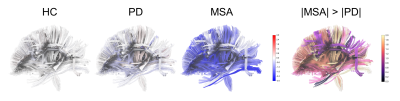 |
12 | Microstructural Deviation in Parkinson’s Disease and Multiple System Atrophy Detected by Spatial Normative Models
Chang-Le Chen1,2, Ming-Che Kuo3, Chun-Hwei Tai3, Yung-Chin Hsu4, Ruey-Meei Wu3, and Wen-Yih Isaac Tseng1,5
1Institute of Medical Device and Imaging, National Taiwan University College of Medicine, Taipei, Taiwan, 2Department of Bioengineering, University of Pittsburgh, Pittsburgh, PA, United States, 3Department of Neurology, National Taiwan University Hospital, Taipei, Taiwan, 4AcroViz Technology Inc., Taipei, Taiwan, 5Molecular Imaging Center, National Taiwan University, Taipei, Taiwan
By applying spatial normative models based on a population-based cohort, we can quantify the microstructural deviation of white matter in patients with Parkinson’s disease (PD) and multiple system atrophy (MSA). We found that microstructural deviation of the frontal aslant tracts, cerebrospinal tracts, and parts of corpus callosum was more profound in MSA compared to that in PD. Also, the uncinate fasciculi were the common degenerative marker in these two diseases. Moreover, the microstructural deviation of WM tracts may reflect the association with the disease-related daily functional deficit in MSA and the timing of disease onset in PD.
|
|
3066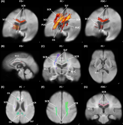 |
13 | Fixel-Based Analysis Identifies White Matter Tract Degeneration in Parkinson’s Disease with Mild Cognitive Impairment
Chih-Chien Tsai1,2, Ting-Wei Liao3, Yih-Ru Wu3,4, Yi-Ming Wu5, and Jiun-Jie Wang2,6,7
1Department of Medical imaging and Radiological Science, Chang Gung University, Taoyuan City, Taiwan, 2Healthy Aging Research Center, Chang Gung University, Taoyuan City, Taiwan, 3Department of Neurology, Chang Gung Memorial Hospital, Taoyuan City, Taiwan, 4College of Medicine, Chang Gung University, Taoyuan City, Taiwan, 5Department of f Medical Imaging and Intervention, Chang Gung Memorial Hospital, Taoyuan City, Taiwan, 6Department of Medical imaging and Radiological Science, Chang-Gung University, Taoyuan City, Taiwan, 7Medical Imaging Research Center, Institute for Radiological Research, Chang Gung University/Chang Gung Memorial Hospital, Taoyuan City, Taiwan
Cognitive decline is a common non-motor symptom in Parkinson's disease (PD) patients, described as mild cognitive impairment (PD-MCI). PD-MCI is identified as a risk for PD with Dementia. Previous studies indicated that PD-MCI patients showed gray matter volume decrease and white matter tract degeneration. We investigate the utility of fixel based analysis as a biomarker for disease progression. The results show a significant degeneration of white matter in PD-MCI patients. Our findings indicated the white matter tract degeneration in PD-MCI patients and shed light on the value of the fiber-specific method to the clinical diagnosis of PD-MCI.
|
||
3067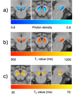 |
14 | Relaxometry of the Substantia Nigra Pars Compacta and Subsegment Analysis: A Healthy Volunteer Study
Yasuhiro Fujiwara1, Shota Ishida2, Yuki Matta3, Masayuki Kanamoto3, and Hirohiko Kimura4
1Department of Life Sciences, Fuculty of Medical Image Sciences, Kumamoto University, Kumamoto, Japan, 2Department of Radiological Technology, Faculty of Medical Sciences, Kyoto College of Medical Science, Kyoto, Japan, 3Radiological Center, University of Fukui Hospital, Fukui, Japan, 4Department of Radiology, University of Fukui, Fukui, Japan
An anatomical atlas of the three regions (medial, dorsal, and ventrolateral) of the substantia nigra pars compacta (SNpc) were created based on neuromelanin (NM)-sensitive images, and Proton density, T1, and T2 values for each region were characterized in healthy volunteers. The T1 value of the ventrolateral group was higher than the other SNpc regions, which may be correlated with the NM concentration. The measured relaxation times can be used as baseline values for the SNpc.
|
||
3068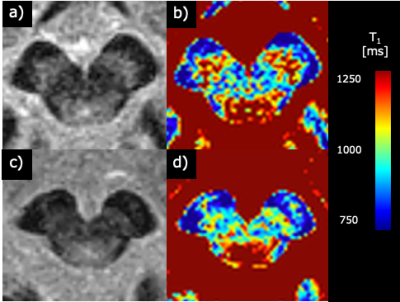 |
15 | Quantitative Evaluation of Substantia Nigra Degeneration in Parkinson’s Disease using T1 mapping
Ayaka Yokota1, Yasuhiro Fujiwara2, Tetsuyoshi Hirai3, Nobutaka Sakae4, Naokazu Sasagasako4, Takahisa Izumi5, Kuniharu Ohi5, and Hiroyuki Kumazoe5
1Graduated School of Health Sciences, Kumamoto University, Kumamoto, Japan, 2Department of Medical Image Sciences, Faculty of Life Sciences, Kumamoto University, Kumamoto, Japan, 3Faculty of Medicine, Saga University, Saga, Japan, 4Department of Neurology, National Hospital Organization Omuta National Hospital, Omuta, Japan, 5Department of Radiology, National Hospital Organization Omuta National Hospital, Omuta, Japan
We evaluated the T1 values of the whole and subregions of the substantia nigra (SN) in patients with Parkinson's disease (PD) to identify an imaging biomarker for the early diagnosis of PD. T1 values of the SN of patients with PD were significantly shorter than those of healthy controls and tended to be longer in the lateral part for both groups. T1 values may reflect the pathology of the SN and help assess the degeneration of the SN caused by PD.
|
||
 Back to Meeting Home
 Back to the Program-at-a-Glance
The International Society for Magnetic Resonance in Medicine is accredited by the Accreditation Council for Continuing Medical Education to provide continuing medical education for physicians.
 Back to Meeting Home
 Back to the Program-at-a-Glance
 View the Presentation