Online Gather.town Pitches
Cerebrovascular, Stroke, Ischemia, Atherosclerosis I
Joint Annual Meeting ISMRM-ESMRMB & ISMRT 31st Annual Meeting • 07-12 May 2022 • London, UK

Online Gather.town Pitches
Cerebrovascular, Stroke, Ischemia, Atherosclerosis I
| Booth # | ||||
|---|---|---|---|---|
4168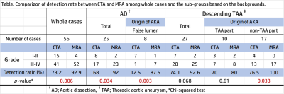 |
1 | Comparative study between the Slow Infusion MR angiography and CT angiography in detection of the Adamkiewicz artery
Shohei Mizushima1, Takahiko Mine1, Masashi Abe1, Hiromitsu Hayashi2, Masahiro Fujii3, Tetsuro Sekine4, Taro Yokoyama1, Shinpei Ikeda1, Seigoh Happoh1, and Shin-ichiro Kumita2
1Radiology, Nippon Medical School Chiba Hokusoh Hospital, Inzai, Chiba, Japan, 2Radiology, Nippon Medical School Hospital, Tokyo, Japan, 3Cardiovascular surgery, Nippon Medical School Chiba Hokusoh Hospital, Inzai, Chiba, Japan, 4Radiology, Nippon Medical School Musashikosugi Hospital, Kawasaki, Kanagawa, Japan
The Slow-Infusion MRA with SPGR provided the higher detectability of the AKA with whole patients, all AD patients, and with the AD patients whose AKA originating from false lumen in comparison with CTA.
|
||
4169 |
2 | Small-patch CNN and random forest to model and quantify the lenticulostriate artery from 7T time-of-flight MR angiography
Zhixin Li1,2,3, Dongbiao Sun1,2,3, Yue Wu1,2,3, Chen Ling4,5, Jing An6, Yun Yuan4,5, Zhaoxia Wang4,5, Rong Xue1,2,3, Yan Zhuo1,2,3, and Zihao Zhang1,2,3
1State Key Laboratory of Brain and Cognitive Science, Institute of Biophysics, Chinese Academy of Sciences, Beijing, China, Beijing, China, 2University of Chinese Academy of Sciences, Beijing, China, Beijing, China, 3The Innovation Center of Excellence on Brain Science, Chinese Academy of Sciences, Beijing, China, Beijing, China, 4Department of Neurology, Peking University First Hospital, Beijing, 100034, China, Beijing, China, 5Beijing Key Laboratory of Neurovascular Disease Discovery, Peking University First Hospital, Beijing, 100034, China, Beijing, China, 6Siemens Shenzhen Magnetic Resonance Ltd., Shenzhen, China, Shenzhen, China
Time-of-flight MR angiography at 7T allowed noninvasive visualization of the lenticulostriate artery (LSA). However, vasculature modeling of LSA remained challenging due to limited signal-to-noise ratio and pulsation artifact. In this study, we introduced an automated vascular segmentation and tracing method based on small-patch convolutional neural network (CNN), random forest, and multiple filtering. This method outperformed existing U-NET based methods and found radius changes of LSA branches in patients with cerebral small vessel diseases (cSVD). The automated quantification of LSA vasculatures will potentially facilitate diagnosis and clinical studies of cSVD.
|
||
4170 |
3 | Fast 3D Wheel Acquisition: Comparison of Efficacy for Cerebral MR Angiography with Conventional Parallel Imaging
Satomu Hanamatsu1, Kazuhiro Murayama2, Yoshiharu Ohno3, Kaori Yamamoto4, Yuki Obama1, Hirotaka Ikeda1, Hiroyuki Nagata1, Masato Ikedo4, Masao Yui4, Akiyoshi Iwase5, and Hiroshi Toyama1
1Radiology, Fujita Health University, School of Medicine, Toyoake, Japan, 2Joint Research Laboratory of Advanced Medical Imaging, Fujita Health University, School of Medicine, Toyoake, Japan, 3Radiology, Joint Research Laboratory of Advanced Medical Imaging, Fujita Health University, School of Medicine, Toyoake, Japan, 4Canon Medical Systems Corporation, Otawara, Japan, 5Radiology, Fujita Health University, Hospital, Toyoake, Japan
To the best of our knowledges, there are no major papers that assess the influence of Fast 3D wheel to cerebral MR angiography in patients with cerebrovascular diseases. We hypothesize that “Fast 3D wheel (Fast 3Dw)” has a potential to reduce examination time without degradation of image quality and aneurysm or vascular evaluations, when compared with PI. The purpose of this study was to directly compare the efficacy of Fast 3Dw for cerebral MR angiography with conventional PI in patients with cerebral aneurysm.
|
||
4171 |
4 | Comparison analysis of imaging techniques about routine, different acceleration factor coefficients of compressed sensing and PETRA TOF-MRA
Xiangcheng Hao1, Shaoyu Wang2, Huapeng Zhang2, He Zhao1, and Yang Gao1
1Affiliated Hospital of Inner Mongolia Medical University, Hohhot, China, 2MR Scientific Marketing, Siemens Healthineers, Shanghai, China
We compare three MRA techniques between CS-TOF, PI-TOF and PETRA-TOF about imaging quality and speed, and also compared the effects of different acceleration factor coefficients (4.6×, 7.2×, 10.3×) on CS-TOF-MRA images on a 3T MR scanner.
|
||
4172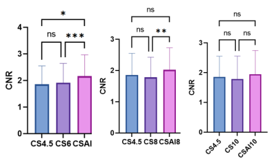 |
5 | Non-contrast-enhanced mDixon water-fat separation whole-heart coronary MRA with a deep learning constrained Compressed SENSE reconstruction
Wenyun Liu1, Lei Zhang1, Cheng Li2, Yuejiao Sun1, Ying Qiu1, Yi Zhu3, Ke Jiang3, Shuo Wang1, and Huimao Zhang1
1Department of Radiology, The First Hospital of Jilin University, Changchun, China, 2Department of Cardiovascular center, The First Hospital of Jilin University, Changchun, China, 3Philips Healthcare, Beijing, China
The conventional 3D whole-heart free-breathing coronary MR angiography suffers from a long scan time. However, using very high acceleration factors leads to degradation of image quality due to insufficient noise removal. In this study, we use Compressed-SENSE Artificial Intelligence (CS-AI) framework to acquire highly accelerated 3D non-contrast-enhanced mDixon water-fat separation whole-heart CMRA. The result shows that CS-AI reconstruction can significantly decrease scan time with sufficient image quality compared to Compressed-SENSE(CS) and might be clinically useful in assessment of coronary artery disease.
|
||
4173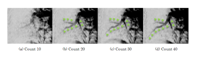 |
6 | Non-contrast enhanced spatially-selective and time-resolved vessel imaging by using cylinder-shaped pre-saturation pulse train in the Lung Video Permission Withheld
Masahiro Takizawa1, Takashi Nishihara1, and Chikako Moriwake1
1Radiation Diagnostic System Division, FUJIFILM Healthcare Corporation, Kashiwa-shi, Japan
Cylinder-shaped pre-saturation pulse train is developed for non-contrast enhanced spatially-selective and time-resolved vessel imaging. The target vessel is selected by cylinder-shaped pre-saturation, and the dynamics of blood flow in the target vessel is observed by changing the number of applied pre-saturation pulses. The developed pulse train was demonstrated to visualize dynamics of a target pulmonary vessel in the lung.
|
||
4174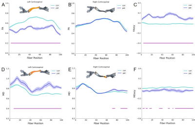 |
7 | Characterization of Corticospinal Tract Injury along Fibers by Automated Fiber Quantification in Stroke
Fan Liu1, Yu Wei1, Qiurong Yu1, Hewei Wang2, Dazhi Yin3, Guojun Xu1, Miao Guo1, Ling Liu1, Qianwen Li1, and Mingxia Fan1
1Shanghai Key Laboratory of Magnetic Resonance, School of Physics and Electronic Science, East China Normal University, Shanghai, China, 2Department of rehabilitation, Huashan Hospital, Fudan University, Shanghai, China, 3School of Psychology and Cognitive Science, East China Normal University, Shanghai, China Previous DTI studies mainly detected partial ROIs or mean diffusion profiles of corticospinal tract (CST), few revealed changes along CST trajectory. We used Automated Fiber Quantification (AFQ) derived from DTI to investigate whether diffusion parameters changes were localized to its specific regions or spread throughout. The results showed AFQ traced out a clearer picture of the damaged CST along its trajectory in patients with hemiplegia after stroke. Furthermore, the severity of several damaged CST segments was correlated with motor scores, possibly providing more accurate and reliable "target" regions for assessing clinical prognosis and the efficacy of motor rehabilitation. |
||
4175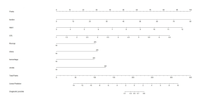 |
8 | Atherosclerotic ischemic recurrent stroke patients: a nomogram model of risk factors and risk prediction Video Not Available
Niane Ma1, XiaoLing Zhang1, Xiaoyan Lei1, Min Tang1, Ling Li1, Xuejiao Yan1, and Kai Ai2
1Shaanxi Provincial People's Hospital, Xi’an, China, 2Philips healthcare, Xi’an, China
The aim of this study was to construct a diagnostic model of recurrent stroke with the combination of high resolution MRI imaging characteristics and clinical parameters. The vessel wall characteristics of plaque and clinical data were compared between stroke and recurrent stroke patients. Multivariate logistic regression analysis was explored, seven risk factors (plaque burden, fibrous cap, hyperdense, hemorrhage, HbA1, low density lipoprotein and smoke) were independent risk factors for recurrent stroke patients. Then a nomogram model was established for risk prediction.
|
||
4176 |
9 | Venous Response in Brains with Ischemic Stroke Assessed with Susceptibility-Based MR Imaging
Shihui Zhou1,2, Zhiqiang Wei3, Yulong Qi4, Li Yi3, Siqi Cai1,2, Chunxiang Jiang1,2, and Lijuan Zhang*1
1Shenzhen Institutes of Advanced Technology, Chinese Academy of Sciences, Shenzhen, China, 2University of Chinese Academy of Sciences, Beijing, China, 3Department of Neurology, Peking University Shenzhen Hospital, Shenzhen, China, 4Department of Radiology, Beijing University Shenzhen Hospital, Shenzhen, China
Venous response has been overlooked in the characterization of ischemic stroke (IS). In this study, vein density (VD) and oxygen extraction fraction (OEF) were assessed as indices of venous response for both the ipsi- and contra-lesional hemispheres in a cohort of patient with corona radiata IS using susceptibility MR imaging. Relative VD and OEF (ratios between the averaged ipsilesional and contralesional measurements) increased from early (1-7 days) to late subacute stages of IS (7-15 days). Patients with lesion in right corona radiata showed more extensive interhemispheric difference in OEF, suggesting an anatomy specific distribution of the venous response after IS.
|
||
4177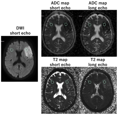 |
10 | Time estimation from stroke onset with diffusion-relaxation matrix-based T2 and ADC simultaneous mapping
Hajime Yokota1, Takayuki Sakai2, Masami Yoneyama3, and Takashi Uno1
1Department of Diagnostic Radiology and Radiation Oncology, Chiba University, Chiba, Japan, 2Department of Radiology, Eastern Chiba Medical Center, Togane, Japan, 3Philips Japan, Tokyo, Japan
Diffusion-relaxation matrix (DRM) sequence consists of dual-echo single-shot DW-EPI. DRM was able to detect acute cerebral infarction as well as conventional DWI, and can simultaneously acquire ADC and T2 values. T2 values in the infarcted area correlated significantly with time since stroke onset, whereas ADC did not. In addition, T2 values could predict whether the infarct was within 4.5, 6, or 16 hours, which are the thresholds for thrombolysis and endovascular treatment.
|
||
4178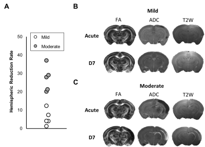 |
11 | Early Dynamics of Regional Volume and Diffusion Characteristics after Experimental Neonatal Hypoxic Ischemia with Different Damage Outcomes
Po-Yu Chan1, Chiao-Ching Huang2, Chia-Feng Lu1, Bao-Yu Hsieh3, and Yu-Chieh Jill Kao1
1Department of Biomedical Imaging and Radiological Sciences, National Yang Ming Chiao Tung University, Taipei, Taiwan, 2Department of Pediatrics, National Cheng Kung University Hospital, College of Medicine, National Cheng Kung University, Tainan, Taiwan, 3Department of Medical Imaging and Radiological Sciences, College of Medicine, Chang-Gung University, Taoyuan, Taiwan
To stratify neonates with different outcomes of hypoxic ischemia (HI) within few hours after birth for the subsequent therapeutic hypothermia, the spatiotemporal profile of MR measurements between two severity outcome groups was depicted as early at 6 h after insult. Changes in volumes, ADC and FA values were observed among different brain regions, suggesting different susceptibility of brain regions to HI in the acute phase of HI.
|
||
4179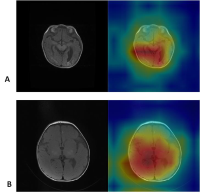 |
12 | Deep learning for term neonate hypoxic ischemic encephalopathy diagnosis on structural brain MR images: a retrospective study Video Not Available
Tongjia Gan1, Wenzhen Zhu1, Jingjing Shi1, and Wenzhi Lv2
1Tongji Hospital, Tongji Medical College, Huazhong University of Science and Technology, Wuhan, China, 2Department of Artificial Intelligence, Julei Technology Company, Wuhan, China This study aims to diagnose objectively term neonate hypoxic ischemic encephalopathy (HIE) by using deep learning network to extract deep information from multiple modalities of conventional magnetic resonance (MR) images. Neonate HIE diagnosis accuracy is restricted to lesion diversity, MR images quality, high interobserver variability. The network got high diagnosis accuracy in the ROC curve. The network could detect severe neonate HIE with characteristic appearance such as basal ganglia injury and periventricular leukomalacia. The network can help diagnosis neonate HIE objectively without the effect of different radiologist experience and contribute to risk stratification and clinical decision making. |
||
4180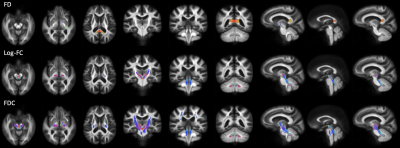 |
13 | Fiber-specific white matter reductions in older adults with metabolic syndrome
Christina Andica1, Koji Kamagata1, Wataru Uchida1, Kaito Takabayashi1, Yuya Saito1, Keigo Shimoji1,2, Hideyoshi Kaga3,4, Yuki Someya3, Yoshifumi Tamura3,4, Ryuzo Kawamori3,4, Hirotaka Watada3,4, Masaaki Hori1,5, and Shigeki Aoki1
1Department of Radiology, Juntendo University Graduate School of Medicine, Tokyo, Japan, 2Department of Radiology, Tokyo Metropolitan Geriatric Hospital and Institute of Gerontology, Tokyo, Japan, 3Sportology Center, Juntendo University Graduate School of Medicine, Tokyo, Japan, 4Department of Metabolism & Endocrinology, Juntendo University Graduate School of Medicine, Tokyo, Japan, 5Department of Radiology, Toho University Omori Medical Center, Tokyo, Japan We performed fixel-based analysis, which implements the Multi-Shell Multi-Tissue Constrained Spherical Deconvolution method, to investigate the effect of metabolic syndrome (MetS) on fiber-specific white matter (WM) integrity and assess its association with MetS-related components or cognitive or motor functions. Our findings provide evidence of WM degeneration, as reflected by reduced microstructural fiber density and macrostructural fiber bundle cross-section, in regions associated with cognitive and motor functions in MetS. We also observed reduced fiber density in the corticospinal tract in preMetS. Finally, our findings clarified visceral fat accumulation, insulin resistance, and arterial stiffness as risk factors for WM alterations in MetS. |
||
4181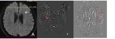 |
14 | Evaluation of Secondary Prevention Effectiveness of Ginkgo Biloba Extract (EGb 761) in Acute Ischemic Stroke by QSM and Venous Oxygen Saturation Video Not Available
Wen-Ting Lan1, Yue-Fei Wu2, Hui Zhang1, Xin-Zhong Ruan1, Yi Huang3, Xiao-Jing Li4, and Yunzhu Wu5
1Department of Radiology, Ningbo First Hospital, Ningbo Hospital of Zhejiang University, Ningbo, China, 2Department of Neurology, Ningbo First Hospital, Ningbo, China, 3Department of Neurosurgery, Ningbo First Hospital, Ningbo, China, 4Department of Gerontology, Ningbo First Hospital, Ningbo, China, 5MR Scientific Marketing, SIEMENS Healthineers, Shanghai, China
In this study, we applied quantitative susceptibility mapping (QSM) and venous oxygen saturation measurement in acute ischemic stroke patients who took ginkgo biloba extract (EGb 761) treatment. EGb 761’s secondary prevention effectiveness of ischemic stroke patients with cerebral microbleeds was similar to Aspirin. Furthermore, Ginkgo biloba extract could not only improve neuroprotective efficiency and prognosis by increasing oxygen saturation of the vein near infarction, but also could reduce the incidence of adverse side effects such as gastrointestinal ulcers and bleeding usually caused by Aspirin.
|
||
4182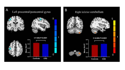 |
15 | Aberrant Spontaneous Brain Activity in Coronary Heart Disease: A Preliminary Resting-State Functional MRI Study.
Simin Lin1, PuYeh Wu2, and Hengyu Zhao1
1Xiamen Cardiovascular Hospital of Xiamen University, Xiamen, China, 2GE Healthcare, Beijing, China
Coronary heart disease is an urgent, rapidly-developing disease with high disability and mortality. Previous studies indicated that CHD patients exhibited an increased risk of mild cognitive and emotional dysfunction. Here we collected rs-fMRI data to assess global brain activity in CHD patients and controls by measuring fALFF. We demonstrated that CHD patients occur abnormal brain activity in left precentral/postcentral gyrus and right inferior cerebellum, which is mainly related to sensorimotor network and pain processing. Spontaneous brain activity abnormalities may contribute to understanding underlying neurological mechanisms of CHD.
|
||
 Back to Meeting Home
 Back to the Program-at-a-Glance
The International Society for Magnetic Resonance in Medicine is accredited by the Accreditation Council for Continuing Medical Education to provide continuing medical education for physicians.
 Back to Meeting Home
 Back to the Program-at-a-Glance
 View the Presentation