Online Gather.town Pitches
Neurological Conditions II
Joint Annual Meeting ISMRM-ESMRMB & ISMRT 31st Annual Meeting • 07-12 May 2022 • London, UK

Online Gather.town Pitches
Neurological Conditions II
| Booth # | ||||
|---|---|---|---|---|
3069 |
1 | Effect of 1 Hz rTMS on functional connectivity in Parkinson’s Disease.
Priyanka Bhat1, S Senthil Kumaran2, Vinay Goyal3, and Achal K Srivastava2
1IIT Delhi, Delhi, India, 2All India Institute of Medical Sciences, Delhi, India, 3Neuroscience Institute, Medanta-The Medicity, Delhi, India
Repetitive transcranial magnetic stimulation is known to render clinical benefits in subjects with Parkinson’s Disease. Because of dopaminergic depletion the cortical connectivity patterns are affected in PD. This study aimed to inhibit the primary motor area which is an important node in the movement control pathway. The effect was studied using clinical scales and task based functional connectivity. The results reveal the ability of rTMS to facilitate the movement by inducting cortical as well as subcortical brain regions.
|
||
3070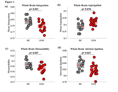 |
2 | Decreased the stability of brain hubs and the heterogeneity of whole brain dynamics in early onset schizophrenia
Mukesh Kumar1, S Senthil Kumaran1, Pankaj Pankaj1, and Rajesh Sagar2
1Department of NMR & MRI Facility, All India Institute of Medical Sciences, New Delhi, India, 2Department of psychiatry, All India Institute of Medical Sciences, New Delhi, India
The whole-brain effective connectivity (EC) was estimated to interpret the brain dynamics, time-dependent communicability and propagation of neuronal activity within the brain state in patients with early onset schizophrenia. We observed decreased effective connectivity (p<0.05) and global integration of local endogenous events in early onset schizophrenia (EOS) as compared to controls. EOS suffer decreased propagation of neuronal activity and responsiveness to events.
|
||
3071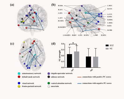 |
3 | Linked brain connectivity patterns with psychopathological and cognitive phenotypes in drug-naïve first-episode schizophrenia
Hui Sun1, Wenjing Zhang1, Chengmin Yang1, Qiyong Gong1, and Su Lui1
1Huaxi MR Research Center (HMRRC), Department of Radiology, West China Hospital of Sichuan University, Chengdu, China
Schizophrenia is characterized by abnormal functional integration between distinct brain regions but whether a common deficit in functional connectivity in relation to both clinical symptoms and cognitive impairments would present in drug-naïve first-episode patients remains elusive. A connectome-wise analysis on resting-state functional MRI in never-treated patients with first-episode schizophrenia. Using the principal component analysis, we found a trans-symptomatic pattern of functional connectivity associated with both psychopathological and cognitive manifestations in never-treated first-episode schizophrenia characterized as the dysconnections involving frontal and visual cortices, suggesting a core deficit of brain functional connectivity that might underpin the psychopathology of schizophrenia.
|
||
3072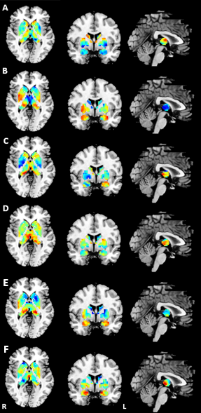 |
4 | The disrupted subcortical functional gradient in schizophrenia and the normalization effects of antipsychotic treatment
CHENGMIN YANG1, WENJING ZHANG1, JIAJUN LIU2, ZHIPENG YANG2, and SU LUI1
1Huaxi MR Research Center (HMRRC), Functional and Molecular Imaging Key Laboratory of Sichuan Province, Department of Radiology, West China Hospital of Sichuan University, Chengdu, China., Chengdu, China, 2College of Electronic Engineering, Chengdu University of Information Technology, Chengdu, P.R. China., Chengdu, China
To characterize the connectome gradient changes of subcortical system in drug-naive first-episode schizophrenia and the longitudinal treatment effect, we calculated the functional gradient and conducted the group comparisons. The principal gradient of subcortex demonstrated the fundamental functional segregation differences between patients at baseline and healthy controls, and the longitudinal analyses indicated that the treatment would gradually normalize the altered gradients with significant improvement of symptoms. This study provided the new perspective on the abnormal subcortical hierarchy organization in schizophrenia and its longitudinal gradient changes could be sensitive to reflect the antipsychotic treatment effect.
|
||
3073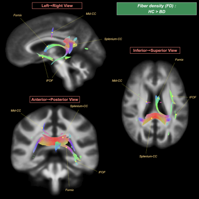 |
5 | White matter fiber-specific alterations in bipolar disorder: a fixel-based analysis
Paola Magioncalda1,2, Chun-Hung Yeh3, Hsiang-Yuan Lin4, Rung-Yu Tseng3, Mario Amore5, and Matteo Martino1,2
1Graduate Institute of Mind, Brain, and Consciousness, Taipei Medical University, Taipei, Taiwan, 2Brain and Consciousness Research Centre, Taipei Medical University - Shuang Ho Hospital, New Taipei City, Taiwan, 3Institute for Radiological Research, Chang Gung University and Chang Gung Memorial Hospital, Taoyuan, Taiwan, 4Azrieli Adult Neurodevelopmental Centre, Campbell Family Mental Health Research Institute, and Adult Neurodevelopmental and Geriatric Psychiatry Division, Centre for Addiction and Mental Health, Toronto, ON, Canada, 5Department of Neuroscience, Rehabilitation, Ophthalmology, Genetics, Maternal and Child Health, Section of Psychiatry, University of Genoa, Genoa, Italy
The recent unified model of the pathophysiology of bipolar disorder (BD) proposed that micro/macrostructural disturbance in limbic white matter (WM) fibers is important in the pathophysiological mechanism of BD. To gain more insights into this model, preprocessed diffusion MRI data of 67 BD and 125 healthy participants were analyzed using fixel-based analysis. We found a reduction of fiber density and/or cross-section of tracts, including the fornix and cingulum bundle that are core elements of the limbic system. These findings robustly support an association of BD with a specific spatial pattern of WM alterations.
|
||
3074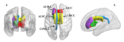 |
6 | Myelin abnormalities in patients with bipolar disorder: an inhomogeneous magnetization transfer study Video Not Available
Zhifeng Zhou1, Long Qian2, Wentao Lai1, Wentao Jiang1, Gangqiang Hou1, and Yingli Zhang1
1Shenzhen Mental Health Center/Shenzhen Kangning Hospital, Shenzhen, China, 2MR Research, GE Healthcare, Beijing, China
Previous DTI studies have revealed widespread increased radial diffusivity, which tend to be associated with myelin alteration, in patients with bipolar disorder (BD). This study utilizes a surrogate measure of myelin content termed inhomogeneous magnetization transfer (ihMT) to investigate myelin abnormalities in patients with BD. Our results demonstrate that regions with myelin deficits are mainly concentrated in the premiddle corpus callosum and left anterior white matter tracts, suggesting that interhemispheric communication and information transmission of left anterior brain areas may be most affected in patients with BD. Overall, this study provides new insight into the neuropathological mechanisms underlying BD.
|
||
3075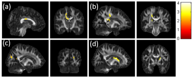 |
7 | Disrupted white matter network of brain structural connectomes in bipolar disorder patients revealed by q-ball imaging
Pei-Ti Lin1, Huai-Hsuan Tseng2,3, Po See Chen2,3, and Jun-Cheng Weng1,4,5
1Department of Medical Imaging and Radiological Sciences, Graduate Institute of Artificial Intelligence, Chang Gung University, Taoyuan, Taiwan, 2Department of Psychiatry, National Cheng Kung University Hospital, College of Medicine, National Cheng Kung University, Tainan, Taiwan, 3Institute of Behavioral Medicine, College of Medicine, National Cheng Kung University, Tainan, Taiwan, 4Department of Psychiatry, Chang Gung Memorial Hospital, Chiayi, Taiwan, 5Medical Imaging Research Center, Institute for Radiological Research, Chang Gung University and Chang Gung Memorial Hospital at Linkou, Taoyuan, Taiwan
Bipolar disorder (BD) is a major psychiatric disorder associated with structural and functional brain alterations and cognitive deficits. This study used q-ball imaging and graph theoretical analysis to investigate both neurological structural change and network alterations between BD patients and healthy controls. The results showed the alterations in several brain regions including the corpus callosum and cingulate gyrus, which are associated with executive, cognitive, emotional function, and memory. We found the BD group demonstrated higher global integrity than the HC group, but they remained small-world properties. It indicated that white matter integrity and network alterations were associated with bipolar disorder.
|
||
3076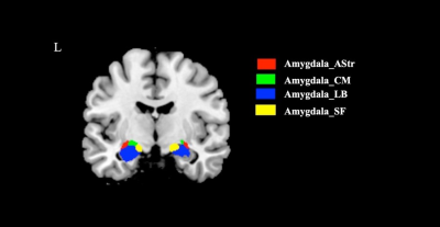 |
8 | Anomalous functional connectivity of amygdala subregional networks in adolescents with major depressive disorder
Mengyue Tang1, Zilin Zhou1, Lingxiao Cao1, Yingxue Gao1, Lianqing Zhang1, Hailong Li1, Yingying Wang1, Qiyong Gong1, and Xiaoqi Huang1
1Huaxi MR Research Center (HMRRC), Functional and molecular imaging Key Laboratory of Sichuan Province, Department of Radiology, West China Hospital, Sichuan University, Chengdu, China
Using seed based FC analysis, we investigated alterations in functional connectivity of amygdala subregional networks in a relatively large sample of adolescent MDD. Our findings identified adolescent MDD patients, compared to HC, have anomalous amygdala subregional functional network connections to several brain regions that play a pivotal role in emotional and cognitive processing, which may contribute to revealing the pathophysiology underlying MDD. These findings were not detected when using the whole amygdala as seeds, further verify the significance of analyses at the subregion level.
|
||
3077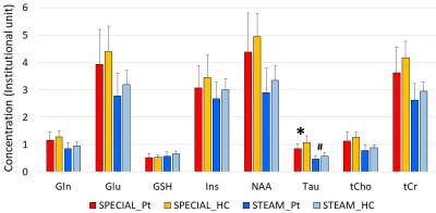 |
9 | Changes in neurochemicals of patients with depression observed using two MRS sequences at 7T
Tomohisa Okada1, Yujiro Yoshihara2, Manabu Kubota2, Taro Suwa2, Jun Miyata2, Yuhei Takado3, Jamie Near4, Masoumeh Dehghani4, Thai Akasaka1, Dinh Ha Duy Thuy1, Ravi Teja Seethamraju5, Sinyeob Ahn6, Tadashi Isa1, and Toshiya
Murai2
1Human Brain Research Center, Kyoto University, Kyoto, Japan, 2Department of Psychiatry, Kyoto University, Kyoto, Japan, 3Department of Functional Brain imaging, National Institutes for Quantum and Radiological Science and Technology, Chiba, Japan, 4Physical Sciences Platform, Sunnybrook Research Institute, Toronto, ON, Canada, 5Siemens Healthcare USA, Boston, MA, United States, 6Siemens Healthcare USA, Berkeley, CA, United States
Major depressive disorder (MDD) afflicts up to 20% of the total population. MRS findings are different among studies. One of the reasons is differences in MRS sequences used. This study compared two sequences: sSPECIAL and STEAM, which consistently found significant and marginally significant reduction of taurine in the anterior cingulate cortex in MDD compared with healthy controls, although concentration of neurochemicals in sSPECIAL and STEAM was different. The difference was lost when concentration was normalized by total creatine due to its significant positive correlation to Taurine. Differences between the two sequences did not result in different neurochemical findings for MDD.
|
||
3078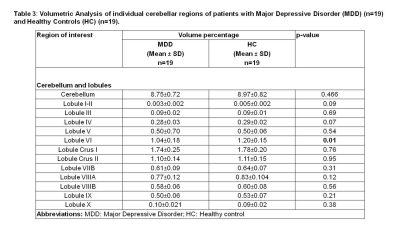 |
10 | Cerebellar atrophy in Major Depressive Disorder
Vishwa Rawat1, Ritu Tyagi1, Gagan Hans2, Pratap sharan2, S Senthil Kumaran1, and Uma Sharma1
1Department of NMR, All India Institute of Medical Sciences (AIIMS), New Delhi, India, 2Department of Psychiatry, All India Institute of Medical Sciences (AIIMS), New Delhi, India
Present study investigated volumetric changes in the brain of Indian patients suffering from Major Depressive Disorder (MDD) using Magnetic Resonance Imaging (MRI). These patients were seen to have significantly reduced brain and cerebellar volumes. Volume of caudate, putamen and globus pallidus were seen to be reduced but no significant changes were seen in hippocampus and amygdala. Volume of cerebellum and lobule VI of cerebellum was also reduced. Our results suggest that cerebellum as well as VI lobule of cerebellum may be associated with MDD.
|
||
3079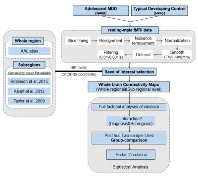 |
11 | Aberrant Intrinsic Functional Connectivity of Hippo-Orbitofrontal-Insular Circuit in Drug-naive Adolescents with Major Depressive Disorder
Zilin Zhou1, Yingxue Gao1, Weijie Bao1, Hui Qiu1, Lianqing Zhang1, Ruohan Feng1,2, Yingying Wang1, Mengyue Tang1, Qiyong Gong1, and Xiaoqi Huang1
1Department of Radiology, Huaxi Magnetic Resonance Research Centre (HMRRC),Functional and Molecular Imaging Key Laboratory of Sichuan Province, Chengdu, China, 2Department of Radiology, the Third Hospital of Mianyang, Sichuan Mental Health Center, Mianyang, China Through seed-based functional connectivity (SBFC) analysis, we investigated the altered intrinsic connectivity patterns of hippocampus, orbitofrontal cortex (OFC) and insula in adolescents with major depressive disorder(aMDD) comparing to typically developing control(TDC). We found distinct network alterations of those three structures at regional and subregional level. Moreover, we located the aberrant connectivity in hippo-orbitofrontal-insular circuit with family conflict and depressvie symptoms in aMDD which may underline the neural mechanism for depressvie symptoms in adolescents. |
||
3080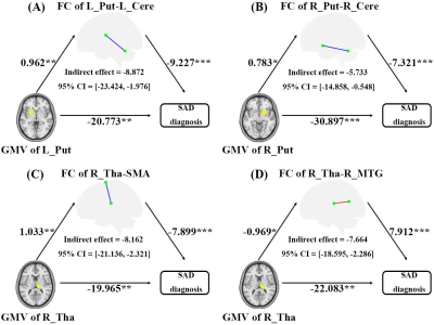 |
12 | Functional Deficits in the Large-scale Circuitry Mediate the Links of Structural Atrophy to Social Anxiety Disorder Outcomes
Xun Zhang1, Song Wang1, and Qiyong Gong1
1Huaxi MR Research Center (HMRRC), West China Hospital of Sichuan University, Chengdu, China Social anxiety disorder (SAD) is a prevalent and disabling psychiatric disorder, for which the underlying neurobiological mechanisms remain poorly understood. Herein, whole-brain voxel-based morphometry and coupled functional connectivity analyses were conducted to investigate structural and functional alterations, while correlation and mediation analyses were performed to probe the potential roles of structural-functional couplings in SAD diagnosis. As a result, we observed significant subcortical grey matter atrophy and widespread dysconnectivity in cortico-striato-thalamo-cerebellar circuitry in SAD; functional dysconnectivity could partially mediate the effects of structural atrophy on SAD diagnosis, which may contribute to clarifying the underlying mechanisms of structure-functional couplings for SAD. |
||
 Back to Meeting Home
 Back to the Program-at-a-Glance
The International Society for Magnetic Resonance in Medicine is accredited by the Accreditation Council for Continuing Medical Education to provide continuing medical education for physicians.
 Back to Meeting Home
 Back to the Program-at-a-Glance
 View the Presentation