Online Gather.town Pitches
Cardiac III
Joint Annual Meeting ISMRM-ESMRMB & ISMRT 31st Annual Meeting • 07-12 May 2022 • London, UK

| Booth # | ||||
|---|---|---|---|---|
4923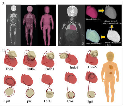 |
1 | A novel patient-adjustable coil technology substantially reduces RF heating of cardiac implantable electronic devices in pediatric patients
Bhumi Bhusal1, Fuchang Jiang1, Pia Panravi Sanpitak1, Boris Keil2, Gurinder Kaur Multani2, Nicolas Kutscha2, Giorgio Bonmassar3, Gregory Webster1, Andrada Popescu4, Daniel Kim1, and Laleh Golestanirad1
1Northwestern University, Chicago, IL, United States, 2TH Mittelhessen University of Applied Sciences, Giessen, Germany, 3Massachusetts General Hospital, Charlestown, MA, United States, 4Lurie Children's Hospital, Chicago, IL, United States
Infants and children with congenital heart disease may require cardiovascular implantable electronic devices (CIEDs), which typically precludes future evaluation by MRI due to risks associated with RF heating of implants. Here we demonstrate that a novel concept based on patient-adjustable rotating RF coil technology that was originally developed to reduce RF heating of brain implants can be adopted in pediatric patients with CIEDs to substantially reduce RF heating. Our simulations in a cohort of children with both epicardial and endocardial devices showed an 87% reduction in average SAR compared to conventional body coils.
|
||
4924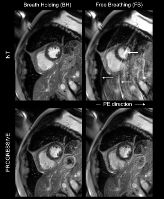 |
2 | Motion Robust Segmented Delayed Enhancement MRI by Partially Reversed ReOrdering for GeneRal SupprESSion of Motion Induced Variable Errors
Wolfgang G Rehwald1,2, Rafael Rojas2, Nestor Mena2, Ryan Seward2, George Gamoneda2, Jeana Dement2, Elizabeth Jenista2, David Wendell2, Han Kim2, Enn-Ling Chen2, Igor Klem2, Vera Kimbrell2, and Raymond Kim2
1Siemens Healthineers, Durham, NC, United States, 2Duke Cardiovascular MR Center, Duke University, Durham, NC, United States
Standard segmented delayed enhancement (DE) is particularly motion sensitive owing to its interleaved reordering required for inversion recovery (IR). Ghosting artifacts frequently result from imperfect breath holding. The sequence cannot run free-breathing (FB). We present a novel DE technique, PROGRESSIVE, using a partially continuous reordering and dummy pulses for simultaneous motion robustness and IR-compatibility. We tested PROGRESSIVE in simulations, a motion phantom, and a cohort of patients. PROGRESSIVE images were devoid of ghosting. When acquired FB, quality was significantly better than standard DE, and equally good during perfect breath holding. PROGRESSIVE has the potential for FB self-navigated DE imaging.
|
||
4925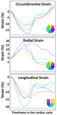 |
3 | Comprehensive MRI reveals compromised baseline cardiac function in lung cancer patients undergoing radiation therapy treatment
El-Sayed H Ibrahim1, Elizabeth Gore1, Sherry-Ann Brown1, and Carmen Bergom2
1Medical College of Wisconsin, Milwaukee, WI, United States, 2Washington University, St Louis, MO, United States
Along with systemic therapies, radiation therapy (RT) plays a key role in treating lung cancer, despite high incidence of RT-induced cardiac complications. Most lung cancer patients present with cardiac risk factors that result in compromised baseline cardiac function that puts the patients at higher risk of developing cardiac complications. In this study, we investigate characteristics of baseline cardiovascular function in lung cancer patients undergoing RT. The results showed that the patients had borderline cardiac function and reduced myocardial strain measurements. Myocardial T1/T2 values in the patients were slightly high and the hemodynamic measurements showed different pattern than that in volunteers.
|
||
4926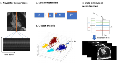 |
4 | ECG-free 2D cardiac cine MRI with data-driven clustering
Zhengyang Ming1,2, Caroline M. Colbert1,2,3, Ruan Dan1,4, Holden H. Wu1,2, Anthony G. Christodoulou5, J. Paul Finn1,2, Peng Hu1,2, and Kim-Lien Nguyen1,2,3
1Physics and Biology in Medicine Graduate Program, University of California, Los Angeles, Los Angeles, CA, United States, 2Department of Radiological Sciences, David Geffen School of Medicine at UCLA, Los Angeles, CA, United States, 3Division of Cardiology, David Geffen School of Medicine at UCLA and VA Greater Los Angeles Healthcare System, Los Angeles, CA, United States, 4Department of Radiation Oncology, David Geffen School of Medicine at UCLA, Los Angeles, CA, United States, 5Biomedical Imaging Research Institute, Cedars-Sinai Medical Center, Los Angeles, CA, United States Conventional 2D cardiac cine imaging relies on ECG-gating whereas self-gated approaches mitigate ECG-dependency by assuming that cardiac motion is periodic with well-defined frequencies. This assumption breaks down when patients have irregular cardiac rhythm. We propose a segmented Cartesian golden step balanced steady-state free precession sequence (bSSFP) with motion navigators and a clustering algorithm to alleviate the dependency on regular motion assumptions and to emphasize the intrinsic similarity of mechanical motion. Compared to standard ECG-gated 2D bSSFP cine performed in normal sinus rhythm, initial validation using our approach achieved similar image quality and quantitative metrics for cardiac function.
|
||
4927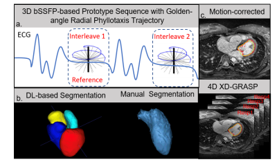 |
5 | DL-Based LV Volume Segmentation to Compare Respiratory-Corrected and 4D XD-GRASP Respiratory Motion-Resolved Whole-Heart Reconstructions
Yitong Yang1, Zahraw Shah2, Athira Jacob3, Jackson Hair1, Teodora Chitiboi3, Tiziano Passerini3, Jerome Yerly4, Lorenzo Di Sopra4, Davide Piccini5, Zahra Hosseini6, Puneet Sharma3, Anurag Sahu7, Matthias Stuber4, and John
Oshinski1,8
1Biomedical Engineering, Emory University/Georgia Institute of Technology, Atlanta, GA, United States, 2Biomedical Engineering, Georgia Institute of Technology, Atlanta, GA, United States, 3Siemens Medical Solutions USA, Princeton, NJ, United States, 4Diagnostic and Interventional Radiology, Lausanne University Hospital, Lausanne, Switzerland, 5Siemens Healthcare, Lausanne, Switzerland, 6Siemens Medical Solutions USA, Atlanta, GA, United States, 7Cardiology, Emory University School of Medicine, Atlanta, GA, United States, 8Radiology, Emory University School of Medicine, Atlanta, GA, United States
Rapid and accurate quantification of left ventricular volume is essential for studying cardiac function. In this work, two reconstruction techniques for free-breathing, ECG-gated, radial, golden-angle phyllotaxis acquisition of whole-heart CMR are assessed for their accuracy in quantifying left ventricular volume using deep learning (DL)-based automatic cardiac chamber segmentation. The off-line reconstruction that resolves 4D (3D+respiratory) images had better agreement between the DL-based segmentation and an expert’s manual segmentation than the in-line reconstruction that corrects for respiratory motion, as assessed by the average volume difference (p=0.026) and 3D Dice coefficients (p=0.032).
|
||
4928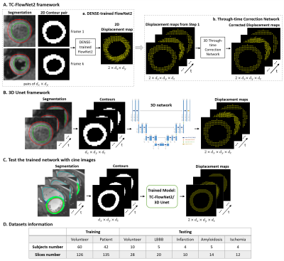 |
6 | Improved strain analysis of cine images by deep learning from DENSE: Comparison of a 3D Unet and an optical-flow net
Yu Wang1, Changyu Sun1, Sona Ghadimi1, Auger C. Daniel1, Pierre Croisille2,3, Magalie Viallon2,3, Jie Jane Cao4, Yang Joshua Cheng4, Andrew D. Scott5,6, Pedro F. Ferreira5,6, John N. Oshinski7, Daniel B. Ennis8, Kenneth C. Bilchick9,
and Frederick H. Epstein1,10
1Biomedical Engineering, University of Virginia, Charlottesville, VA, United States, 2University of Lyon, UJM-Saint-Etienne, INSA, CNRS UMR 5520, INSERM U1206, CREATIS, Saint-Etienne, France, 3Department of Radiology, University Hospital Saint-Etienne, Saint-Etienne, France, 4St. Francis Hospital, DeMatteis Center for Research and Education, Cardiac Imaging, Greenvale, NY, United States, 5Cardiovascular Magnetic Resonance Unit, The Royal Brompton Hospital, London, United Kingdom, 6National Heart and Lung Institute, Imperial College, London, United Kingdom, 7Department of Radiology and Imaging Sciences, Emory University School of Medicine, Atlanta, GA, United States, 8Department of Radiology, Stanford University, Stanford, CA, United States, 9Cardiovascular Division, Department of Medicine, University of Virginia Health System, Charlottesville, VA, United States, 10Radiology, University of Virginia, Charlottesville, VA, United States
Cine DENSE provides both myocardial contours and intramyocardial displacements. We propose to use DENSE to train deep networks to predict intramyocardial motion from contour motion. Two workflows were implemented: a two-step FlowNet2-based framework with a through-time correction network and a 3D (2D+t) Unet framework. Both networks depicted cardiac contraction and abnormal motion patterns. The 3D Unet showed excellent reliability for global circumferential strain (Ecc) and good reliability for segmental Ecc, and it outperformed commercial FT for both global and segmental Ecc.
|
||
4929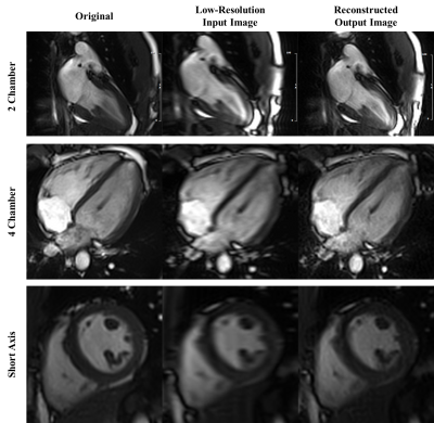 |
7 | Accelerated Cardiac MR Imaging with Inline Cascaded Parallel Imaging and Generative Adversarial Neural Network (PI-GAN) Framework
Siyeop Yoon1, Xiaoying Cai1,2, Eiryu Sai1, Salah Assana1, Amine Amyar1, Kelvin Chow3, Amanda Paskavitz1, Julia Cirillo1, Warren J. Manning1, and Reza Nezafat1
1Department of Medicine, Cardiovascular Division, Beth Israel Deaconess Medical Center and Harvard Medical School, Boston, MA, United States, 2Siemens Medical Solutions USA, Inc., Boston, MA, United States, 3Siemens Medical Solutions USA, Inc., Chicago, IL, United States
We developed and evaluated an inline cascaded parallel imaging and generative adversarial network for cardiac MRI. The preliminary results show excellent image quality of the cardiac cine images in four heat-beat.
|
||
4930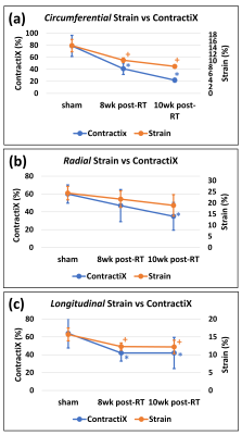 |
8 | MRI-derived contractility index as an early measure of thoracic radiation therapy-induced subclinical cardiotoxicity
El-Sayed H Ibrahim1, John Baker1, and Carmen Bergom2
1Medical College of Wisconsin, Milwaukee, WI, United States, 2Washington University, St Louis, MO, United States
Radiation therapy (RT) plays an integral role in treating a number of thoracic cancers, despite risks of radiation-induced cardiotoxicity. The purpose of this study is to investigate the capability of advanced the MRI-generated contractility index (ContractiX) for early detection of RT-induced cardiotoxicity in a pre-clinical rat model of thoracic cancer RT. While EF increased post-RT, peak systolic strain and ContractiX significantly decreased post-RT, with more relative reduction in ContractiX compared to strain. Therefore, ContractiX is a sensitive early marker for detection of subclinical cardiac dysfunction post-RT in pre-clinical models.
|
||
 Back to Meeting Home
 Back to the Program-at-a-Glance
The International Society for Magnetic Resonance in Medicine is accredited by the Accreditation Council for Continuing Medical Education to provide continuing medical education for physicians.
 Back to Meeting Home
 Back to the Program-at-a-Glance
 View the Presentation