Online Gather.town Pitches
Data Acquisition & Artefacts I
Joint Annual Meeting ISMRM-ESMRMB & ISMRT 31st Annual Meeting • 07-12 May 2022 • London, UK

Online Gather.town Pitches
Data Acquisition & Artefacts I
| Booth # | ||||
|---|---|---|---|---|
3648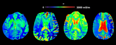 |
1 | The Role of Electric Properties Tomography in Biochemical Analysis of the Cerebrospinal Fluid and Parameter Optimization for Improved Accuracy Video Permission Withheld
Khin Khin Tha1, Ulrich Katscher2, Hiroyuki Hamaguchi3, Xinnan Li3, Tomohiro Kawasaki4, Shigeru Yamaguchi5, Ichiro Yabe5, and Hideki Hyodoh5
1Global Center for Biomedical Science and Engineering, Hokkaido University Faculty of Medicine, Sapporo, Japan, 2Philips Research, Hamburg, Germany, 3Hokkaido University Graduate School of Biomedical Science and Engineering, Sapporo, Japan, 4Hokkaido University Hospital, Sapporo, Japan, 5Hokkaido University Faculty of Medicine, Sapporo, Japan
This study was aimed to evaluate if electrical conductivity (σ) by electric properties tomography (EPT) could detect variations in the cerebrospinal fluid (CSF) biochemical composition and if optimization of scan and analysis parameters improved the accuracy of in vivo σ measurements. σ values varied among patients and CSF samples with varying CSF biochemical composition, and showed significant correlation with CSF albumin concentration and total cell count. σ may be sensitive to CSF abnormalities. Optimization of scan and analysis parameters is important for the accuracy of in vivo σ measurements.
|
||
3649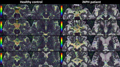 |
2 | Quantification of fine CSF dynamics using Intra-Voxel Incoherent Motion (IVIM) MRI
Shigeki Yamada1,2, Yoshiyuki Watanabe3, Tomohito Otani4, Shinnosuke Hiratsuka3, Masahiro Yoshimura3, Naoki Takeishi4, Shigeo Wada4, Marie Oshima2, and Kazuhiko Nozaki1
1Neurosurgery, Shiga University of Medical Science, Otsu, Japan, 2Interfaculty Initiative in Information Studies, Institute of Industrial Science, The University of Tokyo, Tokyo, Japan, 3Radiology, Shiga University of Medical Science, Otsu, Japan, 4Mechanical Science and Bioengineering, Graduate School of Engineering Science, Osaka University, Osaka, Japan
The f value calculated by the Intra-Voxel Incoherent Motion (IVIM) MRI was was high in the areas where the CSF flow was reported to be fast, specifically in the cerebral aqueduct and subarachnoid spaces around the major intracranial arteries. Therefore, f value was considered to reflect flow velocity of CSF, regardless of its direction. The mean f values in the whole lateral ventricles, anterior part of the third ventricle, ambient cisterns, central sulci, and marginal sulci were significantly lower and that in the foramina of Luschka was significantly higher among 28 iNPH patients, compared with 57 healthy volunteers.
|
||
3650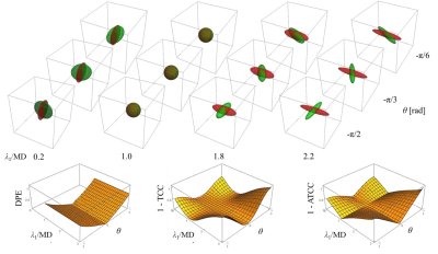 |
3 | Evaluating Repeatability of Low b-value DTI of CSF using Novel Angular Tensor-Correlation-Coefficient
Yoshitaka Bito1,2, Hisaaki Ochi1,2, Kuniaki Harada2, Ryuji Shirase1, and Kohsuke Kudo2
1FUJIFILM Healthcare Corporation, Tokyo, Japan, 2Hokkaido University Graduate School of Medicine, Sapporo, Japan Low b-value DTI (Low-b DTI) has been recently proposed for investigating the CSF pseudorandom flow. High repeatability of Low-b DTI is essential for the investigation; however, an efficient index evaluating repeatability of both shape and orientation of DT has not been presented. In this study, a new index, angular tensor-correlation-coefficient (ATCC), was proposed and was demonstrated to have good features in understanding the degree of repeatability by Monte Carlo simulation. ATCC was used to evaluate the repeatability of an experiment about Low-b DTI of CSF and showed high repeatability of this experiment. |
||
3651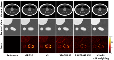 |
4 | Improved Motion Correction in Dynamic Contrast Enhancement MRI Using Low Rank with Soft Weighting
Jichang Zhang1, Xinpei Wang1, Faisal Najeeb2, Pengfei Xu1, Hammad Omer2, Penny Gowland3, Sue Francis3, Paul Glover3, Richard Bowtell3, and Chengbo Wang1
1SPMIC, The University of Nottingham Ningbo China, Ningbo, China, 2COMSATS University Islamabad, Islamabad, Pakistan, 3SPMIC, The University of Nottingham, Nottingham, United Kingdom
This work presents a robust motion corrected free breathing Dynamic Contrast Enhanced MRI (DCE-MRI) reconstruction method called Low rank plus sparse (L+S) with soft weighting which compressed motion blurring by employing a soft weighting matrix. The motion correction performance was quantified in this work. A computer simulation framework based on a modified shepp-logan model was developed with the ground truth to quantify the reconstruction errors. The proposed method achieved better motion correction and high reconstruction efficiency simultaneously.
|
||
3652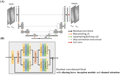 |
5 | Accelerate Single-Channel MRI by Exploiting Uniform Undersampling Aliasing Periodicity through Deep Learning
Christopher Man1,2, Zheyuan Yi1,2, Vick Lau1,2, Jiahao Hu1,2, Yujiao Zhao1,2, Linfang Xiao1,2, Alex T.L. Leong1,2, and Ed X. Wu1,2
1Laboratory of Biomedical Imaging and Signal Processing, The University of Hong Kong, Hong Kong SAR, China, 2Department of Electrical and Electronic Engineering, The University of Hong Kong, Hong Kong SAR, China
Conventional parallel imaging methods mostly utilize the spatial encoding by array of receiver coils to unfold the periodic aliasing artifact resulted from uniformly undersampled k-space data. In scenarios such as low- or ultra-low-field MRI where effective receiver arrays do not exist and SNRs are low, these methods are not generally applicable. This study presents a U-Net based deep learning approach to single-channel MRI acceleration that unfolds the aliasing by exploiting its periodicity. The results demonstrate the aliasing unfolding capability of this method for single-channel MRI even at very high acceleration and in presence of pathologies.
|
||
3653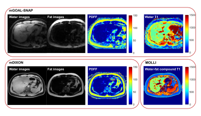 |
6 | Free-breathing Water-fat Separation and T1 Mapping of the Whole Liver with Isotropic Resolution using 3D Golden Angle Radial Trajectory
Yajie Wang1, Haikun Qi2, Yishi Wang3, and Huijun Chen1
1Center for Biomedical Imaging Research, Department of Biomedical Engineering, School of Medicine, Tsinghua University, Beijing, China, 2School of Biomedical Engineering, ShanghaiTech University, Shanghai, China, 3Philips Healthcare, Beijing, China
T1 mapping of the liver has been used for the diagnosis and grading of the liver disease, and the evaluation of the liver function, while conventional liver T1 mapping techniques need breath-holding, have limited slice coverage or need multiple acquisitions. In addition, the accuracy of the T1 quantification is affected by the presence of fat in the liver. In this study, free-breathing water-fat separation and T1 mapping quantification of the whole liver was achieved within one scan using the proposed mGOAL-SNAP sequence. The quantitative accuracy and the in-vivo feasibility of mGOAL-SNAP have been demonstrated in phantom and volunteers studies.
|
||
3654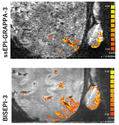 |
7 | Comparison of BOLD detectability of ‘multi shot EPI’ and ‘single shot EPI with GRAPPA’ at 7T
Guoxiang Liu1,2, Adnan Shah1,2, Takashi Ueguchi1,2, and Seiji Ogawa1,3
1NICT, Osaka, Japan, 2Graduate School of Frontier Biosciences, Osaka University, Osaka, Japan, 3Tohoku Fukushi University, Sendai, Japan
We compare the detectability of multi shot EPI (msEPI) and single shot EPI with GRAPPA (ssEPI-GRAPPA) using D-value maps to show the benefit of BISEPI (Block-Interleaved Segmented EPI) [1] for the same total acquisition time. D-values were derived based on the relationship between target effect, the significance level of detection, the number of time points (volumes), and tSNR [2]. The proposed D-value maps can be a useful quality assurance measure for determining the tSNR gain and activity detection performance of msEPI in comparison to ssEPI-GRAPPA in high-resolution fMRI at 7T.
|
||
3655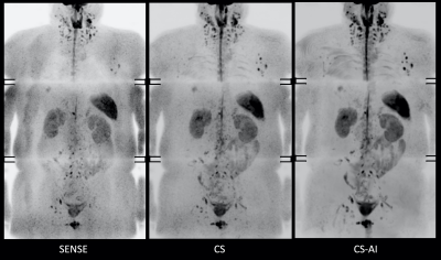 |
8 | SNR boost in whole-body DWIBS utilizing deep learning constrained Compressed SENSE reconstruction
Masami Yoneyama1, Takashige Yoshida2, Johannes M Peeters3, Jihun Kwon1, Yasutomo Katsumata3, Shuo Zhang4, and Marc Van Cauteren3
1Philips Japan, Tokyo, Japan, 2Tokyo Metropolitan Police Hospital, Tokyo, Japan, 3Philips Healthcare, Best, Netherlands, 4Philips GmbH Market DACH, Hamburg, Germany
Diffusion weighted whole body imaging with background body signal suppression (DWIBS) is an important tool for whole body imaging to e.g. visualize malignant tumor and abnormal lymph nodes. Next to the low intrinsic SNR of DWIBS, direct coronal DWIBS can suffer from another SNR penalty due to the high acceleration factors to minimize distortion. In this work, we compare SENSE, C-SENSE and C-SENSE-AI (CS-AI) accelerated direct coronal DWIBS scans. Results indicate that CS-AI clearly reduces the noise artifacts, boosts the overall image SNR, and improves lesion conspicuity.
|
||
 Back to Meeting Home
 Back to the Program-at-a-Glance
The International Society for Magnetic Resonance in Medicine is accredited by the Accreditation Council for Continuing Medical Education to provide continuing medical education for physicians.
 Back to Meeting Home
 Back to the Program-at-a-Glance
 View the Presentation