Online Gather.town Pitches
Image Reconstruction II
Joint Annual Meeting ISMRM-ESMRMB & ISMRT 31st Annual Meeting • 07-12 May 2022 • London, UK

Online Gather.town Pitches
Image Reconstruction II
| Booth # | |||
|---|---|---|---|
3499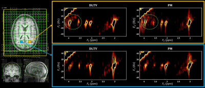 |
1 | Accelerated 5D Echo Planar J-Resolved Spectroscopic Imaging and Dictionary Learning Reconstruction: Pilot Findings in Obstructive Sleep Apnea
Ajin Joy1, Paul M Macey2, Manoj K Sarma1, Andres Saucedo1, and M Albert Thomas1
1Radiological Sciences, University of California-Los Angeles, Los Angeles, CA, United States, 2School of Nursing and Brain Research Institute, University of California-Los Angeles, Los Angeles, CA, United States
Obstructive sleep apnea (OSA) affects 10% of the population, and is associated with brain injury. Neurochemical changes in the brain of OSA patients can be recorded using 5D echo-planar J-resolved spectroscopic imaging. Accelerated acquisition is achieved with non-uniform undersampling, which requires data reconstruction that can be done with compressed sensing (CS). We implemented CS with a hybrid DLTV reconstruction method combining dictionary learning (DL) and total variation (TV), and compared its performance with Perona-Malik (PM) reconstruction. The metabolite ratios were consistent with both DLTV and PM while DLTV recovered the metabolite peaks near residual water better in many voxels.
|
|
3500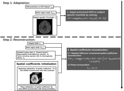 |
2 | Generative Image Prior Constrained Subspace Reconstruction for High-Resolution MRSI
Ruiyang Zhao1,2, Zepeng Wang1,3, and Fan Lam1,3
1Beckman Institute for Advanced Science and Technology, Urbana, IL, United States, 2Department of Electrical and Computer Engineering, University of Illinois Urbana-Champaign, Urbana, IL, United States, 3Department of Bioengineering, University of illinois Urbana-Champaign, Urbana, IL, United States
We propose a novel method that integrates generative image priors with subspace constrained MRSI reconstruction. A sufficient and flexible image representation was first generated by adapting a pretrained StyleGAN to subject-specific anatomical images. We validate that StyleGAN can be flexibly adapted to accurately represent different contrast of the same subject. The adapted GAN prior is then used to model the spatial coefficients in the subspace-based reconstruction. Improved performance over the original subspace reconstruction in the SPICE framework is demonstrated using simulation and in vivo data.
|
|
3501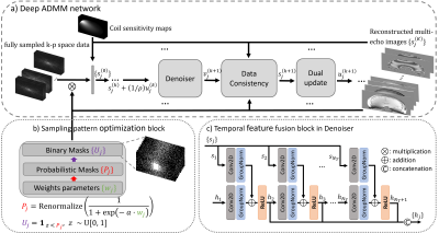 |
3 | Fast Quantitative Susceptibility Mapping with Temporal Feature Augmented and Spatiotemporal Sampling Pattern Optimized Network
Jinwei Zhang1, Hang Zhang1, Pascal Spincemaille2, Chao Li1, Thanh Nguyen2, Mert Sabuncu1, and Yi Wang2
1Cornell University, New York, NY, United States, 2Weill Cornell Medicine, New York, NY, United States
A multi-echo sampling pattern optimization and temporal fusion network for MRI reconstruction is proposed to accelerate data acquisition for quantitative susceptibility mapping (QSM). Experiments show that both features help improve multi-echo image reconstruction. The proposed method was applied to a prospectively under-sampled mGRE acquisition demonstrating better image quality over all comparison methods.
|
|
3502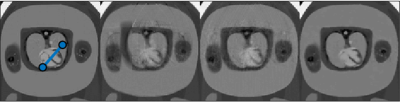 |
4 | Compressed sensing for motion-robust real-time fetal cardiac imaging
Nicholas Rubert1, Quin Lu2, Gaurav Jategaonkar1, and Luis Goncalves1,3
1Radiology, Phoenix Children's Hospital, Phoenix, AZ, United States, 2Philips, Best, Netherlands, 3University of Arizona, Phoenix, AZ, United States
We examined undersampled real-time bSSFP reconstructions for fetal cardiac imaging. Two compressed sensing reconstructions, ICTGV and L plus S, were compared to k-t SENSE with respect to spatial and temporal blurring with and without fetal motion. Results with a digital fetal cardiac MRI phantom demonstrated ICTGV was able to capture expansion and contraction of the fetal heart even during periods of fetal motion while k-t SENSE and L plus S were not. Digital phantom results were confirmed on a 1.5T MRI scanner with implementation of ICTGV and acquisitions demonstrating pulsation of the fetal heart during periods of fetal motion.
|
|
3503 |
5 | Joint recovery of time aligned multi-slice dynamic speech MR images from under-sampled data using a deep generative manifold model
Rushdi Zahid Rusho1, Qing Zou2, Mathews Jacob2, and Sajan Goud Lingala1,3
1Roy J. Carver Department of Biomedical Engineering, The University of Iowa, Iowa City, IA, United States, 2Electrical and Computer Engineering, The University of Iowa, Iowa City, IA, United States, 3Department of Radiology, The University of Iowa, Iowa City, IA, United States
Dynamic speech MRI is a powerful tool to characterize complex speech articulations. Current accelerated 2D dynamic speech MRI schemes can achieve high time resolutions of the order of (10-20 ms) while sequentially acquiring multiple 2D slices. However, the complex articulatory motion is difficult to interpret jointly across the slices due to mis-alignment of motion patterns. Here, we apply a novel generative manifold model which reconstructs and generates a time aligned multi-slice 2D speech dataset at 18 ms/frame from under-sampled k-space v/s time data sequentially acquired from multiple 2D slices. We evaluate this scheme on two speakers producing repeated speech tasks.
|
|
3504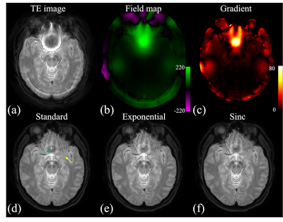 |
6 | Deblurring for Spiral Imaging with Intra-Voxel Dephasing Modeling
Dinghui Wang1 and James G. Pipe1
1Department of Radiology, Mayo Clinic, Rochester, MN, United States
Advantages of spiral imaging include fast imaging speed, high scan efficiency and low sensitivity to motion. However, spiral imaging remains challenging in regions where the field map Δf0 changes rapidly in space, especially with long spiral readouts. The goal of this work is to address this issue by modeling the signal decay in deblurring procedure. Volunteer data demonstrated that image quality can be improved by the proposed method.
|
|
3505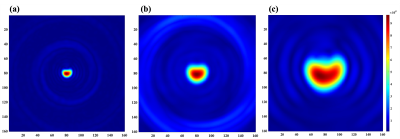 |
7 | Flexible FOV image reconstruction using multi-echo 13C imaging with rotating spiral arms
Sung-Han Lin1, Junjie Ma1, Leon S. Khalyavin2, and JaeMo Park1,2,3
1Advanced Imaging Research Center, UT Southwestern Medical Center, Dallas, TX, United States, 2Electrical and Computer Engineering, UT Dallas, Richardson, TX, United States, 3Department of Radiology, UT Southwestern Medical Center, Dallas, TX, United States
Metabolic imaging with hyperpolarized 13C substrates often detects signals only from small subregions within the prescribed FOV, primarily due to lacking background signals and limited perfusion or metabolic activities in the adjacent tissues. In this study, we propose an imaging method that provides flexible reconstruction options with variable FOV. The proposed pulse sequence was implemented, and the feasibility of the reconstruction method was validated with a 13C phantom.
|
|
3506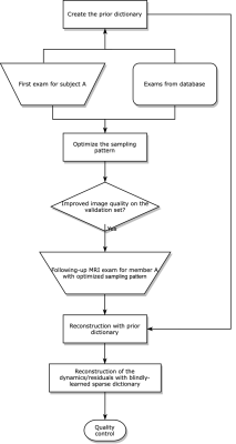 |
8 | Delta Scan: Reconstruction and Protocol Design for Ultra-fast Longitudinal MRI
Guanhua Wang1, Alessandro Francavilla2, Sen Ma2, Jeffrey Kaditz2, and Thomas Witzel2
1Biomedical Engineering, University of Michigan, Ann Arbor, MI, United States, 2Q Bio, Inc., San Carlos, CA, United States
Delta Scan proposes a joint design of scan protocols and image reconstruction to accelerate longitudinal MRI, using image priors from previous exams. The method models the static and dynamic structural information as two adaptive dictionaries, and optimizes sampling patterns using fully-sampled or mildly undersampled historical scans, via a stochastic optimization approach. Extensive experiments show that the proposed method led to improved reconstruction quality under a high acceleration factor, with reduced resolution loss than conventional compressed sensing-based methods. In comparison with previous reference-based reconstruction methods, the proposed method is less overfitted to past exams, truthfully reflecting the anatomical changes between exams.
|
|
3507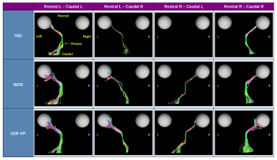 |
9 | Once upon a chiasm: Reconstruction of mouse optic pathways with ODF-Fingerprinting
Patryk Filipiak1, Thajunnisa A. Sajitha2, Timothy Shepherd1, Ying-Chia Lin1, Dimitris G. Placantonakis3, Kevin C. Chan2, Fernando E. Boada1, and Steven H. Baete1
1Center for Advanced Imaging Innovation and Research (CAI2R), Department of Radiology, NYU Langone Health, New York, NY, United States, 2NeuroImaging and Visual Science Laboratory, Departments of Ophthalmology and Radiology, NYU Langone Health, New York, NY, United States, 3Department of Neurosurgery, Perlmutter Cancer Center, Neuroscience Institute, Kimmel Center for Stem Cell Biology, NYU Langone Health, New York, NY, United States We reconstruct mouse optic pathways with ODF-Fingerprinting in a challenging crossing area of the optic chiasm. Our method replaces the commonly used peak finding approach with pattern matching when reconstructing white matter fiber directions from orientation distribution functions. As reference, we perform analogous reconstructions using Radial Diffusion Spectrum Imaging and Generalized Q-space Imaging. To obtain ground truth information, we inject intravitreally a 0.1M solution of manganese chloride into four C57BL/6N mice. The optic pathways reconstructed with deterministic tractography based on directional information calculated with our approach reach higher agreement with the ground truth than the other two methods. |
|
3508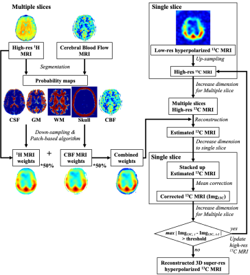 |
10 | Volumetric patch-based image reconstruction for enhancing hyperpolarized 13C MRI with hybrid image guidance
Sung-Han Lin1, Junjie Ma1, and JaeMo Park1,2,3
1Advanced Imaging Research Center, UT Southwestern Medical Center, Dallas, TX, United States, 2Department of Radiology, UT Southwestern Medical Center, Dallas, TX, United States, 3Electrical and Computer Engineering, UT Dallas, Richardson, TX, United States
This study presents a volumetric extension of the previously proposed patch-based algorithm for enhancing spatial resolution of 13C MRI. Feasibility of integrating structural and perfusion images as guidance for the patch-based reconstruction is also demonstrated with a digital phantom. The performance of the method was further tested for a hyperpolarized 13C lactate brain image using hybrid probability maps, created from T1-weighted 1H MRI and perfusion images.
|
|
3509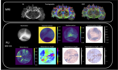 |
11 | Backscattering Mueller Matrix polarimetry shows promise for validation of diffusion MRI microstructural features in thick tissue specimens
Rhea L Carlson1, Justina M Bonaventura2, Courtney J Comrie1, Elizabeth Hutchinson1, and Travis W Sawyer2
1Biomedical Engineering, University of Arizona, Tucson, AZ, United States, 2Optical Sciences, University of Arizona, Tucson, AZ, United States
Ground truth validation methods are essential to improve the accuracy of biophysical representation by diffusion MRI methods. The objective of this study is to advance PLI methodology for thick tissue imaging of fiber distributions by application of multispectral backscattering polarimetry, and of the Mueller matrix mathematical framework for unmixing contributions from depolarization, diattenutation, and retardance. Our results indicate that orientation features derived from PLI are in concordance with known brain physiology. Due to the consistent presence of retardance even in areas of crossing fibers, the results suggest that retardance angle is most sensitive to microstructural features rather than macroscopic geometry.
|
|
3510 |
12 | Measurements of cell size and density using temporal diffusion spectroscopy distinguish hepatocellular carcinoma
xiaoyu jiang1,2, John C. Gore1,3, and junzhong xu1,4
1Vanderbilt University Institute of Imaging Science, nashville, TN, United States, 2Department of Radiology and Radiological Sciences, Vanderbilt University Institute of Imaging Science, nashville, TN, United States, 3Department of Radiology and Radiological Sciences, Vanderbilt University Medical Center, nashville, TN, United States, 4Department of Radiology and Radiological Sciences, Vanderbilt University Medical Center, Nashville, TN, United States
There is an unmet need to develop a non-invasive and reliable method for detecting hepatocellular carcinoma (HCC) at early stages in high-risk patients (e.g., patients with cirrhosis). We hypothesized that temporal diffusion spectroscopy (TDS), which reports histopathological information such as mean cell size in vivo, can improve current Liver Imaging Reporting and Data System (LI-RADS) criteria for assessment of HCC and reduce the need for biopsies. To test this hypothesis, we applied TDS to distinguish cell sizes and densities in HCC and other liver conditions, such as benign/dysplastic nodules, fibrosis, and cholangiocarcinoma (iCCA), in ex vivo studies.
|
|
3511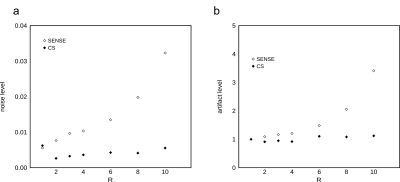 |
13 | Application of compressed sensing in High Spectral and Spatial resolution (HiSS) MRI – evaluation of effective resolution and image quality
Milica Medved1, Marco Vicari2, and Gregory S Karczmar1
1Department of Radiology, The University of Chicago, Chicago, IL, United States, 2Fraunhofer MEVIS, Bremen, Germany
Compressed sensing (CS) acceleration was evaluated in high spectral and spatial resolution (HiSS) MRI, at acceleration factors up to R=10. Effective spatial resolution was maintained in the readout direction, and decreased with R in the phase encoding direction, although acceleration factors of up to R = 4 are realistic. Noise and artifact level amplification were not observed. CS could improve diagnostic utility of HiSS MRI in breast by allowing longer echo trains and thus heavier T2* weighting in a fewer number of k-space lines. CS could also facilitate use of HiSS MRI in geometrically constrained applications, such as prostate MRI.
|
|
 Back to Meeting Home
 Back to the Program-at-a-Glance
The International Society for Magnetic Resonance in Medicine is accredited by the Accreditation Council for Continuing Medical Education to provide continuing medical education for physicians.
 Back to Meeting Home
 Back to the Program-at-a-Glance