Online Gather.town Pitches
Cancer III
Joint Annual Meeting ISMRM-ESMRMB & ISMRT 31st Annual Meeting • 07-12 May 2022 • London, UK

| Booth # | ||||
|---|---|---|---|---|
4293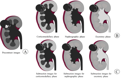 |
1 | Bosniak II-III Cystic Renal Masses: Subtraction MR Images for Improvement of Interobserver Agreement with Bosniak Classification, Version 2019 Video Permission Withheld
Huanhuan Kang1, Xu Bai2, Wei Xu1, Mengqiu Cui1, Shaopeng Zhou1, Baichuan Liu1, and Haiyi Wang1
1Radiology, the First Medical Center of Chinese PLA General Hospital, Beijing, China, 2Radiology, the Fifth Medical Center of Chinese PLA General Hospital, Beijing, China
The purpose of this study was to investigate whether subtraction MR images can improve the interobserver agreement for Bosniak II, IIF and III cystic renal masses (CRMs) with Bosniak classification, version 2019. The results showed that the interobserver agreement was higher with subtraction MR images than without subtraction MR images (weighted k = 0.73 vs 0.60, respectively; P=.011), which may contribute to the popularization and application of Bosniak classification, version 2019.
|
||
| 4294 | 2 | Correlation between changes of pelvic bone marrow fat content and hematological toxicity in concurrent chemoradiotherapy for cervical cancer Video Permission Withheld
XIAOHANG QIN1,2, CONG WANG3, and YONG YIN2
1Department of Graduate, Shandong First Medical University and Shandong Academy of Medical Sciences, Shandong Ji’ nan, China, 2Department of Radiation Physics, Shandong Cancer Hospital and Institute, Shandong First Medical University and Shandong Academy of Medical Sciences, Shandong Ji’ nan, China, 3Department of Fourth Ward of Gynecological Tumor, Shandong Cancer Hospital and Institute, Shandong First Medical University and Shandong Academy of Medical Sciences, Shandong Ji’ nan, China Bone marrow is a heterogeneous mixed tissue, which can be divided into red bone marrow with hematopoietic activity and yellow bone marrow with high fat content. Concurrent chemoradiotherapy can induce red bone marrow transfer into yellow bone marrow, increasing the bone marrow fat content and inhibiting hematopoietic function. The study used the MRI IDEAL IQ sequence to quantify the changes of pelvic bone marrow fat content receiving different doses of concurrent chemoradiotherapy for cervical cancer, and to determine associations with peripheral blood cell counts. |
||
4295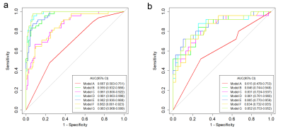 |
3 | MRI-based radiomics model for preoperative prediction of extramural venous invasion of rectal adenocarcinoma
Huijie Jiang1, Xue Lin1, Sheng Zhao1, and Hongbo Hu1
1Harbin Medical University, Harbin, China
Radiomics features were extracted through MRI images and different prediction models (clinical model, logistic regression [LR], random forest [RF], support vector machine [SVM], clinical-LR model, clinical-RF model, and clinical-SVM model) for extramural venous invasion (EMVI) were constructed on the basis of radiomics features and clinical factors, respectively. The optimal model, which had the best predictive efficiency, was screened out for predicting the EMVI status of rectal adenocarcinoma patients.
|
||
4296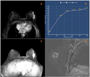 |
4 | Diagnostic evaluation of the Kaiser scoring combined with breast vascular assessment for the characterization of breast lesions
Xin-zhu Zhou1, Hua Lai1, Lian-hua Liu1, Shuang He1, Hui-fang Yao1, Li-ping Chen1, Chen Deng1, Shuang-Ling Li1, and Xiao-yong Zhang2
1Department of Radiology, Chengdu Women's and Children's Central Hospital, School of Medicine, University of Electronic Science and Technology of China, Chengdu, China, 2Clinical Science, Philips Healthcare, Chengdu, China
Kaiser scoring (KS) system for breast magnetic resonance imaging (bMRI) is a new clinical decision-making tool for the diagnosis of breast lesions. This study proposed and investigated the feasibility of Kaiser score combined with breast vascular assessment, defined as KS* , for its diagnostic performance in breast magnetic resonance imaging (bMRI). The results showed that the additional used ipsilateral breast vascularity and positive adjacent vessel sign (AVS) can accurately evaluate breast lesions, and KS* has higher diagnostic efficiency for breast lesions than conventional KS, especially for non-mass lesions.
|
||
4297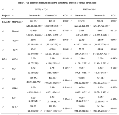 |
5 | Value of ESWAN combined with DTI in the differential diagnosis of solid pseudopapillary tumor and neuroendocrine tumor of pancreas
Lu Wang1, Yi Wang2, Xinqi Wang1, Longshuang Wang1, Qinhe Zhang3, Lizhi Xie4, and Ailian Liu3
1School of Medical Imaging, Dalian Medical University, Dalian, China, 2DepartmentofRadiology,DalianFriendshipHospital, Dalian, China, 3Department of Radiology, the First Affiliated Hospital of Dalian Medical University, Dalian, China, 4GE Healthcare, MR Research China, Beijing, China
It is difficult to differentiate the solid pseudopapillary tumor of the pancreas (SPTP) from pancreatic neuroendocrine tumor (PNET). Enhanced T2* weighted angiography (ESWAN) technique can simultaneously obtain amplitude, phase, R2* and T2* values, which can quantitatively assess the magnetic sensitivity characteristics of tissues. DTI can evaluate the diffusion of water molecules in living tissues. This study founded that R2* and T2* of ESWAN, fractional aniso (FA) and volume ratio aniso (VRA) of DTI were significantly different (P<0.05) and the AUC was 0.860(sensitivity: 100%; specificity: 79.2%). Therefore, ESWAN combined with DTI may be an effective method to identify SPTP and PNET.
|
||
4298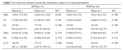 |
6 | Differentiation of solid pseudopapillary tumor and neuroendocrine tumor of pancreas using DTI
Xinqi Wang1, Yi Wang2, Lu Wang1, Qinhe Zhang3, Lizhi Xie4, and Ailian Liu3
1School of Medical Imaging, Dalian Medical University, Dalian, China, 2Department of Radiology, Dalian Friendship Hospital, Dalian, China, 3Department of Radiology, the First Affiliated Hospital of Dalian Medical University, Dalian, China, 4GE Healthcare, MR Research China, Beijing, China
It is difficult to differentiate the solidpseudopapillary tumer of the pancreas(SPTP) from pancreatic neuroendocrine tumers (PNET), due to their similar imaging characteristics. There was significant difference in DTI quantitative parameters Fractional Aniso (FA) and Volume ratio Aniso (VRA) between SPTP and PNET. The results showed that the FA and VRA values of DTI sequence of SPTP were significantly higher than that of PNET. Therefore, DTI sequences may be an effective method to identify SPTP and PNET.
|
||
4299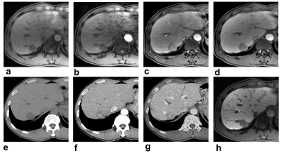 |
7 | New Diagnosis Criteria for Hepatocellular Carcinoma: Gadoxetic Acid-enhanced MRI Integrating CT-Vascular Features May Facilitate Evaluation Video Permission Withheld
Shan Yao1, Yi Wei1, Hehan Tang1, Lisha Nie2, Xiaocheng Wei2, and Bin Song1
1Department of Radiology, West China Hospital, Sichuan University, Chengdu, China, 2GE Healthcare China, Beijing, China
Gadoxetic acid-enhanced magnetic resonance imaging (MRI) has been widely used in detection and diagnosis for hepatocellular carcinoma (HCC), while transient severe motion artifact that affects the arterial phase (AP) images quality and reduces diagnosis performance remains a concern. A new diagnosis criterion for HCC integrating CTAP and MRAP images was introduced in this study and achieved a high sensitivity and comparable specificity when compared with the gadoxetic acid-enhanced MRI only. It may be able to improve the diagnostic performance of gadoxetic acid-enhanced MRI in patients with HCCs and further affect the clinical treatment plan.
|
||
4300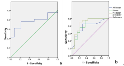 |
8 | Value of Amide Proton Transfer Combined with Dynamic Contrast-Enhanced MRI in Predicting Pathological Features of cervical cancer Video Not Available
Qingling Song1, Ailian Liu2, Changjun Ma3, and Qingwei Song2
11. Department of Radiology, Department of Radiology, The First Affiliated Hospital, Dalian Medical University, Dalian, China, 21.Department of Radiology, The First Affiliated Hospital, Dalian Medical University 2. Dalian Engineering Research Center for Artificial Intelligence in Medical Imaging, Dalian, China, 3Department of Radiology, The First Affiliated Hospital, Dalian Medical University, Dalian, China
The proper evaluation of pathological features including differentiation degree, deep stromal invasion (DSI) and vascular space invasion (VSI) is vital for patients with cervical cancer. Amide Proton Transfer (APTW) and Dynamic Contrast-Enhanced MRI (DCE-MRI) were used to predict tumor differentiation, DSI and VSI. Results showed that combined APTW and DCE-MRI predicted VSI with good performance. Therefore, combination of APTW and DCE-MRI is potentially a promising and valuable non-invasive method in detection for predicting VSI.
|
||
 Back to Meeting Home
 Back to the Program-at-a-Glance
The International Society for Magnetic Resonance in Medicine is accredited by the Accreditation Council for Continuing Medical Education to provide continuing medical education for physicians.
 Back to Meeting Home
 Back to the Program-at-a-Glance
 View the Presentation