Online Gather.town Pitches
Cerebrovascular, Stroke, Ischemia, Atherosclerosis II
Joint Annual Meeting ISMRM-ESMRMB & ISMRT 31st Annual Meeting • 07-12 May 2022 • London, UK

Online Gather.town Pitches
Cerebrovascular, Stroke, Ischemia, Atherosclerosis II
| Booth # | ||||
|---|---|---|---|---|
4931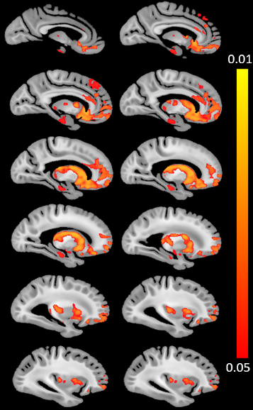 |
1 | Cerebral Amyloid Angiopathy (CAA) is associated with higher R2 relaxation rate: An ex-vivo MRI and pathology study
Md Tahmid Yasar1, Mahir Tazwar 1, Ashish A. Tamhane2, Arnold M. Evia2, David A. Bennett2, Julie A. Schneider2, and Konstantinos Arfanakis1,2
1Department of Biomedical Engineering, Illinois Institute of Technology, Chicago, IL, United States, 2Rush Alzheimer's Disease Center, Rush University Medical Center, Chicago, IL, United States Cerebral amyloid angiopathy (CAA) is characterized by deposition of amyloid-β protein in the walls of cortical and leptomemingeal small vessels. CAA is common in community-based older adults and has been associated with cognitive decline and dementia. To date, the association of CAA with the transverse relaxation rate, R2, remains unknown. This study in a large number of autopsied brains from community-based older adults showed for the first time that CAA is associated with higher R2, independent of other neuropathologies and demographic factors. The spatial pattern for this association derived from voxel-wise analysis included subcortical and frontal lobe structures. |
||
4932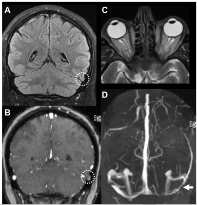 |
2 | Evaluating the clinical significance of MRI findings of Brain Herniations into Arachnoid Granulations in patients with pulsatile tinnitus
Justin Remer1, Eric Smith1, Javier Villanueva-Meyer1, Laura B Eisenmenger2, M. Travis Caton1, Amanda Baker1, Vinil Shah1, Karl Meisel3, Adelyn Tu-Chan3, and Matthew Amans1
1Radiology, UCSF, San Francisco, CA, United States, 2Radiology, University of Wisconsin, Madison, WI, United States, 3Neurology, UCSF, San Francisco, CA, United States
Pulsatile tinnitus, a severely debilitating condition where a “whooshing” sound is heard with each heart beat, affects over 3 million Americans, leading to anxiety, depression and even suicide. While many there are many diseases associated with pulsatile tinnitus one common and serious condition is idiopathic intracranial hypertension. Our study is the first to show that the presence of brain herniations through arachnoid granulations into dural sinuses on MRI is significantly associated with idiopathic intracranial hypertension in patient's with pulsatile tinnitus.
|
||
4933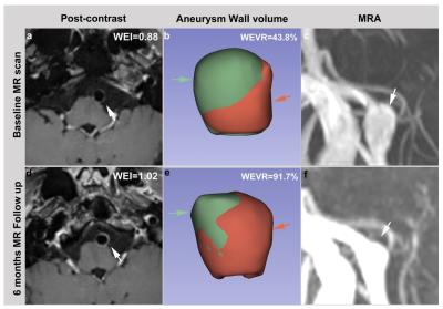 |
3 | A randomized controlled trial of statins to reduce inflammation in cerebral aneurysms using vessel wall MRI
Chengcheng Zhu1, Huibin Kang2, Decai Tian3, Mahmud Mossa-basha1, Michael Levitt4, David Hasan5, Xinjian Yang2, and Yisen Zhang2
1Radiology, University of Washington, Seattle, WA, United States, 2Neurosurgery, Tiantan Hospital, Beijing, China, 3Neurology, Tiantan Hospital, Beijing, China, 4Neurosurgery, University of Washington, Seattle, WA, United States, 5Neurosurgery, University of Iowa, Iowa, IA, United States
Aneurysm wall enhancement as shown on contrast-enhanced vessel wall MRI (VW-MRI) is a surrogate biomarker of intracranial aneurysm (IA) wall inflammation, and it is associated with IA symptoms, growth, and rupture. However, it hasn't been used in clinical trails for UIA treatment. We performed the first prospective randomized controlled trial on the effect of statins for changes in aneurysm wall enhancement as shown in VW-MRI. We found that statins decreased aneurysm wall enhancement of UIAs in a short term of 6 months. Our results suggest VWI-MRI has great potential in future image-guided clinical trails to investigate novel treatment of IAs.
|
||
4934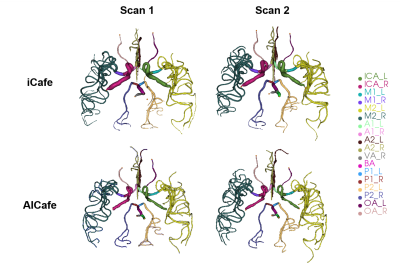 |
4 | Vascular map assessment - the dual use of an analysis tool to evaluate imaging technique reproducibility and AI technique’s performance
Kaiyu Zhang1, Anders Gould2, Li Chen1, Zhensen Chen3, Gador Canton1, Niranjan Balu1, Thomas Hatsukami1, and Chun Yuan1
1Vascular Imaging Lab and BioMolecular Imaging Center, Department of Radiology, University of Washington, Seattle, WA, United States, 2Carle Illinois College of Medicine, University of Illinois at Urbana-Champaign, Urbana-Champaign, IL, United States, 3Institute of Science and Technology for Brain-Inspired Intelligence, Fudan University, Shanghai, China
Intracranial artery feature extraction (iCafe), a custom-made semi-automatic vascular map construction software, can quantitatively measure intracranial vascular features on time of flight (TOF) MRA with good scan-rescan and inter/intra operator reproducibility. To fully extend the use of iCafe to a novel MRA technique named simultaneous non-contrast angiography and intraplaque hemorrhage (SNAP), we will first evaluate its vascular feature measurements reproducibility and then compare these measurements with those acquired with a newly developed AICafe (AI + iCafe) technique on the same dataset. Good reproducibility was obtained on SNAP MRA using iCafe, as well as the AICafe technique.
|
||
4935 |
5 | Characterization of a novel rodent model of arteriovenous malformations (AVMs) with time-of-flight magnetic resonance angiography
Chul Han1, Gregory Harrison Turner2, Candice Nguyen1, Michael Lawton1, and S. Paul Oh1
1Barrow Aneurysm and AVM Research Center, Barrow Neurological Institute, Phoenix, AZ, United States, 2Neuroimaging Research, Barrow Neurological Institute, Phoenix, AZ, United States
A mouse model of arteriovenous malformations was developed with a genetics-based approach that conditionally deleted the causative activin receptor-like kinase 1 (ACVRL1 or ALK1) gene. This model was characterized in vivo using time-of-flight angiography (TOF_MRA). We correlated radiographic and histopathologic findings and determined that the 55 Tagln-Cre (+);Alk12f/2f mouse was a promising experimental brain AVM model for future studies of AVM pathophysiology, growth, rupture, and therapeutic regression.
|
||
4936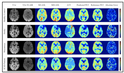 |
6 | PET Synthesis from Multi-Contrast MRI with Attention-Based 3D Encoder-Decoder Networks
Ramy Hussein1, David D. Shin2, Moss Zhao1, Jia Guo3, Michael Moseley1, and Greg Zaharchuk1
1Radiology, Stanford University, Stanford, CA, United States, 2Global MR Applications & Workflow, GE Healthcare, Menlo Park, CA, United States, 3Department of Bioengineering, University of California Riverside, Riverside, CA, United States
We present an attention-based 3D convolutional encoder-decoder network to synthetize PET Cerebral Blood Flow (CBF) maps from multi-parametric MRI images without using radioactive tracers. Inputs to the prediction model are structural MRI (T1 and T2 fluid-attenuated inversion recovery [FLAIR]), and arterial spin labeling (ASL) perfusion MRI images. Results show that encoder-decoder networks, with attention mechanisms and customized loss functions, can adequately combine multiple MRI image types and predict the gold-standard oxygen-15-water PET CBF maps. Adequate quantification of PET from MRI has a great potential for increasing the accessibility of cerebrovascular diseases assessment for underserved populations, underprivileged communities, and developing nations.
|
||
4937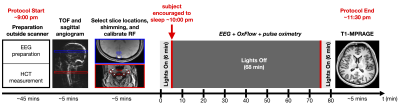 |
7 | Implementation of OxFlow MRI with concurrent EEG for tracking the cerebral metabolic rate of oxygen during extended-duration scans of sleep
Alexander M Barclay1, Alessandra S Caporale1, Hengyi Rao1, Hyunyeol Lee2, Michael C Langham1, and Felix W Wehrli1
1University of Pennsylvania, Philadelphia, PA, United States, 2Kyungpook National University, Daegu, Korea, Republic of
OxFlow was used to track the global cerebral metabolic rate of oxygen (CMRO2) during wakefulness and sleep, with concurrent EEG, in eleven healthy subjects scanned continuously for 80 minutes. CMRO2 was derived from phase-contrast measurements of cerebral blood flow (CBF) in the neck with susceptometry-based oximetry measurements of venous oxygenation (SvO2) in the superior sagittal sinus (SSS). Results reveal that there is negligible bias between total CBF (tCBF), obtained by upscaling the value measured in the SSS, and CBF measured at the neck, suggesting that single-slice OxFlow can be implemented to simplify data acquisition and processing without sacrificing accuracy.
|
||
4938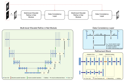 |
8 | CAMWARE: Cascaded Multi-level Wavelet Refine Neural Network for Accelerated Whole-brain MR Vessel Wall Imaging
Zhehao Hu1,2, Alexander Lerner1, Roy Poblete3, and Zhaoyang Fan1,4,5
1Department of Radiology, Keck School of Medicine, University of Southern California, Los Angeles, CA, United States, 2Department of Bioengineering, University of California, Los Angeles, Los Angeles, CA, United States, 3Department of Neurology, Keck School of Medicine, University of Southern California, Los Angeles, CA, United States, 4Department of Radiation Oncology, Keck School of Medicine, University of Southern California, Los Angeles, CA, United States, 5Department of Biomedical Engineering, University of Southern California, Los Angeles, CA, United States
3D MR vessel wall imaging (VWI) is a non-invasive imaging modality for directly assessing intracranial arterial wall diseases. A typical intracranial VWI protocol requires 6-12 minutes per scan to obtain adequate spatial coverage and resolution. Such a long scan time hinders widespread use of VWI in clinical settings. We have developed a novel intracranial vessel-dedicated CAscaded Multi-level WAvelet REfine (CAMWARE) network that enables a VWI scan within 4 minutes. The proposed network achieved significant improvement in vessel wall delination over conventional compressed sensing reconstruction, and several state-of-the-art deep neural networks, such as U-Net and multi-level wavelet U-Net.
|
||
 Back to Meeting Home
 Back to the Program-at-a-Glance
The International Society for Magnetic Resonance in Medicine is accredited by the Accreditation Council for Continuing Medical Education to provide continuing medical education for physicians.
 Back to Meeting Home
 Back to the Program-at-a-Glance
 View the Presentation