Online Gather.town Pitches
Quantitative Image Acquisition & Analysis I
Joint Annual Meeting ISMRM-ESMRMB & ISMRT 31st Annual Meeting • 07-12 May 2022 • London, UK

Online Gather.town Pitches
Quantitative Image Acquisition & Analysis I
| Booth # | ||||
|---|---|---|---|---|
3811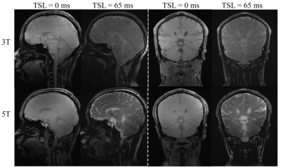 |
1 | Brain T1ρ quantification at the first 5T whole-body MR system: a pilot study
Yuanyuan Liu1,2, Lanlan Gao3, Shuheng Zhang3, Zhuoxu Cui4, Qingyong Zhu4, Haifeng Wang1, Dong Liang1, and Yanjie Zhu1
1Paul C. Lauterbur Research Center for Biomedical Imaging,Shenzhen Institute of Advanced Technology, Chinese Academy of Sciences, Shenzhen, China, 2National Innovation Center for Advanced Medical Devices, Shenzhen, China, 3United Imaging Healthcare, Shanghai, China, 4Research Center for Medical AI,Shenzhen Institute of Advanced Technology, Chinese Academy of Sciences, Shenzhen, China
MR quantitative T1ρ mapping has gained increasing attention due to its capability in studying low-frequency motional processes and chemical exchange in biological tissues. At ultra-high fields, the chemical exchange and proton diffusion in biological tissues should be more prominent. In this study, for the first time, we aim to test the feasibility of T1ρ mapping of brain at 5T and evaluate the T1ρ values estimated from datasets using both 3T and 5T scanners.
|
||
3812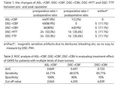 |
2 | Systematic investigation of gamma knife in the treatment of brain tumors: an MR perfusion imaging study Video Not Available
ling huang1, hui bai1, HAI ZHONG1, and WEI QIANG DOU2
1the second hospital of shandong university, JINAN, China, 2GE Healthcare, MR Research, BEIJING, China
The main purpose was to evaluate the gamma knife radiosurgery(GKRS) treatment effect of multiple kinds of brain tumors by MR perfusion imaging. We collected 45 patients with three kinds of brain tumors treated with GKRS. Perfusion imaging, including 3D-ASL and DSC-PWI, was performed before and after treatment. ASL-rCBF, DSC-rCBF and DSC-rCBV decreased significantly after GKRS treatment, and ASL-rCBF before GKRS treatment showed the most robust performance with high AUC in predicting GKRS treatment effect. With these findings, MR perfusion imaging can be considered a time and effective method understanding the treatment effect of GKRS for brain tumor patients.
|
||
3813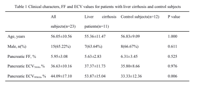 |
3 | Pancreatic extracellular volume fraction using T1 mapping in patients with liver cirrhosis Video Not Available
Qinhe Zhang1, Nan Wan1, Xue Ren1, Peng Sun2, Xu Dai3, Liangjie Lin4, Zhigang Wu5, Jiazheng Wang4, and Ailian Liu1
1The first affiliated hospital of Dalian Medical University, Dalian, China, 2Philips health, Wu han, China, 3Philips health, Shenyang, China, 4Philips health, Beijing, China, 5Philips health, Shen zhen, China
Extracellular volume (ECV) fraction might represent a useful parameter for the noninvasive quantification of pancreatic fibrosis, and the proton density fat fraction (FF) of pancreas can be obtained by mDxion Quant. Here, we hypothesized that pancreatic ECV and FF, for evaluation of pancreatic fibrosis and fat content, can be used to assess patients with liver cirrhosis. Results indicated that pancreatic ECV at 60 mins after contrast agent administration showed a statistical difference between liver cirrhosis patients and controls. A significant correlation was observed between pancreatic FF and ECV at 5 mins after administration.
|
||
3814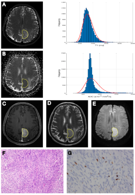 |
4 | Histogram analysis of T1 Mapping and Diffusion-Weighted Imaging in the Prediction of Grades, Subtypes and Proliferation Status of Meningiomas
Tiexin Cao1, Lin Lin1, Yunjing Xue1, Pu-Yeh Wu2, Rufei Zhang1, and Lingmin Zheng1
1Fujian Medical University Union Hospital, Fuzhou, China, 2GE Healthcare, Beijing, China
In daily clinical practice, preoperative grading of meningioma is particularly important for selecting the best treatment strategy. Our hypothesis is that a combination of T1 mapping and diffusion weighted imaging (DWI) based on histogram analysis would be useful for differentiating between low-grade and high-grade meningiomas. Our study, for the first time, provides evidence that T1 mapping may be an imaging biomarker for differentiating grades and predicting the proliferation potential of meningiomas. The combination of T1 mapping and DWI showed the highest diagnostic performances in grading meningiomas.
|
||
3815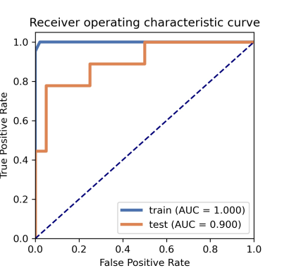 |
5 | A machine learning method to identify grade Ⅱ/Ⅲ glioma based on perfusion parameters derived from DCE-MRI and DSC-MRI
Qiaoli Yao1, Kan Deng2, Zhiyu Liang1, and Yikai Xu1
1Medical Image Center, Nanfang Hospital, Southern Medical University, Guangzhou, China, 2Philips Healthcare, Guangzhou, China As grade Ⅱ and Ⅲ gliomas are difficult to distinguish in preoperative, this study attempted to find the best perfusion parameters for identifying grade Ⅱ/Ⅲ glioma by machine learning model. The machine learning model showed robust performance when using the parameters of volume transfer coefficient (Ktrans) and mean transit time (MTT) derived from the dynamic contrast-enhanced (DCE) and dynamic susceptibility contrast (DSC) imaging, which indicated that the combination of DCE and DSC perfusion techniques is expected to further improve the differential diagnosis of grade Ⅱ and Ⅲ gliomas. |
||
3816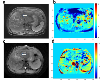 |
6 | Amide proton transfer weighted imaging in differential diagnosis of hepatocellular carcinoma from intrahepatic cholangiocarcinoma
Jingcheng Huang1, Junfei Chen1, Weiqiang Dou2, Jing Ye1, Wei Xia1, and Xianfu Luo1
1Clinical Medical School of Yangzhou University, Northern Jiangsu People’s Hospital, Yangzhou, China, Yangzhou City, China, 2GE Healthcare, MR Research China, Beijing, P.R. China, Beijing City, China
In this study, we aimed to investigate the feasibility of amide proton transfer weighted (APTw) imaging for differentiating hepatocellular carcinoma(HCC) from intrahepatic cholangiocarcinoma(ICC) patients. With the APTw imaging metric of magnetization transfer ratio asymmetry (MTRasym), significantly different magnetization transfer ratio asymmetry (MTRasym) values, a typical metric of APTw imaging, were shown between hepatocellular carcinoma and intrahepatic cholangiocarcinoma. With this finding, APTw imaging could be considered an effective imaging biomarker for differentiating HCC from ICC.
|
||
3817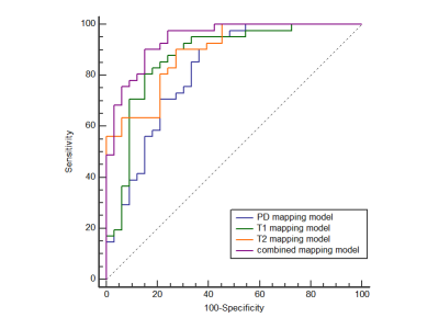 |
7 | The role of histogram analysis in MAGiC sequence in the differential diagnosis of early-stage endometrial adenocarcinoma and normal endometrium Video Not Available
Shuang Chen1, Xiaoduo Yu1, Qi Zhang1, Jieying Zhang1, Lizhi Xie2, and Han Ouyang1
1National Cancer Center/National Clinical Research Center for Cancer/Cancer Hospital, Chinese Academy of Medical Sciences and Peking Union Medical College, Beijing, China, 2GE Healthcare, MR Research, Beijing, China
To discuss the histogram parameters diagnostic value of Magnetic Resonance Image Compilation (MAGiC) sequence for differentiation of early-stage endometrial carcinoma (EC) from normal endometrium of health control (HC).
|
||
3818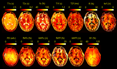 |
8 | Fast parameters Mapping in the brain 4-pool model from tsDESPOT acquired data within 10 seconds using deep learning.
Yuuzo Kamiguchi1 and Sadanori Tomiha2
1Advanced Technology Research Dept., CANON MEDICAL SYSTEMS CORPORATION, Kawasaki-shi, Japan, 2Advanced Technology Research Dept., CANON MEDICAL SYSTEMS CORPORATION, Ootawara-shi, Japan
In quantitative imaging of the brain, it is common that tissues having different T1 and T2 values are mixed in a voxel and exchange proton each other. So multiparametric imaging method of brain microstructure is attracting much attention. In this study, we demonstrated that quantitative maps of 12 parameters in the brain 4-pool model (2 free water pools with exchange and 2 semi-solid water pools) were estimated within 10 seconds using deep learning from the data acquired by the tsDESPOT sequence.
|
||
3819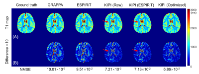 |
9 | Acceleration of Relaxation Time Mapping by Joint K-space and Image-space Parallel Imaging (KIPI)
Tao Zu1, Yi-Cheng Hsu2, Yi Sun2, Dan Wu1,3,4, and Yi Zhang1,3,4
1Key Laboratory for Biomedical Engineering of Ministry of Education, Department of Biomedical Engineering, College of Biomedical Engineering & Instrument Science, Zhejiang University, Hangzhou, China, 2MR Collaboration, Siemens Healthcare Ltd., Shanghai, China, 3Department of Neurology, The First Affiliated Hospital, Zhejiang University, Hangzhou, China, 4Cancer Center, Zhejiang University, Hangzhou, China
The relaxation time mapping has proven to be an important diagnostic tool, but it is limited by the prolonged scan time due to the measurements of multiple frames at the same location. In this study, the recently proposed auto-calibrated reconstruction method by joint k-space and image-space parallel imaging (KIPI) is utilized for the acceleration of relaxation time mapping. Combined with the ESPIRiT method, KIPI generates improved coil sensitivity maps and allows an acceleration factor of up to 4-fold for acquiring source images, yielding the accurate parameter map without obvious errors or artifacts.
|
||
 |
3820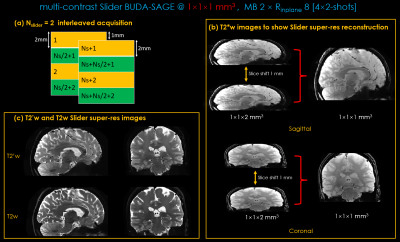 |
10 | BUDA-SAGE with Slider encoding and self-supervised denoising enables fast, distortion-free, high-resolution T2 and T2* mapping
Zijing Zhang1,2, Huihui Ye1, Long Wang3, Kawin Setsompop4, Huafeng Liu1, and Berkin Bilgic2,5,6
1State Key Laboratory of Modern Optical Instrumentation, College of Optical Science and Engineering, Zhejiang University, Hangzhou, China, 2Athinoula A. Martinos Center for Biomedical Imaging, Massachusetts General Hospital, Boston, MA, United States, 3Subtle Medical Inc, Menlo Park, CA, United States, 4Radiological Sciences Laboratory, Stanford University, Stanford, CA, United States, 5Department of Radiology, Harvard Medical School, Boston, MA, United States, 6Harvard-MIT Health Sciences and Technology, Massachusetts Institute of Technology, Cambridge, MA, United States
BUDA-SAGE is an efficient multi-shot echo‐planar imaging (EPI) sequence that provides multiple distortion-free T2*-, T2’- and T2-weighted contrasts for quantitative mapping. In this work, we employ Slider encoding to reach 1 mm isotropic resolution by performing super-resolution reconstruction on volumes acquired with 2 mm slice thickness. A self-supervised neural network MR-Self2Self (MR-S2S) was utilized to perform denoising to boost SNR. Estimated quantitative maps showed consistent values with conventional mapping methods in phantom and in-vivo measurements. We demonstrate that BUDA-SAGE acquisition with self-supervised denoising and Slider encoding enables rapid, distortion-free, whole-brain T2/T2* mapping at 1 mm isotropic resolution under 90 seconds.
|
|
3821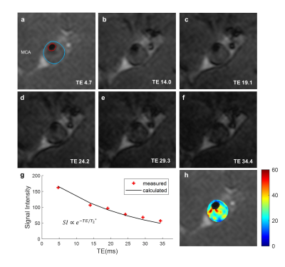 |
11 | In Vivo T2* Mapping of Intracranial Atherosclerotic Plaque Distinguishes Symptom-Producing Plaques: a 7T MRI study
Ziming Xu1, Xiaoyan Bai2, Yajie Wang1, Zhiye Li2, Jiaqi Dou1, Binbin Sui3, and Huijun Chen1
1Center for Biomedical Imaging Research, School of Medicine, Tsinghua University, Beijing, China, 2Department of Radiology, Beijing Tiantan Hospital, Capital Medical University, Beijing Neurosurgical Institute, Beijing, China, 3Tiantan Neuroimaging Center of Excellence, China National Clinical Research Center for Neurological Diseases, Beijing, China
In vivo quantitative mapping of intracranial atherosclerotic plaque is very important for plaque characterization. Previous intracranial plaque quantitative studies were usually carried out ex vivo, while studies of in vivo intracranial plaque mapping have rarely been reported. In this study, the in vivo feasibility of quantitative T2* mapping of intracranial plaque at 7T MRI had firstly been demonstrated. Symptomatic patients showed significantly lower intraplaque T2* values than asymptomatic patients (26.33 ± 7.16 vs. 33.47 ± 8.68 ms, p = 0.048). This quantitative and noninvasive assessment may serve as one potential tool to characterize and evaluate intracranial atherosclerotic plaques.
|
||
3822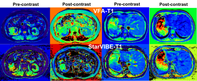 |
12 | Comparison of T1 mapping from StarVIBE and conventional variable flip angle in abdomen: a preliminary study
Junjiao Hu1, Huiting Zhang2, Jun Liu1, Hu Guo2, Shan Jiang1, and Weijun Situ1
1Department of Radiology,The Second Xiangya Hospital, Central South University, Changsha, China, 2MR Scientific Marketing, Siemens Healthineers, Wuhan, China,, Wuhan, China
The purpose of this study to assess image quality and T1 value value between variable flip angle (VFA-T1) and StarVIBE (StarVIBE-T1) methods using breath-holding and free breathing, respectively. Compared with VFA-T1 method, StarVIBE-T1 had better image quality, including overall image quality, blurring, and clearer edge of lesions, especially for the patients without breath-holding. The T1 values from the two methods had high correlation and moderate agreement.
|
||
3823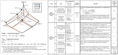 |
13 | Enhancing Analysis Algorithm for T2-based water suppressed diffusion MRI (T2wsup-dMRI) by adding least-square fitting
Tokunori Kimura1
1Radiological Science, Shizuoka College of Medical care Science, Hamamatsu, Japan
A T2-based water suppressed diffusion MRI (T2wsup-dMRI) was proposed to solve the CSF partial volume effects (CSF-PVE) in quantifying several parameters in tissue. There a simple closed form (CF) algorithm was used but errors were increased when tissue T2, T2t is long (> 100ms). In this study, to reduce those errors, several algorithms using least-squares (LSQ) fitting method were assessed by simulation and in vivo data. Through this study, the combined algorithm of single and bi-exponential LSQ applied in 2D (TE, b) space is the best in keeping those accuracies especially when applied in random data pattern.
|
||
3824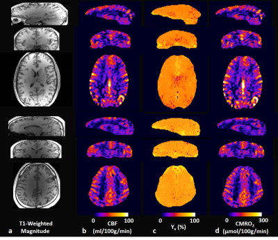 |
14 | Whole-Brain 3D CMRO2 Mapping with Prior-Guided Quantitative BOLD
Hyunyeol Lee1,2 and Felix Wehrli2
1Electronics Engineering, Kyungpook National University, Daegu, Korea, Republic of, 2Radiology, University of Pennsylvania, Philadelphia, PA, United States
qBOLD permits noninvasive measurements of the two critical determinants of the BOLD signal, i.e., deoxygenated-blood-volume (DBV) and venous-oxygen-saturation-level (Yv), and along with CBF imaging, the cerebral metabolic rate of oxygen (CMRO2). Two major challenges in qBOLD are 1) separation of heme-originated R2′ from other signal sources, and 2) subsequent extraction of DBV and Yv. We had previously addressed these issues by developing a prior-guided qBOLD method. Here, we aimed to evaluate the method's utility in 3D CMRO2 mapping. Results suggest feasibility of the new qBOLD method as a practical means for measuring neurometabolic parameters over an extended brain coverage.
|
||
3825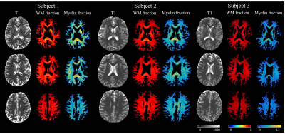 |
15 | Optimized Ultrashort Echo Time – Magnetic Resonance Fingerprinting (UTE-MRF) for Myelin-Proton fraction Imaging
Zihan Zhou1, Qing Li2, Congyu Liao3,4, Xiaozhi Cao3,4, Huihui Ye5, Jianhui Zhong1,6, and Hongjian He1
1Center for Brain Imaging Science and Technology, College of Biomedical Engineering and Instrumental Science, Zhejiang University, Hangzhou, China, 2MR Collaborations, Siemens Healthineers Ltd, Shanghai, China, 3Department of Radiology, Stanford University, Stanford, CA, United States, 4Department of Electrical Engineering, Stanford University, Stanford, CA, United States, 5State Key Laboratory of Modern Optical Instrumentation, College of Optical Science and Engineering, Zhejiang University, Hangzhou, China, 6Department of Imaging Sciences, University of Rochester, Rochester, NY, United States
Optimization of MRF (MR fingerprinting) sequence is important for encoding MR tissue parameters with the maximal SNR for better quantification of representative tissues of WM, GM and CSF. Here, we targeted ultrashort T2 tissues and proposed an optimized acquisition parameter patterns for 3D ultrashort echo time MRF sequence with the goal of achieving higher quantification accuracy for myelin-proton based on Cramér‐Rao Lower Bound. Our results show that the optimized UTE-MRF sequence achieved high accuracy in myelin-proton quantification and whole-brain myelin-proton imaging in 15 min with 1mm isotropic resolution. The optimization also opens the door to further reduce scan time.
|
||
3826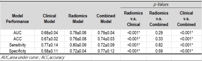 |
16 | MR radiomic predictors of post-GKRS edema in meningiomas
Man-Chin Chen1, Huai-Zhe Yang2, Cheng-Chia Lee2,3, Hsiu-Mei Wu3,4, and Chia-Feng Lu1
1Department of Biomedical Imaging and Radiological Sciences, National Yang Ming Chiao Tung University, Taipei, Taiwan, 2Department of Neurosurgery, Neurological Institute, Taipei Veteran General Hospital, Taipei, Taiwan, 3School of Medicine, National Yang Ming Chiao Tung University, Taipei, Taiwan, 4Department of Radiology, Taipei Veteran General Hospital, Taipei, Taiwan
Several issues concerning the optimal parameters of radiosurgery treatment, the occurrence of radiation-induced edema, and other postoperative complications remain unsolved in treating meningiomas. We aimed to determine whether combining radiomic features with clinical risk factors can improve the prediction of edema occurrence after Gamma-Knife radiosurgery (GKRS). Pre-GKRS MR radiomic features and clinical features were used to construct the prediction model. The model combing radiomic features and clinical features showed the highest performance for prediction of post-GKRS edema (AUC=0.79). The outcomes of this study can provide a risk assessment to facilitate precision medicine in treating meningiomas.
|
||
3827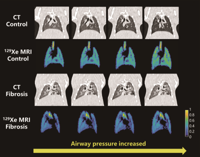 |
17 | Pulmonary Compliance Imaging Using Hyperpolarized Gas MRI Video Permission Withheld
Ming Zhang1, Haidong Li1, Hongchuang Li1, Xiuchao Zhao1, Xiaoling Liu1, Yeqing Han1, Xianping Sun1, Xin Lou2, Chaohui Ye1, and Xin Zhou1
1Key Laboratory of Magnetic Resonance in Biological Systems, State Key Laboratory of Magnetic Resonance and Atomic and Molecular Physics, National Center for Magnetic Resonance in Wuhan, Wuhan Institute of Physics and Mathematics, Innovation Academy for Precision Measurement Science and Technology, Chinese Academy of Sciences- Wuhan National Laboratory for Optoelectronics, Wuhan, China, 2Department of Radiology, Chinese PLA General Hospital, Beijing, China
Pulmonary compliance is generally measured using ventilator and esophageal balloon in clinical practice, and only global compliance could be obtained. In this study, we proposed a method for imaging pulmonary compliance using hyperpolarized (HP) 129Xe MRI. Lung compliance derived by HP 129Xe MRI could be used for quantifying the injuries caused by fibrosis in rats.
|
||
3828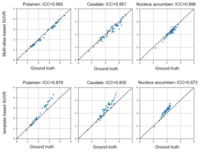 |
18 | Comparison of multi-atlas based and template based method in the quantitative analysis of [18F]-FP-DTBZ PET Video Permission Withheld
Yiwei Pan1, Shuying Liu2, Yao Zeng1, Chenfei Ye3, Hongwen Qiao2, Tianbing Song2, Haiyan Lv4, Piu Chan2, Jie Lu2, and Ting Ma1,2,3
1Department of Electronic and Information Engineering, Harbin Institute of Technology at Shenzhen, Shenzhen, China, 2Xuanwu Hospital Capital Medical University, Beijing, China, 3Peng Cheng Laboratory, Shenzhen, China, 4Mindsgo Life Science Shenzhen Co. Ltd, Shenzhen, China
[18F]-FP-DTBZ PET provides reliable information for the diagnosis of Parkinson’s disease. Quantitative analysis of PET images requires a precise segmentation of the region of interest. Two [18F]-FP-DTBZ PET image quantification methods, multi-atlas based and template based methods were compared in this study. A total of 68 subjects were included, each of them underwent 3D T1-weighted MR imaging and [18F]-FP-DTBZ PET imaging. Twenty subjects were used to build atlases and 48 were used for validation. Using dice coefficient and ICC coefficient for evaluation, we observed that the multi-atlas based method showed better performance than the template based method.
|
||
3829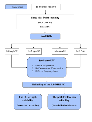 |
19 | The location reliability of the functional connectivity of deep emotion-related regions towards functional connectivity guided rTMS therapy
Na Zhao1,2,3,4,5, Juan Yue1,2,3, Zi-Jian Feng1,2,3, Yang Qiao 1,2,3,5,6, Li-Xia Yuan1,2,3, Jue Wang7, Yong Zhang8, Yu-Tao Xiang 4,5,9, and Yu-Feng Zang 1,2,3
1Center for Cognition and Brain Disorders, The Affiliated Hospital of Hangzhou Normal University, Hangzhou, China, Hangzhou, China, 2Institute of Psychological Sciences, Hangzhou Normal University, Hangzhou, China, Hangzhou, China, 3Zhejiang Key Laboratory for Research in Assessment of Cognitive Impairments, Hangzhou, China, Hangzhou, China, 4Unit of Psychiatry, Department of Public Health and Medicinal Administration, & Institute of Translational Medicine, Faculty of Health Sciences, University of Macau, Macao SAR, China, Taipa, Macau, 5Centre for Cognitive and Brain Sciences, University of Macau, Macao SAR, China, Taipa, Macau, 6Faculty of Health Sciences, University of Macau, Macao SAR, China, Taipa, Macau, 7Institute of sports medicine and health, Chengdu Sport University, Chengdu, China, Chengdu, China, 8MR Research, GE Healthcare, Shanghai, China, Shanghai, China, 9Institute of Advanced Studies in Humanities and Social Sciences, University of Macau, Macao SAR, China, Taipa, Macau The current study systematically investigated multiple factors that affect the reliability of the FC location and strength of the DLPFC with a few emotional related deep regions. The group-level voxel-wise FC strength reliability was low to moderate, indicating its little significance for guiding individualized rTMS treatment. In terms of the FC location reliability, the intra-individual distances of the center of gravity (COG) were 3.8-7.3 mm across different conditions, which suggest that the COG of the seed-based FC might be potential stimulation target for individualized precise rTMS treatment of affective brain disorders. |
||
 Back to Meeting Home
 Back to the Program-at-a-Glance
The International Society for Magnetic Resonance in Medicine is accredited by the Accreditation Council for Continuing Medical Education to provide continuing medical education for physicians.
 Back to Meeting Home
 Back to the Program-at-a-Glance
 View the Presentation