Online Gather.town Pitches
Quantitative Image Acquisition & Analysis II
Joint Annual Meeting ISMRM-ESMRMB & ISMRT 31st Annual Meeting • 07-12 May 2022 • London, UK

Online Gather.town Pitches
Quantitative Image Acquisition & Analysis II
| Booth # | ||||
|---|---|---|---|---|
 |
4402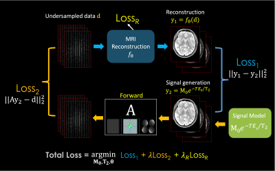 |
1 | Accelerated MR Parameter Mapping with Scan-specific Unsupervised Networks
Tae Hyung Kim1,2, Jaejin Cho1,2, Bo Zhao3, and Berkin Bilgic1,2,4
1Athinoula A. Martinos Center for Biomedical Imaging, Massachusetts General Hospital, Charlestown, MA, United States, 2Radiology, Harvard Medical School, Boston, MA, United States, 3Biomedical Engineering, The University of Texas at Austin, Austin, TX, United States, 4Harvard/MIT Health Sciences and Technology, Cambridge, MA, United States
We introduce a novel framework that jointly performs advanced image reconstruction and model-based MR parameter mapping, where various traditional and modern reconstruction techniques and signal relaxation models (T1, T2, T2*, etc) can be integrated as a plug-and-play manner. Using the proposed framework, we also incorporated model-based parameter mapping with scan-specific deep learning reconstruction (a method named LORAKI). The experiment results with T2, T2* and T1 indicate that this synergistic combination is advantageous, providing improved quantitative imaging over existing methods, e.g. with up to 3.6-fold, 1.7-fold, 2.3-fold NRMSE gain in T2, T2* and T1 estimation, respectively.
|
|
4403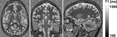 |
2 | T1 mapping and automated segmentation of the brain at 0.55 T
Michael Bach1, Tom Hilbert2,3,4, Francesco Santini5,6, Hanns Christian Breit1, Markus Klarhöfer5,7, Davide Piccini2,3,4, Tobias Kober2,3,4, and Bénédicte Maréchal2,3,4
1Department of Radiology, University Hospital Basel, Basel, Switzerland, 2Advanced Clinical Imaging Technology, Siemens Healthcare, Lausanne, Switzerland, 3Department of Radiology, Lausanne University Hospital and University of Lausanne, Lausanne, Switzerland, 4LTS5, École Polytechnique Fédérale de Lausanne (EPFL), Lausanne, Switzerland, 5Division of Radiological Physics, Department of Radiology, University Hospital Basel, Basel, Switzerland, 6Department of Biomedical Engineering, University of Basel, Basel, Switzerland, 7Siemens Switzerland AG, Healthcare Sector, Zürich, Switzerland
T1 mapping could potentially accurately diagnose microstructural changes due to pathology. In this work, a mp2rage based approach was optimized to perform fast whole-brain T1 mapping at 0.55 T. The mp2rage method was validated by the inversion recovery (IR) method. Finally, a fully automated brain segmentation was applied to perform region-based analysis of T1 values. Both methods (IR and mp2rage) yield comparable T1 values. The fully automated brain segmentation showed a good performance for the mp2rage sequence at 0.55 T. ROI extraction should further be optimized as it showed outliers most likely due to CSF voxels contaminating the ROIs.
|
||
4404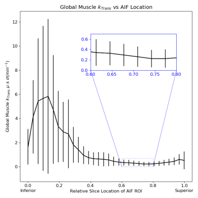 |
3 | Optimization of Arterial Input Function Selection for Quantitative Head and Neck Dynamic Contrast-Enhanced (DCE) MRI
John A Roberts1, Seong-Eun Kim1, Evgueni Kholmovski1,2, Ying Hitchcock3, Tyler John Richards1, and Yoshimi Anzai1
1Radiology and Imaging Sciences, University of Utah, Salt Lake City, UT, United States, 2Biomedical Engineering, University of Maryland, Baltimore, MD, United States, 3Radiation Oncology, Huntsman Cancer Institute, Salt Lake City, UT, United States
Arterial input functions measured in T1 weighted low tip angle SPGRE DCE MRI acquisitions may violate an important assumption used when modeling the conversion of image values to quantitative results: that all tissues of interest have reached steady state within the imaging volume. For arterial flow along the short axis of the imaging volume, blood may not dwell within the imaging volume long enough to reach steady state. We study both the location from which AIF will be measured and the acquisition parameters employed in order to optimize the accuracy of the DCE derived quantitative results.
|
||
4405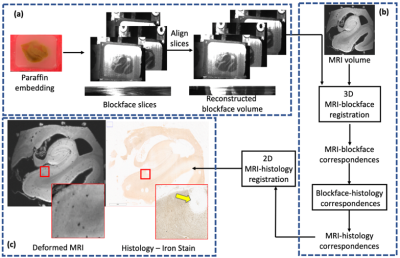 |
4 | A novel blockface imaging pipeline for robust correlative MR-histology of human brain specimens Video Not Available
Phillip DiGiacomo1, Wei Shao1, Marios Georgiadis1, Yi Wang2, Pascal Spincemaille2, Philipp Schlömer3, Markus Axer3, Don Born1, Mirabela Rusu1, and Michael Zeineh1
1Stanford University, Stanford, CA, United States, 2Cornell University, Ithaca, NY, United States, 3Research Centre Jülich, Jülich, Germany
Histology is critical for validating pathology detected in ultra-high-resolution ex-vivo MRI. However, registering histological images with MR is challenging due to 3D deformations between MR and histology, 2D deformations during sectioning, and the fact that histological staining often occurs only in selected slices. Addressing these issues will enable quantification of complex histological features and their relationship with MRI, facilitating the discovery of novel in vivo imaging-based biomarkers of disease. Here, we demonstrate the use of a novel pipeline utilizing ultra-high-resolution specimen MRI, blockface imaging, and a modified tape transfer system for correlative MRI-paraffin-embedded histology of human brain tissue specimens.
|
||
4406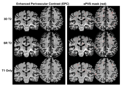 |
5 | Automatic Quantification of Enlarged Perivascular Space aided with Super-resolution 2D T2 images
Jiachen Zhuo1, Muhan Shao2, Steven Roys1, Xiao Liang1, Rosy Linda Njonkou Tchoquessi1, Prashant Raghavan1, Jerry Prince2, and Rao Gullapalli1
1Diagnostic Radiology and Nuclear Medicine, University of Maryland School of Medicine, Baltimore, MD, United States, 2Electrical and Computer Engineering, Johns Hopkins University, Baltimore, MD, United States
The perivascular space (PVS) is key to brain waste clearance and brain metabolic homeostasis. Enlarged PVS (ePVS) can be automatically quantified reliably by combining the 3D T1w and 3D T2w images to produce enhanced PVS contrast followed with frangi filtering and thresholding. However often times only 2D T2w images are available, especially in clinical exams. In this study, we investigate the feasibility of using an innovative deep learning based super-resolution technique (SMORE) to produce 3D T2w images (SR T2) for ePVS quantification. We show that the SR T2 volume provided comparable ePVS estimation as a 3D T2 volume.
|
||
4407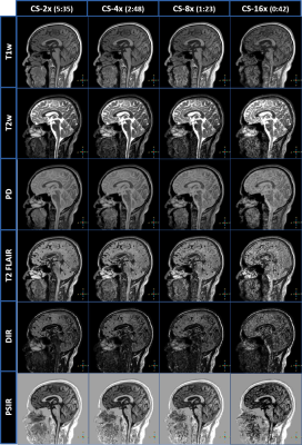 |
6 | Evaluation of 3D-QALAS Multiparametric Imaging with Compressed SENSE Acceleration
Brian Johnson1,2, John Penatzer1, and Sandeep Ganji1,3
1Philips Healthcare, Gainesville, FL, United States, 2University of Texas Southwestern Medical Center, Dallas, TX, United States, 3Mayo Clinic College of Medicine, Rochester, MN, United States
Simultaneous parametric mapping techniques like 3D-QALAS offer means for reliable measurement of T1, T2, and proton density while also offering the generation of standard imaging contrasts. 3D-QALAS has also been shown to provide consistent brain tissue segmentation and volumetric analysis. However, high-resolution 3D-QALAS scans suffer from long scan times. Applying acceleration techniques like compressed SENSE, which are highly effective at reducing 3D scan times, can bring 3D-QALAS scan times under 3-minutes. Here we evaluate the acceleration of 3D-QALAS using multiple compressed SENSE factors on scan times, image quality, multiparametric mapping, and volumetric analysis.
|
||
4408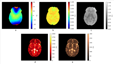 |
7 | Comprehensive 3D quantitative transient response MRI
Gyula Kotek1, Willem van Valenberg1, Laura Nunez-Gonzalez1, Dirk H. J. Poot1, Mika W. Vogel2, and Juan A. Hernandez-Tamames1
1Radiology and Nuclear Medicine, Erasmus MC, Rotterdam, Netherlands, 2GE Healthcare, Hoevelaken, Netherlands
Unlike multi-parametric methods as MR Fingerprinting and quantitative transient imaging we present a quantitative MRI method based on the transient response without variation of experimental parameters (FA…). Our method is based on the exact solution of the recursive equation for the magnetization along an acquisition train. Based on this analytical approach we determine the requirements for the features and parameters of a repeated acquisition block to allow simultaneous estimation of experimental (B0, B1) and intrinsic (PD, T1, T2) parameters. The feasibility of the technique on clinical scanners is demonstrated by acquiring a comprehensive set of 3D in vivo parametric maps.
|
||
4409 |
8 | Improved Harmonization of Renal T2 Mapping Between Vendors using Stimulated Echo Compensation
Hao Li1, Charlotte E Buchanan2, David M Morris3, Alexander J Daniel2, João Sousa4, Steven Sourbron4, David L Thomas5,6,7, Susan T Francis2, and Andrew Nicholas Priest1,8
1Department of Radiology, University of Cambridge, Cambridge, United Kingdom, 2Sir Peter Mansfield Imaging Centre, University of Nottingham, Nottingham, United Kingdom, 3Centre for Cardiovascular Science, University of Edinburgh, Edinburgh, United Kingdom, 4Department of Infection, Immunity and Cardiovascular Disease, University of Sheffield, Sheffield, United Kingdom, 5Neuroradiological Academic Unit, UCL Queen Square Institute of Neurology, University College London, London, United Kingdom, 6Dementia Research Centre, UCL Queen Square Institute of Neurology, University College London, London, United Kingdom, 7Wellcome Centre for Human Neuroimaging, UCL Queen Square Institute of Neurology, University College London, London, United Kingdom, 8Department of Radiology, Addenbrooke’s Hospital, Cambridge, United Kingdom
B1 inhomogeneity and non-ideal slice profiles introduce contributions from stimulated and indirect echoes into the multi-echo spin-echo (MESE) T2 mapping sequence, leading to variations in quantitative T2 values across scanners and vendors despite the use of a harmonised scan protocol. This study used an EPG-based fitting method to include corrections for stimulated-echo effects, applied to phantom and ‘travelling kidney’ data collected on scanners from three different MR vendors (GE, Philips, Siemens). Compared with conventional monoexponential fitting, EPG-based fitting substantially reduced the inter-scanner variations of T2 measurements. This improves the harmonization of the MESE T2 mapping sequence across MR scanner vendors.
|
||
4410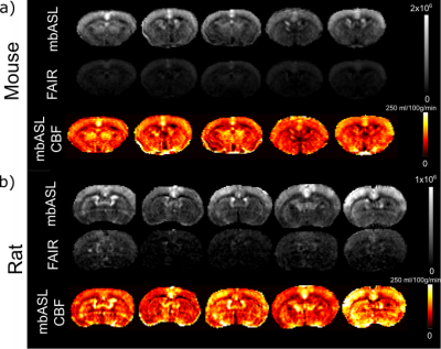 |
9 | Quantitative multiple boli arterial spin labelling
Samantha Paterson1,2, Antoine Vallatos3, Camille Graff4, and William Matthew Holmes2
1EPIC, University of Leeds, Leeds, United Kingdom, 2GEMRIC, University of Glasgow, Glasgow, United Kingdom, 3Centre for Clinical Brain Sciences, University of Edinburgh, Edinburgh, United Kingdom, 4INP Grenoble - Phelma, Grenoble, France
The mbASL sequence is a hybrid between PASL and pCASL. It was shown to produce high SNR perfusion data. Quantification is crucial in order to produce accurate mbASL CBF maps. We have shown successful quantification by modifying the Buxton ASL kinetic model, optimising the number of pulses used for inversion and examining the optimal labelling slab thickness needed to maximise the SNR. We found CBF values that agree with current literature for mice and rats.
|
||
4411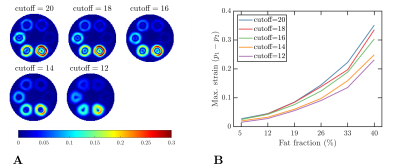 |
10 | The influence of fat in the estimation of strain from tagged MR images
Hernán Arturo Mella1,2,3 and Sergio Uribe2,3,4
1Department of Electrical Engineering, Pontificia Universidad Católica de Chile, Santiago, Chile, 2Biomedical Imaging Center, Pontificia Universidad Católica de Chile, Santiago, Chile, 3ANID - Millennium Science Initiative Program - Millennium Nucleus in Cardiovascular Magnetic Resonance, Santiago, Chile, 4Department of Radiology, School of Medicine, Pontificia Universidad Católica de Chile, Santiago, Chile
In this work, we investigated the influence of fat in the estimation of strain from tagged MR images. The results showed that in a static phantom with sharp and smooth fat fractions, the estimated strain is influenced by the bandwidth of the bandpass filter, the tagging period, and the fat fraction.
|
||
4412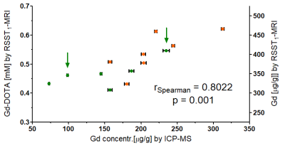 |
11 | Quantitative mapping of Gd-DOTA accumulation in mouse brain by MRI after intraperitoneal administration - Validation by mass spectroscopy. Video Permission Withheld
Anthony Tessier1, Anthony Ruze1, Emilien Royer1, Monique Bernard1, Angele Viola1, and Teodora-Adriana Perles-Barbacaru1
1CRMBM UMR 7339, Aix Marseille University, CNRS, Marseille, France
Maps of the contrast agent concentration over time in mouse brains upon intraperitoneal administration were obtained by a dynamic MRI technique. The mice had different degrees of contrast agent accumulation clearly distinguishable on the maps with a particular distribution resembling the pathways of the glymphatic system. The average brain concentration computed 2 hours post contrast agent administration correlates with the gadolinium dosage in the brain by inductively coupled plasma mass spectroscopy proving that quantification is feasible although the signal analysis can be improved.
|
||
4413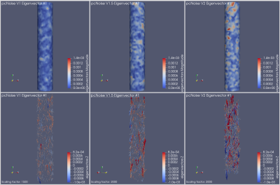 |
12 | In silico uncertainty quantification of the wall shear stress computed from phase-contrast 4d flow measurements.
Constanza Gaínza1,2, Daniele E Schiavazzi3, and Carlos A Sing-Long1,2,4,5,6
1Institute for Mathematical and Computational Engineering, Pontificia Universidad Católica de Chile, Santiago, Chile, 2Millennium Nucleus Center for the Discovery of Structure in Complex Data, Santiago, Chile, 3Department of Applied and Computational Mathematics and Statistics, University of Notre Dame, Notre Dame, IN, United States, 4Institute for Biological and Medical Engineering, Pontificia Universidad Católica de Chile, Santiago, Chile, 5Millennium Nucleus for Cardiovascular Magnetic Resonance, Santiago, Chile, 6Biomedical Imaging Center, Pontificia Universidad Católica de Chile, Santiago, Chile
The wall shear stress (WSS) is a relevant biomarker associated with the rupture of atherosclerotic plaques that can be computed from blood flow velocity measurements. We study the effect of noise velocity on the quantification of the WSS with a focus on the spatial correlations that may arise. We perform in silico experiments in which we consider a Hagen-Poiseuille flow and two noise distributions. Our results show evidence on the existence of spatial correlations in the WSS even when the velocity noise is uncorrelated.
|
||
4414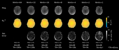 |
13 | Accelerated whole-brain Bloch-Siegert B1-mapping using segmented 3D EPI
Martin Soellradl1, Christina Graf1, and Rudolf Stollberger1
1Institute of Medical Engineering, Graz University of Technology, Graz, Austria, Graz, Austria
To exploit acceleration potential of Bloch-Siegert shift based $$$B_1^+$$$-mapping, we implemented a 3D segmented EPI sequence with variable number of interleaves. Additionally, sampling of the k-space center with variable block-sizes was investigated. Compared to a fully sampled gradient-echo sequence, we show that acceleration by a factor up to 50 is feasible. Depending on the desired accuracy, whole-brain 3D $$$B_1^+$$$-mapping is possible in 5 to 10 seconds.
|
||
 Back to Meeting Home
 Back to the Program-at-a-Glance
The International Society for Magnetic Resonance in Medicine is accredited by the Accreditation Council for Continuing Medical Education to provide continuing medical education for physicians.
 Back to Meeting Home
 Back to the Program-at-a-Glance
 View the Presentation