Online Gather.town Pitches
MRS & Hyperpolarization I
Joint Annual Meeting ISMRM-ESMRMB & ISMRT 31st Annual Meeting • 07-12 May 2022 • London, UK

Online Gather.town Pitches
MRS & Hyperpolarization I
| Booth # | ||||
|---|---|---|---|---|
3524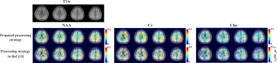 |
1 | Fast Volumetric Diffusion-Weighted MRSI: Improved Acquisition and Data Processing
Zepeng Wang1,2 and Fan Lam1,2,3
1Department of Bioengineering, University of Illinois at Urbana-Champaign, Urbana, IL, United States, 2Beckman Institute for Advanced Science and Technology, Urbana, IL, United States, 3Cancer Center at Illinois, University of Illinois at Urbana-Champaign, Urbana, IL, United States
Diffusion-weighted MRSI (DW-MRSI) is a unique quantitative molecular imaging modality that can potentially provide exclusive cell-type and compartment-specific microstructural information in vivo. However, DW-MRSI studies have been largely limited to single voxels or very low resolutions in basic science and clinical applications due to several fundamental technical challenges. Here, we further enhanced the performance and robustness of the recently proposed subspace imaging method by synergizing improved acquisition and data processing strategies. Higher b-value DE and 3D metabolite mean diffusivity mapping more immune to physiological motions at a 6.9x6.9x8 mm3 nominal resolution were achieved in less than 20-mins.
|
||
3525 |
2 | A Dissolved-Phase 129Xe Phantom for Quantitative Gas-Exchange MRI
Brice Albert1, Joseph W Plummer2, Matthew M Willmering1, Diana M Lindquist3,4,5, Laura L Walkup1,2,6,7, and Zackary I Cleveland1,2,6,7
1Center for Pulmonary Imaging Research, Division of Pulmonary Medicine, Cincinnati Children's Hospital Medical Center, Cincinnati, OH, United States, 2Department of Biomedical Engineering, University of Cincinnati, Cincinnati, OH, United States, 3Imaging Research Center, Cincinnati Children's Hospital Medical Center, Cincinnati, OH, United States, 4Department of Pediatrics, University of Cincinnati, Cincinnati, OH, United States, 5Department of Radiology, University of Cincinnati, Cincinnati, OH, United States, 6Department of Pediatrics, Cincinnati Children's Hospital Medical Center, Cincinnati, OH, United States, 7Department of Radiology, Cincinnati Children's Hospital Medical Center, Cincinnati, OH, United States
Many single-site studies, and increasingly multi-site clinical trials, employ hyperpolarized 129Xe MRI to assess lung structure and function. The lack of reliable imaging 129Xe standards hampers image quantification and rigorous comparisons across research sites. This is particularly true for 129Xe imaging of pulmonary gas-exchange, which must detect the large gaseous resonance and the two, 50-fold-smaller 129Xe dissolved resonances, corresponding to 129Xe dissolved in red bloods cells and adjacent plasma/parenchymal barrier tissues. This work demonstrates the feasibility of constructing practical, thermally-polarized, dissolved-phase 129Xe phantoms that mimic in-vivo resonances and will enable multi-site studies of gas-exchange imaging to be harmonized.
|
||
3526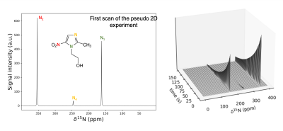 |
3 | Development of Hyperpolarized 15N-labeled Metronidazole as an MR Contrast Agent by d-DNP Technique: A Potential Antibiotic and Hypoxia Probe Video Not Available
David Guarin Bedoya1,2, Erin Elizabeth Hardy1, Anna Samoilenko3, Sameer Joshi3, Jason Stockmann1, Jan Henrik Ardenkjaer-Larsen2,4, Matthew S. Rosen1,5,6, Boyd M. Goodson7, Thomas Theis8, Eduard Y Chekmenev3,9, and Yi-Fen Yen1
1Radiology Department, Athinoula A. Martinos Center for Biomedical Imaging, Massachusttes General Hospital, Boston, MA, United States, 2Polarize ApS, Frederiksberg, Denmark, 3Department of Chemistry, Wayne State University, Detroit, MI, United States, 4Department of Electrical Engineering, Technical University of Denmark, Kongens Lyngby, Denmark, 5Radiology Department, Harvard Medical School, Boston, MA, United States, 6Physics Department, Havard University, Boston, MA, United States, 7School of Chemical and Biomolecular Sciences, Southern Illinois University, Carbondale, IL, United States, 8Department of Chemistry, North Carolina State University, Raleigh, NC, United States, 9Russian Academy of Sciences, Moscow, Russian Federation
In this work, we developed a DNP hyperpolarized 15N-labeled metronidazole contrast agent. The formulation of the sample and the DNP process were optimized to reduce the build-up time constant down to 22 min while reaching liquid state signal enhancements of 104. The T1 relaxation times were measured in liquid-state, and found to be minutes long. Due to the high enhancement and long T1, MNZ hyperpolarized 15N signal could be monitored for several minutes. The contrast agent was tested with MRS in-vivo, in a rat brain. The hyperpolarized MNZ signal remained above the noise level for more than a minute.
|
||
3527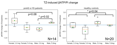 |
4 | A 31P MRS study of Target Engagement of Terazosin in Neurodegenerative Diseases
Jia Xu1, Sarah E Ernst2, Erin E Reasoner3, Stephen A Cross3, Alivia N Brinker3, Cameron A Keomanivong4, Zach Elias4, Daniel R. Thedens1, Nandakumar Narayanan4, Jordan L Schultz3, Michael J Welsh2, and Vincent A Magnotta1
1Radiology, University of Iowa, Iowa City, IA, United States, 2Internal Medicine, University of Iowa, Iowa City, IA, United States, 3Psychiatry, University of Iowa, Iowa City, IA, United States, 4Neurology, University of Iowa, Iowa City, IA, United States
Terazosin is an FDA-approved medicine that has been used primarily for men and data for women are missing. For the first time, the target engagement of Terazosin in neurodegenerative diseases was assessed for both sexes. We chose the βATP/Pi ratio as a biomarker to study the brain energy metabolism and found strong correlations between the in vivo brain ATP levels to whole blood ATP levels. We observed clear sex differences in response to different TZ doses in both mice and humans. Our data suggest that the optimal dose for females may need to be lower as compared to males.
|
||
3528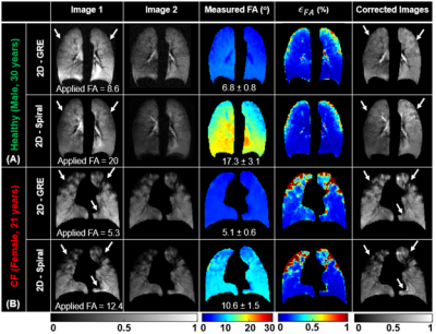 |
5 | B1-inhomogeneity Correction of Hyperpolarized 129Xe Lung Ventilation Imaging Using Spiral
Abdullah S. Bdaiwi1,2, Mariah L. Costa1,2, Joseph W. Plummer1,2, Matthew M. Willmering1, Laura L. Walkup1,2,3,4, and Zackary L. Cleveland1,2,3,4
1Center for Pulmonary Imaging Research, Cincinnati Children’s Hospital Medical Center, Cincinnati, OH, United States, 2Department of Biomedical Engineering, University of Cincinnati, Cincinnati, OH, United States, 3Department of Radiology, Cincinnati Children’s Hospital Medical Center, Cincinnati, OH, United States, 4Department of Pediatrics, University of Cincinnati, Cincinnati, OH, United States
Hyperpolarized 129Xe MRI can non-invasively measure regional ventilation by mapping the spin-density of inhaled gas, thus providing insights into regional disease pathophysiology. However, the quantitative accuracy of 129Xe ventilation imaging is reduced by B1-inhomogeneity causing spatial variations in both coil sensitivity and nonequilibrium magnetization decay. These can cause lung function impairment to be either over or underestimated. We demonstrate these artifacts can be mitigated by generating flip angle maps from paired 2D-spiral images acquired in the same held breath. This yields quantitatively comparable results to those obtained with a conventional, 2D gradient recalled echo sequence in substantially reduced acquisition times.
|
||
3529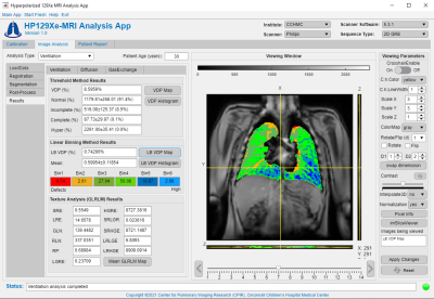 |
6 | User-Friendly Application for Consistent Multi-Vendor Hyperpolarized 129Xe MRI Analysis
Abdullah S. Bdaiwi1,2, Joseph W. Plummer1,2, Matthew M. Willmering1, David J. Roach1, Jason C. Woods1,3,4, Laura L. Walkup1,2,3,4, and Zackary L. Cleveland1,2,3,4
1Center for Pulmonary Imaging Research, Cincinnati Children’s Hospital Medical Center, Cincinnati, OH, United States, 2Department of Biomedical Engineering, University of Cincinnati, Cincinnati, OH, United States, 3Department of Radiology, Cincinnati Children’s Hospital Medical Center, Cincinnati, OH, United States, 4Department of Pediatrics, University of Cincinnati, Cincinnati, OH, United States Advances hyperpolarized 129Xe production, data acquisition and image analysis have allowed 129Xe MRI to emerge as a leading pulmonary imaging modality. As the number of sites capable of 129Xe MRI and quantitative image-derived metrics grow, the need for an intuitive, organized analysis tool increases. Here, we provide a user-friendly application, designed as MATLAB language-based interface, to analyze HP 129Xe data. This open-source, research tool enables standardized analysis of hyperpolarized 129Xe calibration, ventilation, diffusion, and gas-exchange data. Furthermore, the modular design allows developers to easily implement additional functions and analysis procedures, providing a framework for growth and sharing of analysis techniques. |
||
3530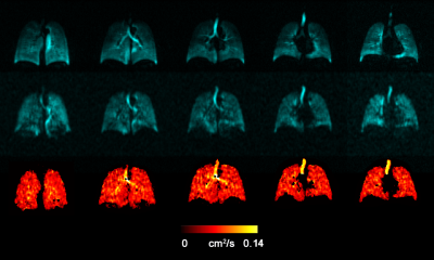 |
7 | Feasibility of the Ventilatory ADC Approach Using Hyperpolarized 129Xe Pulmonary MRI
Elnaz Parniyany1, Elise Woodward 1, Tingting Wu1, Matthew S Fox1,2, and Alexei Ouriadov1,2,3
1Physics and Astronomy, The University of Western Ontario, London, ON, Canada, 2Lawson Health Research Institute, London, ON, Canada, 3School of Biomedical Engineering, Faculty of Engineering, The University of Western Ontario, London, ON, Canada
Hyperpolarized 129Xe lung MRI is an efficient technique used to investigate and assess pulmonary diseases. However, the longitudinal observation of the emphysema progression using hyperpolarized gas MRI-based ADC can be problematic, as the disease-progression can lead to increasing unventilated-lung areas, which likely excludes the largest ADC estimates. One solution to this problem is to combine static-ventilation and ADC measurements following the idea of 3He MRI ventilatory ADC (vADC). We have demonstrated this method adapted for 129Xe MRI to help overcome the above-mentioned shortcomings and provide an accurate assessment of the emphysema progression.
|
||
3531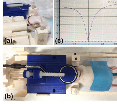 |
8 | Inductively Coupled Floating Coil for Hyperpolarized 13C Imaging of Mouse Brain
Atsushi M. Takahashi1, David Guarin2,3, Erin E. Hardy3, Julie Miller4, Hiroaki Wakimoto5, Daniel P. Cahill5, and Yi-Fen Yen3
1Department of Brain and Cognitive Sciences, McGovern Institute for Brain Research, Massachusetts Institute of Technology, Cambridge, MA, United States, 2Polarize ApS, Frederiksberg, Denmark, 3Department of Radiology, Athinoula A. Martinos Center for Biomedical Imaging, Massachusetts General Hospital, Charlestown, MA, United States, 4Department of Neurology, Massachusetts General Hospital, Boston, MA, United States, 5Department of Neurosurgery, Massachusetts General Hospital, Boston, MA, United States
We report an inductively coupled floating coil designed for hyperpolarized 13C MR spectroscopic imaging of mouse brain, with emphases on the simplicity of the circuit design, the ease of use, whole-brain coverage, and high SNR obtained. The floating coil is designed to “crown” the mouse head for a snug fit to achieve full coverage of the brain with good sensitivity. Here, we demonstrated the coil’s performance in normal mouse and glioblastoma multiforme mouse model at 4.7 T using a next-generation hyperpolarizer. High signal to noise ratio exceeding 70:1 was obtained in the brain with good spatial resolution (1.53mm x 1.53mm).
|
||
3532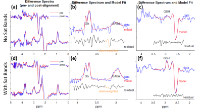 |
9 | Feasibility of Simultaneous Measurements of GABA, Glx and GSH in the Thalamus Using HERMES
Xiao Liang1, Muhammad G Saleh1, Steve Roys1, Rao P Gullapalli1, and Jiachen Zhuo1
1Department of Diagnostic Radiology and Nuclear Medicine, University of Maryland School of Medicine, Baltimore, MD, United States It is desirable to measure GABA, glutamate, and GSH in the thalamus for understanding pathological and pharmacokinetic changes. In this study, we study the feasibility of simultaneous measurements of these metabolites in the thalamus using HERMES. HERMES data were acquired on 20 healthy adult volunteers with and without saturation bands. HERMES data were processed in Gannet with and without spectral alignment. The results demonstrated the feasibility of using HERMES for simultaneous measurements of GABA, Glx and GSH in the thalamus. HERMES combined with saturation bands and retrospective spectral alignment yields high SNR spectra and reproducible measurements in the thalamus. |
||
3533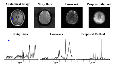 |
10 | Robust Low-rank Denoising of MRSI Data
Wen Jin1,2, Yibo Zhao1,2, Rong Guo1,2, Yudu Li1,2, Jie Luo3, Yao Li3, and Zhi-Pei Liang1,2
1Beckman Institute for Advanced Science and Technology, University of Illinois at Urbana-Champaign, Urbana, IL, United States, 2Department of Electrical and Computer Engineering, University of Illinois at Urbana-Champaign, Urbana, IL, United States, 3School of Biomedical Engineering, Shanghai Jiao Tong University, Shanghai, China
With the increasing use of MRSI in research and clinical applications, we have seen a surge in the use of low-rank models for denoising MRSI data. This paper presents a novel method for robust denoising of MRSI data corrupted by both Gaussian noise and nuisance signals. The proposed method has been validated using both simulated and in vivo data, producing impressive results. This method will further enhance the practical utility of low-rank denoising.
|
||
3534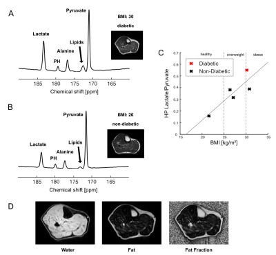 |
11 | Skeletal Muscle Metabolism in Obesity Measured by Hyperpolarized [1-13C]pyruvate
Jun Chen1, Junjie Ma1, James Ratnakar1, Shawn C Burgess2,3, Craig R Malloy1,4,5, and Jae Mo Park1,4,6
1AIRC, UT Southwestern Medical Center at Dallas, Dallas, TX, United States, 2Center for Human Nutrition, UT Southwestern Medical Center at Dallas, Dallas, TX, United States, 3Pharmacology, UT Southwestern Medical Center at Dallas, Dallas, TX, United States, 4Radiology, UT Southwestern Medical Center at Dallas, Dallas, TX, United States, 5Internal Medicine, UT Southwestern Medical Center at Dallas, Dallas, TX, United States, 6Electrical and Computer Engineering, University of Texas at Dallas, Richardson, TX, United States Obesity is associated with insulin resistance and chronic inflammation, both of which are known to affect skeletal muscle metabolism. However, studies in the impact of obesity on metabolism mainly focus on adipose tissue, instead of skeletal muscle. This study analyzes the in vivo pyruvate metabolism in the skeletal muscle of obese volunteers with hyperpolarized [1-13C]pyruvate. The results indicate increased lactate production and decreased pyruvate dehydrogenase activity in the skeletal muscle of obesity. |
||
3535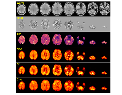 |
12 | Rapid Whole Brain High-Resolution MR Spectroscopic Imaging at 7T
Rong Guo1,2, Yudu Li1,2, Yibo Zhao1,2, Sina Tafti2,3, Aaron Anderson2, Brad Sutton1,2,4, and Zhi-Pei Liang1,2
1Department of Electrical and Computer Engineering, University of Illinois at Urbana-Champaign, Urbana, IL, United States, 2Beckman Institute for Advanced Science and Technology, University of Illinois at Urbana-Champaign, Urbana, IL, United States, 3Siemens Medical Solutions, Inc., Urbana, IL, United States, 4Department of Bioengineering, University of Illinois at Urbana-Champaign, Urbana, IL, United States
Metabolic imaging of the whole brain is desirable in many neuroscience studies and some clinical applications. This work demonstrates whole-brain MRSI (FOV = 240×240×160 mm3) at 7T using a special SPICE-based data acquisition and processing scheme. Experimental results showed that whole brain metabolic imaging at 3.0×3.0×3.2 mm3 resolution could be obtained in an 8-minute scan at 7T.
|
||
3536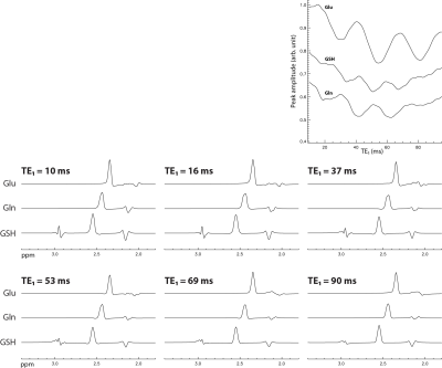 |
13 | Using Strong Coupling Effect to Increase Signal Strength of Glutamate, Glutamine, and Glutathione at 7 T
Li An1, Jennifer Wai Evans1, Courtney Burton1, Jyoti Tomar1, Maria Ferraris Araneta1, Carlos A Zarate1, and Jun Shen1
1National Institute of Mental Health, National Institutes of Health, Bethesda, MD, United States
Unlike weakly coupled spin systems, the signal strength of Glu H4, Gln H4, and GSH glutamyl H4 is affected by the timing of refocusing pulses for a fixed total TE. Using full density matrix simulations, it was found that glutamate H4, glutamine H4, and glutathione glutamyl H4 peak amplitudes can be significantly increased by shortening the echo time of the first spin echo (TE1) in PRESS from a previously reported 69 ms to 16 ms. The improved sequence was tested in vivo and validated using Monte Carlo simulations.
|
||
3537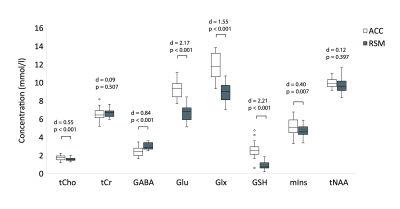 |
14 | Is tissue composition driving regional variation in neurometabolite levels?
Julie M Joyce1,2,3,4, Marilena DeMayo1,2,3,5,6, Chantel T Debert2,3,4,7, and Ashley D Harris1,2,3,4
1Department of Radiology, University of Calgary, Calgary, AB, Canada, 2Hotchkiss Brain Institute, University of Calgary, Calgary, AB, Canada, 3Alberta Children's Hospital Research Institute, University of Calgary, Calgary, AB, Canada, 4Integrated Concussion Research Program, University of Calgary, Calgary, AB, Canada, 5Department of Psychiatry, University of Calgary, Calgary, AB, Canada, 6Mathison Centre for Mental Health Research and Education, University of Calgary, Calgary, AB, Canada, 7Department of Clinical Neurosciences, University of Calgary, Calgary, AB, Canada
Metabolite concentrations vary across brain regions. What remains unclear is the degree to which this variation is due to inherent regional differences or if it is driven by the composition of the underlying tissue. Using magnetic resonance spectroscopy, we show that apparent ‘regional differences’ in metabolite concentrations are driven by tissue composition for glutamate and glutathione. Furthermore, we provide evidence to suggest a need for a tissue correction for glutamate, glutathione and N-acetyl-aspartate that appropriately addresses differences in voxel gray matter and white matter content.
|
||
3538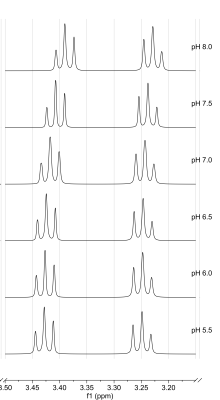 |
15 | pH-dependence of taurine 1H NMR chemical shifts: A potential method for pH determination and imaging Video Permission Withheld
Felizitas Charlotte Wermter1, Wolfgang Dreher1, and Christian Bock2
1in-vivo MR, University Bremen, Bremen, Germany, 2Alfred Wegener Institute Helmholtz Centre for Polar and Marine Research, Bremerhaven, Germany
Magnetic resonance spectroscopy (MRS) allows non-invasive pH measurements via the chemical shift difference between a pH-dependent resonance and a reference. We studied the pH dependence of taurine chemical shifts in 1H MRS for a wide range of pH and temperature. At 37°C and in the physiological range (pH ~7.3), the chemical shifts show a bijective dependence on pH. This approach was evaluated by measurements on taurine phantoms using solutions with different pH and temperatures, demonstrating the possibility of absolute pH determination via the chemical shift of taurine.
|
||
3539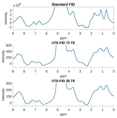 |
16 | Spatially resolved FID spectroscopy using a 3D spiral UTE imaging sequence
Anja Maria Fischer1,2, Petros Martirosian1, and Fritz Schick1
1Section on Experimental Radiology, University Hospital Tübingen, Tübingen, Germany, 2Institute for Diabetes Research and Metabolic Diseases, Helmholtz Center Munich at the University of Tübingen, Tübingen, Germany
This method allows for the calculation of pixel-wise FID spectra based on a 3D spiral UTE imaging sequence. Images were acquired for 36/72 TE and extrapolated. The method is advantageous for rapidly decaying 1H signals such as those of collagen in tendons or fibrotic tissue. A phantom (6 collagen solutions) served for evaluation. Resulting spectra are comparable to a standard non-localized FID spectrum and show positive correlation between collagen concentration and signal. First in-vivo results recorded in the subcutaneous adipose tissue exhibit the typical fat resonances. The presented UTE-FID spectroscopy seems feasible for spatially resolved applications of rapidly decaying signals.
|
||
 |
3540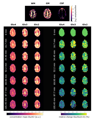 |
17 | Glutamate Metabolism Mapping after Oral Administration of 1-13C-Glc using 1H MRSI in the human brain at 9.4 T
Theresia Ziegs1, Loreen Ruhm1, Andrew Wright1, and Anke Henning1,2
1High-Field MR Center, Max Planck Institute for Biological Cybernetics, Tuebingen, Germany, 2Advanced Imaging Research Center, University of Texas Southwestern Medical Center, Dallas, TX, United States
Glutamate is the major excitatory neurotransmitter in the brain and malfunction of glutamatergic metabolism is associated with various neurological disorders. Respective metabolic rates can be determined via direct 13C or indirect 1H-[13C] MRS after intake of 13C labelled tracers. In this work, labeling effects after oral intake of [13C-1]glucose are observed in human brain using a 1H-FID-MRSI sequence at 9.4T. Spectral time series of GM and WM voxels as well as metabolite maps show regionally distinct and tissue type specific labelling induced changes of Glu and Gln spectral pattern that allow for the determination of 13C label uptake rates.
|
|
3541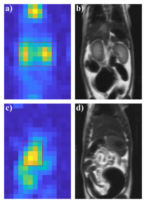 |
18 | Accurate thermometry using hyperpolarized 129Xe free induction decay and spin-echo chemical shift imaging in rats
Agilo L Kern1,2, Marcel Gutberlet1,2, Regina Rumpel3, Inga Brüsch3, Jens M Hohlfeld2,4,5, Frank Wacker1,2, and Bennet Hensen1
1Institute of Diagnostic and Interventional Radiology, Hannover Medical School, Hannover, Germany, 2Biomedical Research in Endstage and Obstructive Lung Disease Hannover (BREATH), German Center for Lung Research (DZL), Hannover, Germany, 3Institute for Laboratory Animal Science and Central Animal Facility, Hannover Medical School, Hannover, Germany, 4Department of Clinical Airway Research, Fraunhofer Institute for Toxicology and Experimental Medicine, Hannover, Germany, 5Department of Respiratory Medicine, Hannover Medical School, Hannover, Germany Thermometry using the proton resonance frequency (PRF) is challenging in inhomogeneous, fatty tissues and moving organs. Purpose of this study was to develop and test a combined free induction decay (FID)/spin-echo chemical shift imaging sequence for accurate hyperpolarized 129Xe MR thermometry in rats exposed to hypothermia. Significant changes of 129Xe lipid chemical shift and good agreement with core body temperature are observed. Bland–Altman analysis shows narrower limits of agreement and reduced mean difference for spin-echo acquisitions, suggesting improved precision and accuracy in fatty tissues. The method may be valuable as reference standard for development of more accurate 1H MR thermometry. |
||
3542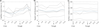 |
19 | Changes in lung microstructure during the cardiac cycle assessed by rapid hyperpolarized 129Xe chemical shift saturation recovery
Agilo L Kern1,2, Marcel Gutberlet1,2, Jens M Hohlfeld2,3,4, Frank Wacker1,2, and Jens Vogel-Claussen1,2
1Institute of Diagnostic and Interventional Radiology, Hannover Medical School, Hannover, Germany, 2Biomedical Research in Endstage and Obstructive Lung Disease Hannover (BREATH), German Center for Lung Research (DZL), Hannover, Germany, 3Department of Clinical Airway Research, Fraunhofer Institute for Toxicology and Experimental Medicine, Hannover, Germany, 4Department of Respiratory Medicine, Hannover Medical School, Hannover, Germany
A rapid chemical shift saturation recovery (CSSR) pulse sequence is developed for reconstructing CSSR uptake curves in different cardiac phases by employing retrospective gating. The sequence is tested in a study with 6 healthy volunteers and compared to conventional CSSR. A significant difference in septal wall thickness between trough and crest of the pulse wave in lung tissue is found. Results from rapid and conventional CSSR are strongly correlated and show good absolute agreement. The proposed rapid CSSR pulse sequence may provide valuable new insights into normal lung function, improve precision of CSSR results and may have clinical applications.
|
||
3543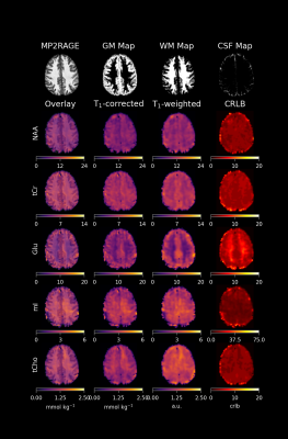 |
1 | Quantitative Metabolite Mapping of 12 Metabolites in the Human Brain Video Not Available
Andrew Martin Wright1,2 and Anke Henning1,3
1MRZ, Max Planck Institute for Biological Cybernetics, Tübingen, Germany, 2IMPRS for Cognitive and Systems Neuroscience, Eberhard-Karls University of Tübingen, Tübingen, Germany, 3Advanced Imaging Research Center, University of Texas Southwestern Medical Center, Dallas, TX, United States
Very short TR (TR < 300) MRSI is a popular method that enables fast acquisition of spectroscopic imaging data; however, raw metabolite signals following short TR acquisitions are influenced by strong T1-weighting. In order to gather more accurate images and to be able to relate MRSI results to SVS studies, it is valuable to correct for T1-weighting and to quantify MRSI images. This study presents results that utilize a voxel-specific, T1-correction method which is an advancement from more traditional methods that utilized average T1-corrections for metabolites. Fully sampled, single-slice 1H MRSI metabolite maps are presented for 12 metabolites.
|
||
 Back to Meeting Home
 Back to the Program-at-a-Glance
The International Society for Magnetic Resonance in Medicine is accredited by the Accreditation Council for Continuing Medical Education to provide continuing medical education for physicians.
 Back to Meeting Home
 Back to the Program-at-a-Glance
 View the Presentation